Step by Step Oral Radiology 1st Edition by Ram Kumar Srivastava ISBN B00INKKBK8 9789350250853
$50.00 Original price was: $50.00.$25.00Current price is: $25.00.
Authors:Jaypee Brothers; 1st edition (2011) , Author sort:edition, Jaypee Brothers; 1st
Step by Step Oral Radiology 1st Edition by Ram Kumar Srivastava – Ebook PDF Instant Download/Delivery. B00INKKBK8, 9789350250853
Full download Step by Step Oral Radiology 1st Edition after payment
Product details:
ISBN 10: B00INKKBK8
ISBN 13: 9789350250853
Author: Ram Kumar Srivastava
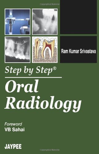
The book “Step by Step® Oral Radiology” elucidates the basic and practical knowledge in the field of dental radiography. It serves as a comprehensive guide for both undergraduate and postgraduate dental students. The book is divided into twenty seven different chapters dealing from the basic of radiography such as atomic structure, X-ray image characteristics, dose units, dosimetry and biological effects. Suitable diagrams and photographs are included with each and every topic for better understanding. Different radiographic techniques such as extraoral radiography, bitewing and occlusal radiography, cephalometric radiography, radiography of the temporomandibular joint and intraoral radiographs are dealt in detail. Benign and malignant tumors associated with jaws, bone related diseases and their radiographic appearances are covered. Cone beam CT, MRI, ultra sonography and scintigraphy are different types of digital imaging which uses electronic sensors for recording the penetration of X-ray photon. Dental caries, periodontal disease and developmental anomalies of teeth, radiolucent lesions of the jaws, radiopaque lesions in the jaws and facial skeleton are elucidated in this book.
Step by Step Oral Radiology 1st Table of contents:
Chapter 1: Introduction to Oral Radiology
- 1.1 History and Evolution of Oral Radiology
- 1.2 The Role of Radiology in Dentistry
- 1.3 Basic Radiologic Principles
- 1.4 The Importance of Radiographs in Diagnosis and Treatment Planning
- 1.5 Radiation Safety: ALARA and Protection Guidelines
Chapter 2: Basic Radiographic Techniques
- 2.1 Understanding Radiographic Equipment
- 2.2 Intraoral Radiography: Periapical and Bitewing Techniques
- 2.2.1 Step-by-Step Guide to Periapical Radiographs
- 2.2.2 Step-by-Step Guide to Bitewing Radiographs
- 2.3 Extraoral Radiography: Panoramic and Cephalometric Techniques
- 2.3.1 Step-by-Step Guide to Panoramic Imaging
- 2.3.2 Step-by-Step Guide to Cephalometric Radiographs
- 2.4 Digital Radiography: Key Concepts and Techniques
Chapter 3: Radiographic Anatomy for the Dental Professional
- 3.1 Overview of Normal Radiographic Anatomy
- 3.2 Identifying Key Structures: Teeth, Bone, and Soft Tissues
- 3.3 Anatomy of the Maxilla and Mandible
- 3.4 Identifying Normal and Abnormal Landmarks in Radiographs
Chapter 4: Radiographic Interpretation and Diagnostic Process
- 4.1 Step-by-Step Guide to Interpreting Radiographs
- 4.1.1 Identifying Normal Structures
- 4.1.2 Recognizing Abnormalities
- 4.2 Common Pathologies and Their Radiographic Features
- 4.2.1 Caries Detection and Progression
- 4.2.2 Periodontal Disease
- 4.2.3 Periapical Pathologies (e.g., Abscesses, Granulomas)
- 4.2.4 Endodontic Issues (e.g., Root Canal Problems)
- 4.2.5 Oral Pathologies (e.g., Tumors, Cysts)
Chapter 5: Advanced Imaging Techniques in Oral Radiology
- 5.1 Cone Beam Computed Tomography (CBCT): Step-by-Step Imaging
- 5.1.1 Basic Principles and Applications
- 5.1.2 Step-by-Step CBCT Protocols
- 5.2 Magnetic Resonance Imaging (MRI) and Its Role in Dentistry
- 5.3 Ultrasound Imaging in Oral Radiology
- 5.4 Emerging Technologies in Digital Imaging
Chapter 6: Radiology in Endodontics
- 6.1 The Role of Radiographs in Endodontic Diagnosis
- 6.2 Step-by-Step Endodontic Radiographic Techniques
- 6.3 Radiographic Indicators of Endodontic Disease
- 6.4 Post-Endodontic Imaging and Treatment Evaluation
Chapter 7: Radiology in Orthodontics
- 7.1 Overview of Orthodontic Imaging Needs
- 7.2 Step-by-Step Guide to Cephalometric Radiographs for Orthodontics
- 7.3 Panoramic Radiographs in Orthodontics
- 7.4 Use of CBCT in Orthodontic Treatment Planning
Chapter 8: Radiology in Periodontics
- 8.1 Radiographic Techniques for Periodontal Disease Diagnosis
- 8.2 Step-by-Step Guide to Periodontal Radiographic Evaluation
- 8.3 Identifying Bone Loss and Assessing Periodontal Health
- 8.4 Radiographs for Implant Assessment
Chapter 9: Pediatric Radiology
- 9.1 Special Considerations for Pediatric Radiographs
- 9.2 Step-by-Step Guide to Pediatric Intraoral Radiography
- 9.3 Common Pediatric Pathologies and Radiographic Features
- 9.4 Managing Pediatric Patients in the Radiology Setting
Chapter 10: Geriatric and Special Needs Radiology
- 10.1 Considerations for Geriatric Dental Radiology
- 10.2 Radiographic Techniques for Special Needs Patients
- 10.3 Managing Complex Cases and Treatment Planning
- 10.4 Considerations for Safe and Effective Radiology in Older Adults
Chapter 11: Radiologic Diagnosis of Common Oral Pathologies
- 11.1 Step-by-Step Identification of Oral Pathologies
- 11.1.1 Tumors and Cysts
- 11.1.2 Infections and Abscesses
- 11.1.3 Bone Diseases and Disorders
- 11.2 Interpreting Radiographs in the Context of Clinical Symptoms
- 11.3 Differential Diagnosis Using Radiographic Findings
Chapter 12: Radiation Safety and Infection Control
- 12.1 Radiation Protection Techniques for Patients and Operators
- 12.2 Infection Control in the Radiology Suite
- 12.3 Proper Handling and Disposal of Radiographic Materials
- 12.4 Legal and Ethical Issues in Radiography
Chapter 13: Future Trends in Oral Radiology
- 13.1 Advances in Digital Imaging
- 13.2 Artificial Intelligence and Its Impact on Radiology
- 13.3 The Future of 3D Imaging and Cone Beam CT
- 13.4 Trends in Radiation Safety and Protection
People also search for Step by Step Oral Radiology 1st:
step by step oral radiology
definition of oral radiology
history of oral radiology
radiology oral boards
radiology steps
You may also like…
eBook PDF
Microsoft Excel 2016 Step by Step 1st Edition by Curtis Frye ISBN 0735697469 9780735697461
eBook PDF
Microsoft ADO Net Step By Step 1st edition by Rebecca Riordan ISBN 0735612366 9780735612365

