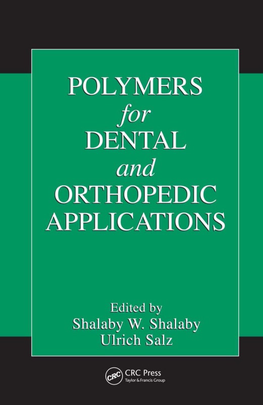
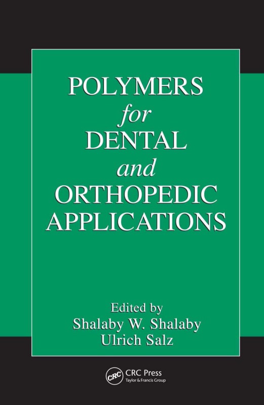
Polymers for Dental and Orthopedic Applications 1st Edition by Shalaby W Shalaby, Ulrich Salz ISBN 1040062962 9781040062968
$50.00 Original price was: $50.00.$25.00Current price is: $25.00.
Authors:Shalaby, Shalaby W. , Author sort:Shalaby, Shalaby W. , Published:Published:Oct 2006
Polymers for Dental and Orthopedic Applications 1st Edition by Shalaby W Shalaby, Ulrich Salz – Ebook PDF Instant Download/Delivery. 1040062962, 9781040062968
Full download Polymers for Dental and Orthopedic Applications 1st Edition after payment
Product details:
ISBN 10: 1040062962
ISBN 13: 9781040062968
Author: Shalaby W Shalaby, Ulrich Salz
Recent advances not only in the creation of new polymers but also in their processing and production have ushered in huge strides in a variety of biomedical and clinical areas. Orthopedics and dentistry are two such areas that benefit immensely from developments in polymer science and technology. Polymers for Dental and Orthopedic Applications.
Polymers for Dental and Orthopedic Applications 1st Table of contents:
Chapter 1 Events Propelling the Use of Polymers in Dental and Orthopedic Applications
Contents
1.1 Introduction
1.2 Technological Events
1.2.1 Development of New Synthetic, Biosynthetic, and Modified Natural Polymers
1.2.2 Development of New Processes to Modulate Properties of Biomedical Polymers and Implants
1.2.3 Advanced Fiber and Microfiber Processing
1.3 Medically Related Events
1.3.1 Clinically Related Events
1.3.2 Biomedical Engineering Driven Events
1.3.3 Damage-Control Associated Events
1.4 Socioeconomic Events
1.5 Conclusion And Perspective on the Future
References
Section B Development and Application of New Systems
Chapter 2 Composites for Dental Restoratives
Contents
2.1 Introduction
2.1.1 General Description of Dental Restoratives
2.1.2 State of the Art of Dental Restoratives
2.1.3 Improvements of Dental Composites
2.2 Filler-Component-Based Improvements of Dental Restoratives
2.2.1 Reduction of Shrinkage Stress
2.2.2 Nanoparticles and Composite Reinforcement
2.2.3 Condensable and Flowable Composites
2.2.4 Bioactive Restoratives
2.2.5 Special Additives
2.3 New Monomers For Dental Restoratives
2.3.1 Ring-Opening Monomers
2.3.1.1 Causes of Polymerization Shrinkage
2.3.1.2 Shrinkage Reduction by Ring-Opening Polymerization of Cyclic Monomers
2.3.1.3 Spiro Orthocarbonates
2.3.1.4 Cationic Ring-Opening Polymerization of SOCs and Cyclic Ethers
2.3.1.5 Cyclic Acetales, Allyl Sulfides, and Vinylcyclopropanes
2.3.2 Liquid-Crystalline Monomers
2.3.2.1 Liquid-Crystalline State and Liquid-Crystalline Polymers
2.3.2.2 Liquid-Crystalline Cross-linkers
2.3.3 Branched and Dendritic Monomers
2.3.3.1 Branched and Dendritic Polymers
2.3.3.2 Branched and Dendritic Methacrylates
2.3.4 Monomers for Compomers
2.3.5 Ormocers
2.3.6 Fluorinated Bis-Gma Analogs and Substitutes
2.3.6.1 Fluorinated Bis-GMA Analogs
2.3.6.2 Bis-GMA substitutes
2.4 Conclusion And Perspective on the Future
References
Chapter 3 Enamel and Dentin Adhesion
Contents
3.1 Introduction
3.1.1 Clinical Requirements
3.1.2 Postoperative Sensitivity
3.1.3 Reasons for the Replacement of Fillings
3.1.4 Evolution of the Adhesive Technique
3.2 Characterization and Pretreatment of Enamel and Dentin
3.2.1 Histology of Enamel and Dentin
3.2.1.1 Enamel
3.2.1.2 Dentin
3.2.1.3 Chemical and Histological Buildup of Sclerotic Modified Dentin
3.2.2 Chemical Composition
3.2.3 Structure of Hydroxyapatite
3.2.4 Structure of Collagen
3.2.5 Physical Properties of Enamel and Dentin
3.2.5.1 Anisotropy of Enamel
3.2.5.2 Anisotropy of Dentin
3.2.5.3 Comparison of Selected Properties of Enamel and Dentin
3.2.6 Pretreatment of Enamel and Dentin
3.2.6.1 Adhesion to Enamel
3.2.6.2 Adhesion to Dentin
3.2.6.2.1 Pretreatment with Acid: Total-Etch Bonding
3.2.6.2.2 Self-Etching Primers
3.3 Physical And Chemical Aspects of Adhesion
3.3.1 Basics of Adhesion
3.3.2 Wetting and Penetration of Dental Substrates
3.3.3 Adhesion by Micromechanical Retention
3.3.4 Adhesion by Chemical Bonding and Physical Forces
3.4 Methods For the Assessment of Enamel–DENTIN ADHESION
3.4.1 Bond Strength Measurements
3.4.1.1 Methods and Variables
3.4.1.1.1 Substrate
3.4.1.1.2 Surface Preparation
3.4.1.1.3 Storage Conditions/Stress Simulation
3.4.1.1.4 Test Performance
3.4.1.2 Shear Bond Strength
3.4.1.3 Push Out Test
3.4.1.4 Tensile Bond Strength
3.4.1.5 Microtensile Bond Strength
3.4.2 Marginal Adaptation
3.4.3 Morphology of the Interface
3.4.4 Interfacial Chemistry
3.5 Chemistry of Enamel And Dentin Adhesives
3.5.1 Introduction
3.5.2 Acidic Bonding Agents
3.5.2.1 Phosphorus-Containing Monomers
3.5.2.2 COOH-Group Containing Monomers and Polymerizable Carboxamides
3.5.2.3 Combinations of Aldehydes and Ketones and Monomers with Active Hydrogen
3.5.2.4 Cyanoacrylates, Isocyanates, and Cyanurate
3.6 Interactions Influencing Enamel–Dentin Adhesion
3.6.1 Saliva or Blood Contamination
3.6.2 Disinfecting Agents
3.6.3 Temporary Materials
3.6.4 Tooth Bleaching
3.7 Conclusion And Perspective on the Future
References
Chapter 4 Application of Polymers in the Therapy for Gingivitis, Periodontitis,and Treatment of Dry Socket
Contents
4.1 Introduction
4.2 General Approaches To the Therapy For Gingivitis And Periodontitis
4.2.1 General Approaches to the Therapy for Gingivitis
4.2.1.1 Chronic Gingivitis
4.2.1.1 Acute Gingival Diseases
4.2.1.3 Gingival Overgrowth
4.2.1.4 Role of Polymers in the Therapy for Gingivitis
4.2.2 General Approaches to Therapy for Periodontitis
4.2.2.1 Scaling and Root Planing
4.2.2.2 Surgical Therapy
4.2.2.3 Pharmacological Therapy
4.2.2.4 Periodontal Regeneration
4.2.2.5 Role of Polymers in the Therapy for Periodontitis
4.3 Evolution of Polymer Applications In the Therapy For Gingivitis And Periodontitis
4.3.1 Polymers for Controlled Drug Delivery
4.3.2 Use of Polymers in Conjunction with Tissue Regeneration
4.3.3 Pertinent Factors to the Effectiveness of Implemented Therapies
4.4 Dry Socket And Use of Polymeric Systems In Its Treatment
4.5 Conclusion And Perspective on the Future
References
Chapter 5 Maxillofacial Bone Augmentation and Replacement
Contents
5.1 Introduction
5.2 Bone Reconstruction And Healing
5.3 Osseous Defects In the Maxillofacial Bone
5.4 Methods For Maxillofacial Bone Augmentation
5.4.1 Procedures For Minor Maxillofacial Bone Augmentation
5.4.2 Autogenous Bone Grafts
5.4.3 Allogenic Bone Grafts
5.4.4 Xenogenic Bone Grafts
5.5 Synthetic Materials For Maxillofacial Bone Augmentation And Replacement
5.5.1 Tricalcium Phosphate And Hydroxyapatite
5.5.2 Active Glass Ceramics
5.5.3 Coral Derived Granules
5.5.4 Tissue Engineering Approaches Using Synthetic Materials
5.6 Other Methods For Maxillofacial Bone Augmentation
5.6.1 Distraction Osteogenesis
5.6.2 Ultrasonic Treatment
5.7 Conclusion And Perspective on the Future
References
Chapter 6 Polymeric Biomaterials for Articulating Joint Repair and Total Joint Replacement
Contents
6.1 Introduction
6.2 Evolution In Articulating Joint Repair And Total Replacement
6.2.1 Nonabsorbable Polymers, Composites, and Their Applications
6.2.2 Absorbable Polymers, Composites, and Their Applications
6.2.3 Injectable, Intra-Articular Polymeric Systems and Their Applications
6.2.4 Tissue Engineering
6.3 Evolution of Support Structures
6.3.1 Ligaments and Tendons
6.3.2 Suture Anchor and Allied Devices
6.4 Conclusion And Prospective on the Future
References
Section C Development in Preparative, Processing, and Evaluation Methods
Chapter 7 Physicochemical Properties of Bioactive Polymeric Composites: Effects of Resin Matrix and the Type of Amorphous Calcium Phosphate Filler
Contents
7.1 Introduction
7.2 Amorphous Calcium Phosphate (Acp): Chemistry And Importance In Dentistry
7.3 Acp Based Composites: Physicochemical And Biocompatibility Aspects
7.4 Methods And Techniques For Physicochemical Evaluation of Copolymers And Acp Composites
7.4.1 X-Ray Diffraction (XRD), Fourier Transform Infrared (FTIR) Spectroscopy, and FTIR-Microspectroscopy (FTIR-M)
7.4.2 Particle Size Distribution (PSD)
7.4.3 Scanning Electron Microscopy (SEM)
7.4.4 Thermogravimetry
7.4.5 Chemical Analysis
7.4.6 Polymerization Shrinkage (PS)
7.4.7 Mechanical Strength
7.4.8 Gravimetry and Water Sorption
7.4.9 Ion Release
7.4.10 Cytotoxicity Testing
7.5 Physicochemical Properties of Acp Fillers, Copolymers, and Composites
7.5.1 ACP Fillers: Structure, Morphology, and Particle Size Distribution
7.5.2 Copolymers and ACP Composites: Effects of Resin Matrix and Filler Type on Chemical and Mechanical Performance
7.5.3 ACP Composites: In Vitro Cytotoxicity
7.6 Conclusion And Perspective on the Future
References
Chapter 8 New Approaches to Improved Polymer Implant Toughness and Modulus
Contents
8.1 Introduction
8.2 New Approaches To Polymer Implants of Improved Toughness And Modulus
8.2.1 Composite Technology
8.2.1.1 Fiber Reinforcement
8.2.1.2 Particulate Reinforcement
8.2.1.3 Nanocomposites
8.2.2 Self-Reinforced Composites
8.2.2.1 Uhmwpe
8.2.2.2 Pmma
8.2.2.3 Biodegradable Polymers
8.2.3 Cross-Linking
8.2.3.1 Radiation-Induced Cross-Linking
8.2.3.2 Chemical-Induced Cross-Linking
8.2.4 Solid-State Orientation
8.3 Conclusion And Perspective on the Future
References
Chapter 9 Physicochemical Modification of Polymers for Bone-Implant Osseointegration
Contents
9.1 Introduction
9.2 Technology Evolution of Physicochemical Surface Modification And Bulk Orientation
9.2.1 Surface Sulfonation of Model Polyolefins
9.2.2 Surface Phosphonylation of Model Polyolefins And Polyether-Ether Ketone (Peek)
9.2.3 Surface Microtexturing of Model Polyolefins And Peek-Based Substrates
9.2.4 Orthogonal Solid-State Orientation of Crystalline Thermoplastic Polymers
9.3 Interaction of Cells With Chemically Modified Surfaces
9.3.1 Interaction of Bacteria
9.3.2 Interaction of Osteoblasts
9.4 Osseointegration of Phosphonylated Polyolefins And Peek Rods As Tibial Implants
9.4.1 Osseointegration of Chemically And Physicochemically Modified Polyethylene And Propylene Rods As Goat Tibial Implants
9.4.2 Osseointegration of Phosphonylated Peek Rods As Rabbit Tibial Implants
9.5 Osseointegration of Chemically And Physicochemically Modified Peek-Based Rods As Goat Mandibular Implants
9.5.1 Preparation of Implantable Sterilized Rods
9.5.2 In Vivo Study of Sterilized Rod Implants Using Goat Mandibles
9.5.2.1 Animal Model and Surgical Protocol
9.5.2.2 Harvesting of Implants in Bone and Their Biomechanical Testing
9.5.2.3 Histological Evaluation
9.5.2.4 Outcome of the In Vivo Study
9.6 Osseointegration of Phosphonylated Peek And Cfr-Peek Endosteal Dental Implants
9.6.1 Development of A New Endosteal Dental Implant Design For Evaluation In Beagles
9.6.2 Development of A Beagle Animal Model And Pilot Study of the Edi
9.6.2.1 Beagle Mandibular Model and Placement Protocols
9.6.2.2 Pilot Study of the EDIs
9.6.3 Biomechanical Evaluation of Osseointegration of Metallic And Peek-Based Edis
9.6.4 Histomorphometric Evaluation of Osseointegration of Metallic And Peek-Based Edis
9.7 Conclusion And Perspective on the Future
References
Chapter 10 Surface Modification of Articulating Surfaces and Tribological Evaluation
Contents
10.1 Introduction
10.2 Total Joint Replacement (TJR) Prostheses — Uhmwpe And Cocr Alloy Components
10.2.1 Cocr Femoral Component
10.2.2 Ultra High Molecular Weight Polyethylene (Uhmwpe) Tibial Component
10.3 Wear of Uhmwpe Component In TJR Prosthesis
10.3.1 Clinical Relevance of Uhmwpe Wear Debris
10.3.2 Factors Related To Uhmwpe Wear Debris Generation
10.4 Friction And Wear of Uhmwpe At A Nanoscale
10.4.1 Nanotribology of Cocr-Uhmwpe Tjr Prosthesis Using Atomic Force Microscopy
10.4.2 Effects of Sample Preparation Temperature on Nanostructure of Uhmwpe
10.4.3 Nanomechanical Properties of Compression Molded Uhmwpe
10.5 Surface Modification of Polymers
10.5.1 Thickness of Surface Modification
10.5.2 Surface Rearrangement
10.6 Classification of Surface Modifications
10.6.1 Chemical Modification
10.6.2 Physical Modification — Ultraviolet Radiation
10.6.3 Physical Modification — Electron Beam Radiation
10.6.4 Physical Modification — Gamma Irradiation
10.6.5 Physical Modification — Ion Beam Implantation
10.6.6 Physical Modification — Plasma
10.6.7 Physical Modification — Glow Discharge
10.7 Biologically Functional Modifications of Polyethylene
10.8 Miscellaneous Surface Modifications
10.9 Concerns of Surface Modification
10.10 Conclusion And Perspective on the Future
References
Chapter 11 Histopathological Evaluation of Hard Tissue Implant Performance
Contents
11.1 Introduction
11.2 General Pathology
11.3 Histopathological Evaluation
11.3.1 Hard Tissue Histological Preparation
11.3.2 Histopathological Evaluation of Hard Tissue Integration With Polymer Implant Device
11.3.2.1 Histopathological Evaluation of Articular Cartilage Repair
11.3.2.2 Histopathology Evaluation of Bone Void Repair
11.4 Recent Advances In Evaluating Hard Tissues
11.4.1 Cartilage Repair
11.4.1.1 Diffraction Enhanced Imaging (DEI)
11.4.1.2 Quantitative Magnetic Resonance Imaging (qMRI)
11.4.1.3 Optical Coherence Tomographic Imaging (OCT)
11.4.2 Bone Void Repair
11.4.2.1 Peripheral Quantitative Computed Tomography (pQCT)
11.4.2.2 Microcomputed Tomography (micro-CT)
11.5 Conclusion And Perspective on the Future
References
Section D Advanced Biomaterials, Technologies, and Sought Applications
Chapter 12 Mechanical Adaptation of Bone: Bioreactors for Orthopedic Tissue Engineering Applications
Contents
12.1 Introduction
12.2 Bone Tissue Development, Homeostasis, And Repair
12.2.1 Skeletal Tissue Development And Maintenance
12.2.2 Skeletal Cells And Their Functions
12.2.3 Relationship Between Physiological Loading And Skeletal Tissue
12.3 Mechanobiology of Bone
12.2.1 In Vivo Studies of Mechanobiology
12.3.2 In Vitro Studies of Mechanobiology
12.3.3 Traditional And Current Theories of Mechanobiology of Bone
12.4 Advances In Bioreactors For Bone Tissue Engineering Applications
12.4.1 Motivations And Functional Requirements
12.4.2 Current Designs of Bioreactors
12.4.3 Assessment of Performance of Bioreactors
12.5 Conclusion And Perspective on the Future
References
Chapter 13 Synthetic Scaffolds Used for Orthopedic Tissue Engineering
Contents
13.1 Introduction
13.2 Orthopedic Tissue Engineering
13.2.1 Bone Scaffolds
13.2.2 Ligament And Tendon Scaffolds
13.2.3 Articular Cartilage Scaffolds
13.3 Conclusion And Perspective on the Future
References
Chapter 14 Polymeric Controlled Release Systems for Management of Bone Infection
Contents
14.1 Introduction
14.1.1 Common Causes of Different Forms of Osteomyelitis And Their Diagnoses
14.1.2 Device-Centered Osteomyelitis
14.2 Critical Review of Current Treatment Options for Osteomyelitis
14.2.1 Systemic Treatment
14.2.2 Significance of Osteomyelitis And Need For New Approaches To Its Management
14.2.3 Local Drug Delivery
14.2.4 Staphylococcus Aureus Osteomyelitis — Special Considerations For Treatment
14.3 Evolution In Bone Infection Management And Development of New Controlled Drug Delivery Systems
14.3.1 Controlled Release Systems Using Traditional Absorbable And Water-Soluble Polymers
14.3.2 Controlled Release of Antibiotics From Absorbable Gel-Forming Formulations
14.3.3 Advances Toward New Approaches To the Treatment of Osteomyelitis
14.4 Conclusion And Perspective on the Future
People also search for Polymers for Dental and Orthopedic Applications 1st:
polymers for dental and orthopedic applications
dental polymers examples
polymer dental implants
polymers in dentistry
polymers used in dentistry
You may also like…
eBook PDF
Bionanomaterials for Dental Applications 1st edition by Mieczyslaw Jurczyk 9789814303842 9814303844
eBook PDF
Dental Functional Morphology How Teeth Work 1st Edition by Peter W Lucas ISBN 9780511735011

