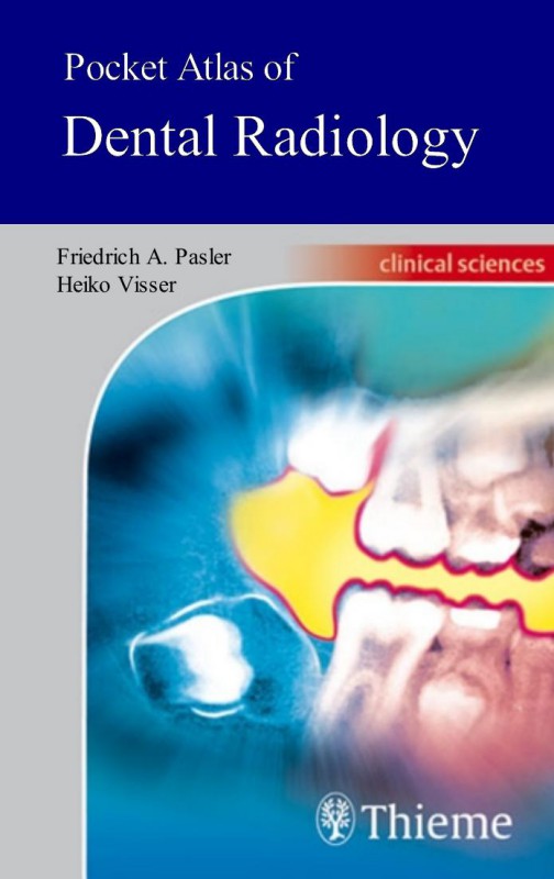
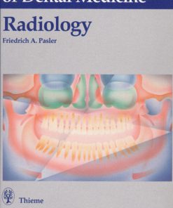
Pocket Atlas of Dental Radiology 1st Edition by Friedrich Pasler, Heiko Visser, Thomas Hassell ISBN 1588903354 9781588903358


Original price was: $50.00.$25.00Current price is: $25.00.
Authors:Thieme; 1st edition (May 23, 2007) , Tags:ANOTHER 3HAXAP RELEASE , Author sort:edition, Thieme; 1st , Published:Published:Oct 2007
Pocket Atlas of Dental Radiology 1st Edition by Friedrich Pasler, Heiko Visser, Thomas M. Hassell – Ebook PDF Instant Download/Delivery. 1588903354, 978-1588903358
Full download Pocket Atlas of Dental Radiology 1st Edition after payment

Product details:
ISBN 10: 1588903354
ISBN 13: 978-1588903358
Author: Friedrich Pasler, Heiko Visser, Thomas M. Hassell
In this age of highly specialized medical imaging, an examination of the teeth and alveolar bone is almost unthinkable without the use of radiographs. This highly informative and easy-to-read book with a collection of 798 radiographs, tables, and photos provides a myriad of problem-solving tips concerning the fundamentals of radiographic techniques, quality assurance, image processing, radiographic anatomy, and radiographic diagnosis. Information is easy to find, enabling the reader to literally get a grasp of essential new knowledge in next to no time. The dental practice team now has a pocket consultant at its fingertips, providing practical ways to incorporate new techniques into daily practice.
A fine-tuned didactic concept:Each topical concept is printed compactly on a double-page spreadOn the left: concise and highly instructive textOn the right: informative, high-quality illustrations
Main topics include:
- Examination strategies, radiation protection, quality assurance
- Conventional and digital radiographic techniques
- Radiographic anatomy: The problems of object localization and how to solve them
- Recent research with conventional radiography, CT, MRI, etc.
- Normal variations and pathologic conditions as viewed with the various imaging techniques
- A concise and up-to-date presentation of modern dental radiology
Pocket Atlas of Dental Radiology 1st Table of contents:
Radiographic Techniques and Imaging Modalities
-
Radiographic Technique, Radiographic Anatomy, Image Processing
- Panoramic Radiography
- Positioning and Positioning Errors
- Additional Programs for Standard Panoramic Radiography
- Radiographic Anatomy in Panoramic Radiographs
- Tooth and Jaw Development as Depicted in Panoramic Radiographs
-
Intraoral Dental Radiographs
- Apical and Periodontal Projections
- Depicting the Third Molars
- Bitewing Radiographs
- Occlusal Radiographs
- Technical Errors that Reduce the Quality of Intraoral Periapical Radiographs
- Radiographic Anatomy in Intraoral Radiographs
-
Conventional Skull Films and Radiographic Anatomy
- Posteroanterior Skull Projection, Overview
- Lateral Skull Projection, Overview
- Axial Skull Projection, Overview
- Waters Projection, Posteroanterior
- Posteroanterior Mandibular Overview (Reverse Towne’s Projection)
- Mandibular Radiograph, Half-Arch
- Lateral Cephalometric Radiograph, Facial Skeleton
- Temporomandibular Joint, Open and Closed Mouth
-
Computed Tomography and Magnetic Resonance Imaging
- Computed Tomography (CT)
- Radiographic Anatomy in CT
- Axial Mandibular CT, Radiographic Anatomy
- Axial Maxillary CT, Radiographic Anatomy
- Coronal CT of the TMJ, Radiographic Anatomy
- Dental CT Programs
- Magnetic Resonance Imaging (MRI)
- MRI: Functional Principles and Structure Signals
- Computed Tomography (CT)
-
Localization and Supplemental Radiographic Projections
- Localization Using Panoramic Radiographs
- Localization Using Intraoral Dental Radiographs
- Localization by Changing the Projection Direction of the Central Ray
- Localization with Supplemental Transversal Projections
- Localization Using Supplemental Skull Projections
- Depiction of Sialoliths
Radiographic Film and Digital Systems
6. Radiographic Film, Conventional Processing, and Processing Errors
- Emulsion Technique and Film Packet Construction
- Conventional Developing of Radiographs
- Film Processing Errors
- Radiography Using Digital Systems
- Digital Radiography
- Digital Image Processing
- Digital Sensors and Storage Phosphor Plates
- Image Quality of Digital Radiographs
- Radiation Exposure of the Patient
Radiographic Pathology
8. Radiographic Pathology and Dysmorphias
- Dysmorphias and Regressive Alterations
- Hypodontia, Hyperodontia
- Malformations of the Jaws
- Dentinogenesis Imperfecta, Taurodontism
- Amelogenesis Imperfecta, Cemental Hyperplasia
- Odontodysplasias
-
Impacted Teeth
- Regressive Alterations, Resorptions
- Calcifications, Concrements, Ossifications
- Dental Calculus, Concrements
- Sialoliths
- Sialoliths, Ossifications
-
Radiographic Diagnosis of Dental and Periodontal Pathology
- Radiographic Diagnosis of Dental Caries
- Radiographic Aspects of Caries Diagnosis
- Rules of Projection, Radiation Effects
- Radiographic Diagnosis of Periodontal Pathology and Inflammation within the Jaws
- Periodontal Bone Loss
- Marginal Periodontal Diseases
- Apical Periodontal Pathology
- Apical Periodontal Pathology, Osteomyelitis
- Osteomyelitis
- Sequestration and Osteoradionecrosis
- Diseases of the Maxillary Sinuses
- Diseases of the Maxillary Sinuses of Dentogenic and Rhinogenic Origin
- Dentogenic Infections
- Dentogenic/Rhinogenic Infections
- Rhinogenic Afflictions
- Rhinogenic Afflictions and Foreign Bodies
- Temporomandibular Joint Disorders
- Primary Arthropathies
- Secondary Arthropathies
Cysts, Tumors, and Other Pathologies
13. Jaw Cysts
- Classification of Cysts
- Developmentally Induced Odontogenic Cysts
- Developmentally Induced Nonodontogenic Cysts
- Inflammation-Induced Radicular Cysts
- Odontogenic and Nonodontogenic Tumors
- Odontogenic Tumors, Hamartoma, Dysplasias
- Ameloblastoma
- Ameloblastic Fibroma, Fibro-Odontoma
- Odontogenic Myxoma
- Calcifying Epithelial Odontogenic Tumor
- Calcifying Odontogenic Cysts, Adenomatoid Odontogenic Tumor
- Odontoma
- Cemento-Osseous Dysplasias
- Cementoblastoma
- Nonodontogenic Tumors and Tumor-Like Lesions
- Fibrous Dysplasia (Jaffé–Lichtenstein)
- Giant Cell Granuloma
- Eosinophilic Granuloma
- Pseudocysts of the Jaws
- Osteochondroma
- Ossifying Fibroma
- Osteoblastoma, Osteoid Osteoma
- Osteoma
- Exostosis, Gardner Syndrome, Paget Disease of Bone
- Osteogenesis Imperfecta, Osteoporosis, Osteopetrosis
- Hyperparathyroidism, Hemangioma
- Carcinoma, Sarcoma
- Sarcoma
- Metastases
Traumatology and Forensic Radiology
15. Traumatology of the Teeth and Jaws
- Tooth and Jaw Fractures
- Foreign Bodies and Forensic Significance of Radiographs
- Foreign Bodies and Forensically Important Radiographic Procedures
Further Readings and Resources
16. Further Reading and Medical Historical Literature
People also search for Pocket Atlas of Dental Radiology 1st:
what is pocket depth of a tooth
pocket atlas of oral diseases
atlas of dental radiography in dogs and cats
dental radiology atlas
pocket atlas of radiographic anatomy
You may also like…


eBook PDF
Pocket Atlas Of Oral Diseases 2nd Edition by George Laskaris ISBN 1588902498 9781588902498