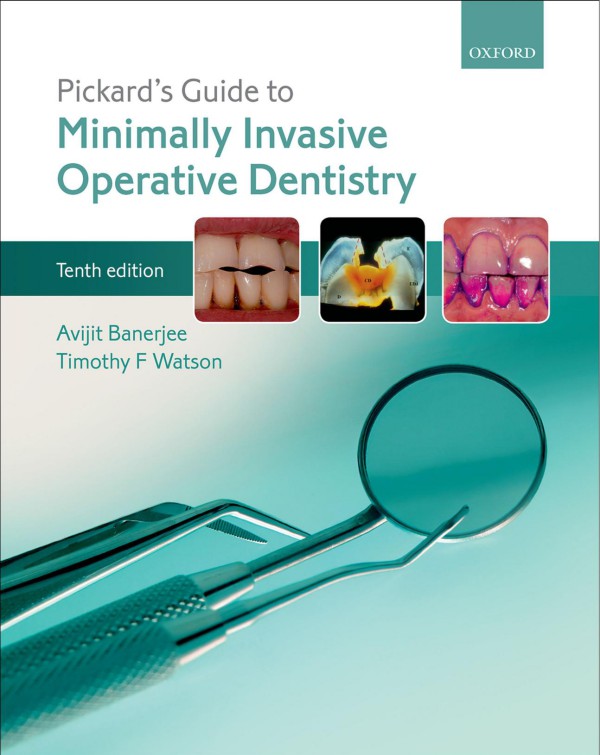
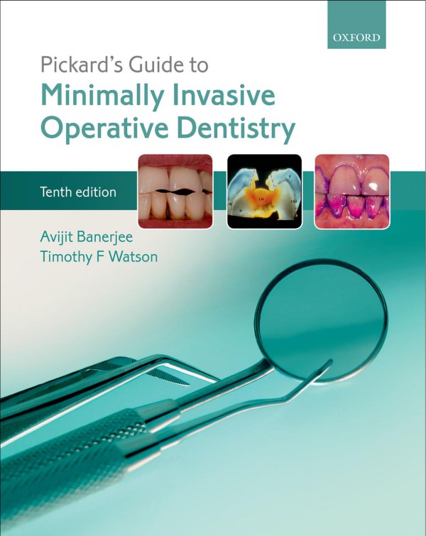
Pickard’s Guide to Minimally Invasive Operative Dentistry 10th Edition by Avit Banerjee, Timothy Watson ISBN 019871209X 9780198712091
$50.00 Original price was: $50.00.$25.00Current price is: $25.00.
Authors:Avijit Banerjee; Timothy F. Watson , Series:Dentistry [184] , Tags:Medical; Dentistry; Oral Surgery , Author sort:Banerjee, Avijit & Watson, Timothy F. , Ids:Google; 9780198712091 , Languages:Languages:eng , Published:Published:Aug 2015 , Publisher:Oxford University Press , Comments:Comments:An ideal introduction to the theory and practical aspects of conservative dentistry, the tenth edition of Pickards’ Guide to Minimally Invasive Operative Dentistry is a must-have text for all dental students, new graduates and oral healthcare professionals alike. Written in an easy tounderstand and concise style, the authors introduce the essentials of dental disease before outlining how to collect patient information clinically in order to detect, diagnose, plan and deliver care. Exploring key topics such as disease prevention and control, the principles of minimally invasive operative dentistry, contemporary restorative materials and procedures, this completely up-to-date revised edition integrates a thorough academic grounding for degree examination with an essentialpreparation for clinical practice for the whole oral healthcare team. Illustrated with step-by-step colour photos, common clinical procedures are clearly set out and labelled for beginners to learn. The tenth edition has been updated to reflect the latest evidence based guidelines forpreventitative management and there is a focus on maintaining existing restorations and follow up/long term care.
Pickard’s Guide to Minimally Invasive Operative Dentistry 10th Edition by Avit Banerjee, Timothy F. Watson – Ebook PDF Instant Download/Delivery. 019871209X, 978-0198712091
Full download Pickard’s Guide to Minimally Invasive Operative Dentistry 10th Edition after payment
Product details:
ISBN 10: 019871209X
ISBN 13: 978-0198712091
Author: Avit Banerjee, Timothy F. Watson
An ideal introduction to the theory and practical aspects of conservative dentistry, the tenth edition of Pickards’ Guide to Minimally Invasive Operative Dentistry is a must-have text for all dental students, new graduates and oral healthcare professionals alike. Written in an easy to understand and concise style, the authors introduce the essentials of dental disease before outlining how to collect patient information clinically in order to detect, diagnose, plan and deliver care.
Exploring key topics such as disease prevention and control, the principles of minimally invasive operative dentistry, contemporary restorative materials and procedures, this completely up-to-date revised edition integrates a thorough academic grounding for degree examination with an essential preparation for clinical practice for the whole oral healthcare team. Illustrated with step-by-step colour photos, common clinical procedures are clearly set out and labelled for beginners to learn. The tenth edition has been updated to reflect the latest evidence based guidelines for preventitative management and there is a focus on maintaining existing restorations and follow up/long term care.
Pickard’s Guide to Minimally Invasive Operative Dentistry 10th Table of contents:
1. Dental Hard Tissue Pathologies, Aetiology, and Their Clinical Manifestations
- 1.1 Introduction: Why Practice Minimally Invasive (Tooth-Preserving) Operative Dentistry?
- 1.2 Dental Caries
- 1.2.1 What is it?
- 1.2.2 Terminology
- 1.2.3 Caries: The Process and the Lesion
- 1.2.4 Aetiology of the Caries Process
- 1.2.5 Speed and Severity of the Caries Process
- 1.2.6 The Carious Lesion
- 1.2.7 Carious Pulp Exposure
- 1.2.8 Dentine-Pulp Complex Reparative Reactions
- 1.3 Tooth Wear (‘Tooth Surface Loss’)
- 1.4 Dental Trauma
- 1.4.1 Aetiology
- 1.5 Developmental Defects
- 1.6 Suggested Further Reading and PubMed Keywords
2. Clinical Detection: ‘Information Gathering’
- 2.1 Introduction
- 2.2 Detection/Identification: ‘Information Gathering’
- 2.3 Taking a Verbal History
- 2.4 Physical Examination
- 2.4.1 General Examination
- 2.4.2 Oral Examination
- 2.4.3 Dental Charting
- 2.4.4 Tooth Notation
- 2.5 Caries Detection
- 2.5.1 Caries Detection Indices
- 2.5.2 Susceptible Surfaces
- 2.5.3 Special Investigations
- 2.5.4 Lesion Activity: Risk Assessment
- 2.5.5 Diet Analysis
- 2.5.6 Caries Detection Technologies
- 2.6 Tooth Wear: Clinical Detection
- 2.6.1 Targeted Verbal History
- 2.6.2 Clinical Presentation of Tooth Wear
- 2.6.3 Summary of the Clinical Manifestations of Tooth Wear
- 2.7 Dental Trauma: Clinical Detection
- 2.8 Developmental Defects
- 2.9 Suggested Further Reading and PubMed Keywords
- 2.10 Answers to Self-Test Questions
3. Diagnosis, Prognosis, and Care Planning: ‘Information Processing’
- 3.1 Introduction
- 3.1.1 Definitions
- 3.2 Diagnosing Dental Pain, or ‘Toothache’
- 3.2.1 Acute Pulpitis
- 3.2.2 Acute Periapical Periodontitis
- 3.2.3 Acute Periapical Abscess
- 3.2.4 Acute Periodontal (Lateral) Abscess
- 3.2.5 Chronic Pulpitis
- 3.2.6 Chronic Periapical Periodontitis
- 3.2.7 Exposed Sensitive Dentine
- 3.2.8 Interproximal Food Packing
- 3.2.9 Cracked Cusp/Tooth Syndrome
- 3.3 Caries Risk/Susceptibility Assessment
- 3.4 Diagnosing Tooth Wear
- 3.5 Diagnosing Dental Trauma and Developmental Defects
- 3.6 Prognostic Indicators
- 3.7 Formulating an Individualized Care Plan
- 3.7.1 Why is a Care Plan Necessary?
- 3.7.2 Structure of the Care Plan
- 3.8 PubMed Keywords
4. Disease Control and Lesion Prevention
- 4.1 Introduction
- 4.1.1 Disease Control
- 4.2 Caries Control (and Lesion Prevention)
- 4.2.1 Categorizing Caries Activity and Risk Status
- 4.2.2 Standard Care (Non-Operative, Preventive Therapy): Low-Risk, Caries-Controlled, Disease Inactive
- 4.2.3 Active Care: High Risk/Uncontrolled, Disease-Active Patient
- 4.3 Tooth-Wear Control (and Lesion Prevention)
- 4.3.1 Process
- 4.3.2 Lesions
- 4.4 Suggested Further Reading and PubMed Keywords
- 4.5 Answers to Self-Test Questions
5. Essentials of Minimally Invasive Operative Dentistry
- 5.1 The Oral Healthcare Team
- 5.2 The Dental Surgery or ‘Dental Clinic’
- 5.2.1 Positioning the Dentist, Patient, and Nurse
- 5.2.2 Lighting
- 5.2.3 Zoning
- 5.3 Infection Control/Personal Protective Equipment (PPE)
- 5.3.1 Decontamination and Sterilization Procedures
- 5.4 Patient Safety and Risk Management
- 5.4.1 Management of Minor Injuries
- 5.5 Dental Aesthetics and Shade Selection
- 5.5.1 Colour Perception
- 5.5.2 Clinical Tips for Shade Selection
- 5.6 Moisture Control
- 5.6.1 Why?
- 5.6.2 Techniques
- 5.6.3 Rubber Dam Placement: The Practical Steps
- 5.7 Magnification
- 5.8 Instruments Used in Operative Dentistry
- 5.8.1 Hand Instruments
- 5.8.2 Rotary Instruments
- 5.8.3 Using Hand/Rotary Instruments: Clinical Tips
- 5.8.4 Dental Air Abrasion
- 5.8.5 Chemo-Mechanical Methods of Caries Removal: Carisolv™ Gel
- 5.8.6 Other Instrumentation Technologies
- 5.9 Minimally Invasive Operative Management of the Carious Lesion
- 5.9.1 Rationale
- 5.9.2 Minimally Invasive Dentistry
- 5.9.3 Enamel Preparation
- 5.9.4 Carious Dentine Removal
- 5.9.5 Peripheral Caries (EDJ)
- 5.9.6 Caries Overlying the Pulp
- 5.9.7 Distinguishing the Zones of Carious Dentine
- 5.9.8 ‘Stepwise Excavation’ and the Atraumatic Restorative Technique (ART)
- 5.10 Cavity Modification
- 5.11 Pulp Protection
- 5.11.1 Rationale
- 5.11.2 Terminology
- 5.11.3 Materials
- 5.12 Dental Matrices
- 5.12.1 Clinical Tips
- 5.13 Temporary (Intermediate) Restorations
- 5.13.1 Definitions
- 5.13.2 Clinical Tips
- 5.14 Principles of Dental Occlusion
- 5.14.1 Definitions
- 5.14.2 Terminology
- 5.14.3 Occlusal Registration Techniques
- 5.14.4 Clinical Tips
- 5.15 Suggested Further Reading and PubMed Keywords
- 5.16 Answers to Self-Test Questions
6. Principles of Management of the Badly Broken Down Tooth
- 6.1 Causes of Broken Down Teeth
- 6.2 Clinical Assessment of Broken Down Teeth
- 6.2.1 Why Restore the Broken Down (or Any) Tooth?
- 6.2.2 Is the Broken Down Tooth Restorable?
- 6.3 Intra-Coronal Core Restoration
- 6.3.1 Direct Core Retention
- 6.4 Clinical Operative Tips
- 6.5 Design Principles for Indirect Restorations
- 6.5.1 Design Features
- 6.6 Answers to Self-Test Questions
7. Restorative Materials and Their Relationship to Tooth Structure
- 7.1 Introduction
- 7.2 Dental Resin Composite
- 7.2.1 History
- 7.2.2 Chemistry
- 7.2.3 The Tooth–Resin Composite Interface
- 7.2.4 Classification of Dentine Bonding Agents (Type 1 – Three-Bottle or Three-Step Bonding Systems)
- 7.2.5 Clinical Issues with Dentine Bonding Agents
- 7.2.6 Developments
- 7.3 Glass Ionomer Cement
- 7.3.1 History
- 7.3.2 Chemistry
- 7.3.3 The Tooth–GIC Interface
- 7.3.4 Clinical Uses of GIC Relating to its Properties
- 7.3.5 Developments
- 7.4 Resin-Modified Glass Ionomer Cement (RM-GIC) and Poly-Acid Modified Resin Composite (‘Compomer’)
- 7.4.1 Chemistry
- 7.4.2 Clinical Indications
- 7.5 Dental Amalgam
- 7.5.1 Chemistry
- 7.5.2 Physical Properties
- 7.5.3 Bonded and Sealed Amalgams
- 7.5.4 Modern Indications for the Use of Amalgam
- 7.6 Temporary (Intermediate) and Provisional Restorative Materials
- 7.6.1 Characteristics
- 7.6.2 Chemistry
- 7.7 Calcium Silicate-Based Cements
- 7.7.1 History
- 7.7.2 Chemistry and Interactions with the Tooth
- 7.7.3 Clinical Applications
- 7.8 Materials and Techniques for Restoring the Endodontically Treated Tooth
- 7.8.1 Materials
- 7.8.2 Root Canal Post Cementation
- 7.9 Suggested Further Reading
- 7.10 Answers to Self-Test Questions
8. Clinical Operative Procedures: A Step-by-Step Guide
- 8.1 Introduction
- 8.1.1 Cavity/Restoration Classification
- 8.1.2 Restoration Procedures
- 8.2 Resin-Based Fissure Sealant
- 8.3 Preventive Resin Restoration (PRR); Type 3 Adhesive (Selective Enamel Etch)
- 8.4 Posterior Occlusal Resin Composite Restoration (Class I); Type 3 Adhesive
- 8.5 Posterior Proximal Resin Composite Restoration (Class II)
- 8.5.1 Posterior Proximal Restoration – Type 3 Adhesive (Selective Enamel Etch)
- 8.5.2 Posterior Proximal Restoration – Type 2 Adhesive, “Moist Bonding”
- 8.6 Buccal Cervical Resin Composite Restoration (Class V); Type 2 Adhesive
- 8.7 Anterior Proximal Resin Composite Restoration (Class III); Type 2 Adhesive
- 8.8 Anterior Incisal Edge/Direct Labial Resin Composite Veneer (Class IV); Type 3 Adhesive (Selective)
- 8.9 Large Posterior Bonded Amalgam Restoration (Courtesy of Dr G Palmer)
- 8.10 ‘Nayyar Core’ Restoration
- 8.11 Direct Fibre-Post/Resin Composite Core Restoration
- 8.12 Types of Dental Adhesives (Dentine Bonding Agents) – A Step-by-Step Practical Guide
- 8.13 Checking the Final Restoration
- 8.14 Patient Instructions
- 8.15 PubMed Keywords
9. Long-Term Management of Direct Restorations
- 9.1 Introduction
- 9.2 Restoration Failure
- 9.2.1 Aetiology
- 9.2.2 Choice of Restorative Material
- 9.2.3 How May Restoration Outcome Be Assessed?
- 9.2.4 How Long Should Restorations Last?
- 9.3 Tooth Failure
- 9.4 Monitoring the Patient/Course of the Disease
- 9.4.1 Recall Assessment and Frequency
- 9.4.2 Points to Consider (Especially for a Previously High Caries Risk Patient)
- 9.4.3 Monitoring Tooth Wear
- 9.5 Managing the Failing Tooth–Restoration Complex: The ‘5 Rs’
- 9.5.1 Dental Amalgam
- 9.5.2 Resin Composites/GIC
- 9.6 Answers to Self-Test Questions
People also search for Pickard’s Guide to Minimally Invasive Operative Dentistry 10th:
pickard’s guide to minimally invasive operative dentistry 10th edition
minima guides review
a pocket guide to clinical midwifery the efficient midwife
michael pickard orthodontics
minimums or minima
You may also like…
eBook PDF
Textbook of Operative Dentistry 1st Edition by Nisha Garg, Amit Garg ISBN 8184487754 9788184487756

