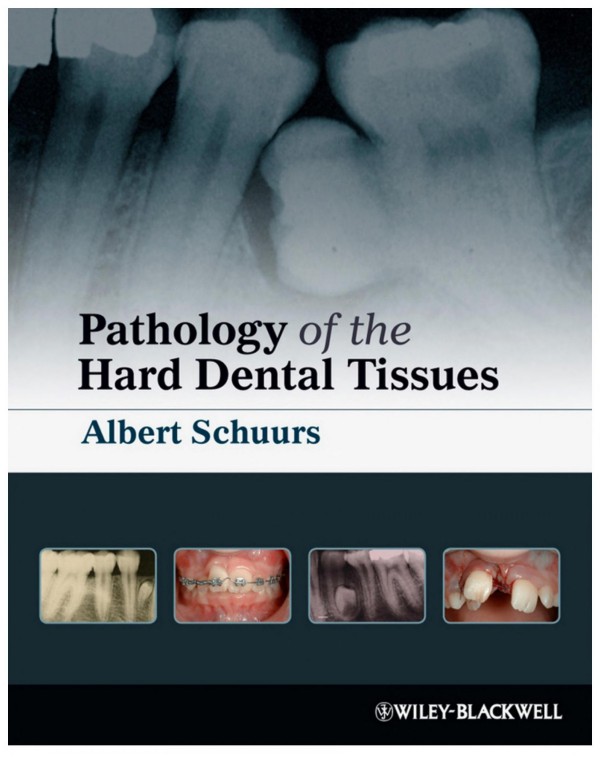
Pathology of the Hard Dental Tissues 1st edition by Albert Schuurs 9781118970430 1118970438
$50.00 Original price was: $50.00.$25.00Current price is: $25.00.
Authors:Albert Schuurs , Series:Dentistry [17] , Tags:Medical; Dentistry; Oral Surgery; General , Author sort:Schuurs, Albert , Ids:Goodreads; Google; 9781118381342 , Languages:Languages:eng , Published:Published:Sep 2012 , Publisher:Wiley-Blackwell , Comments:Comments:This is a seminal text uniquely dedicated to oral hard tissue pathology, presenting the growth of clinical knowledge and advancement in the field in recent years. Starting with a discussion of numerical and formative anomalies and unusual eruption, the book goes on to consider caries, erosion, resorption and toothwear, as well as tooth fractures and discolouration, and ends with a chapter on congenital syndromes with dental anomalies. Pathology of the Hard Dental Tissues is an invaluable reference for specialist practitioners and researchers as well as dental students, combining a scholarly overview of the field with clinical management protocols. Includes prevention techniques as well as treatment regimes Contains many colour clinical photographs Accompanied by a large number of references Provides helpful tables to categorise the causes and characteristics of lesions Written by a leading expert in the field
Pathology of the Hard Dental Tissues 1st edition by Albert Schuurs – Ebook PDF Instant Download/Delivery.9781118970430,1118970438
Full download Pathology of the Hard Dental Tissues 1st edition after payment
Product details:
ISBN 10:1118970438
ISBN 13:9781118970430
Author:Albert Schuurs
Pathology of the Hard Dental Tissues is an invaluable reference for specialist practitioners and researchers as well as dental students, combining a scholarly overview of the field with clinical management protocols.
Pathology of the Hard Dental Tissues 1st Table of contents:
Prior to and during the development of the teeth within the jaws
During the eruption of teeth
After the eruption of the teeth
Genetics
Interpretation of family trees
Table 0.1 Autosomal inherited hypocalcified enamel (A)
Table 0.2 Sex-chromosome (X-bound) inherited hypocalcified enamel
Table 0.3 Polygenetic heredity
Section I Developmental Anomalies
1 Anomalies of Number
1.1 Introduction
1.2 Hypodontia
1.2.1 Isolated dental agenesis
Epidemiology
Primary dentition
Permanent dentition
Third molars
Mandibular second premolar
Maxillary lateral incisor
Figure 1.1 Agenetic right permanent maxillary lateral incisor; the contralateral tooth is underdeveloped.
Maxillary second premolar
Mandibular central incisor
Figure 1.2 Agenetic permanent central incisors – the deciduous teeth are retained.
Figure 1.3 Four retained deciduous incisors in the mandible; the permanent successors are agenetic.
Other teeth
Aetiology
Evolution
Table 1.1 Teeth per quadrant of the archetypal permanent dentition, the teeth remaining in modern humans according to Bolk, and present point of view628
Heredity
Figure 1.4 Mother (A) and daughter (B) with agenetic maxillary permanent lateral incisors.
Relationships between teeth
Tooth germ-related causes
Other causes
Figure 1.5 (A) Agenetic lower second premolar with retained deciduous second molar. (B) The development of the contralateral premolar is arrested.
Consequences
Prevention and treatment
Figure 1.6 Patient with missing maxillary lateral incisors.
Figure 1.7 The canines of the patient in Figure 1.6 have been moved orthodontically into the locations of the agenetic maxillary lateral incisors. The morphology of the canines may be improved by grinding the cusps and building the lateral margins with composite so that the teeth resemble lateral incisors. Note the presence of only three lower incisors.
1.2.2 Oligodontia
Figure 1.8 Oligodont dentition with hypoplastic and conical teeth. (Courtesy of Department of Oral Surgery, University of Groningen.)
Heredity
1.2.3 Anodontia
Treatment of oligodontia and anodontia
1.3 Hyperdontia
1.3.1 Hypodontia with hyperdontia
Epidemiology
Primary dentition
Permanent dentition
Location
Sex, ethnicity
Syndromes
Non-syndromic multiple supernumeraries
Time of development
1.3.2 Supernumerary permanent teeth
Mesiodens
Figure 1.9 Two mesiodentes in the apical region of the permanent maxillary central incisors. They will not erupt because of their inverted direction.
Figure 1.10 A mesiodens between the maxillary permanent central incisors.
Figure 1.11 Inverted atypical extra tooth in the mandible. The small radiopacity visible on the root of the canine increased over time and resembled a late developing atypical extra tooth.
Table 1.2 Characteristics of conical and tuberculate supernumerary teeth251
Distomolar
Figure 1.12 Distomolar situated behind the third maxillary molar.
Paramolar
Figure 1.13 Paramolar (root and crown) attached to a decayed permanent second lower molar.
Figure 1.14 Paramolar or an atypical third premolar in the maxilla. (Courtesy of J.P. Nolte.)
1.3.3 Supplemental permanent teeth
Incisors
Figure 1.15 Supplemental incisor in the right upper quadrant.
Figure 1.16 Two supplemental teeth in the mandible.
Premolars
Figure 1.17 Late developing supplemental mandibular premolar.
Figure 1.18 Extra premolar considered to be atypical because of its small size.
Fourth molars
Figure 1.19 (A) A supplemental (fourth) molar. (B) Inverted eumorphic fourth molar in the ramus of the mandible.
Canines
Eruption issues related to hyperdontia
Aetiology
Heredity
Morphology
Consequences
Figure 1.20 An erupted mesiodens has resulted in lack of space for the lateral incisor, which has erupted palatally.
Figure 1.21 Malalignment of the teeth in the upper right quadrant as a consequence of an extra premolar.
Treatment
1.3.4 “Extra dentitions”
Natal and neonatal teeth
Figure 1.22 Neonatal teeth.
Epidemiology
Appearance
Aetiology
Consequences
Treatment
Third dentition
1.4 Fusion and partial schizodontia
Figure 1.23 Schematic presentation (lower row) of partial schizodontia (A), complete schizodontia or twinning (B), fusion (C) and concrescence (D). The top panel shows a partially split tooth germ (A), a tooth germ split in two (B), two tooth germs pressed together, presumably resulting in fusion (C), and two tooth germs situated too close together (D).
Figure 1.24 Two broad central double teeth; the incisal notches indicate existence of two components.
1.4.1 Diagnosis
Figure 1.25 Possible fusion of a mesiodens with a regular maxillary central incisor.
Diagnostic criteria: fusion versus partial schizodontia
Morphology
Figure 1.26 (A, B) Examples of permanent posterior double teeth from the upper and lower dentitions. The mirror image-like appearances of the occlusal left and right halves of the tooth (Figure 1.26A) possibly indicate partial schizodontia.
Anatomy
Location by jaw
Crowding
Number of teeth in the dentition
Conclusion
1.4.2 Epidemiology
Figure 1.27 Bilateral mandibular fused anterior teeth, suggestive of fusion between the lateral incisors and canines (see text).
Sex
Syndromes
Bilateral double teeth
Posterior double teeth
Triple and quadruple teeth
Figure 1.28 Two mandibular triple teeth (A). The maxillary triple tooth (B) was functional until its shedding. (Courtesy of M.A.C.J. Wevers.)
Solitary symmetrical median central incisor
1.4.3 Aetiology
Evolution
Trauma
Environment
Heredity
Twins
1.4.4 Pathogenesis
1.4.5 Consequences
1.4.6 Treatment
Figure 1.29 (A–D) A double tooth, consisting of conjoined crowns with a common pulp chamber. After hemisection, the mesial part of the tooth was removed and the exposed pulp was treated with direct pulp capping. The resulting diastema was closed orthodontically. (Courtesy of M.H. Ree.)
Figure 1.30 Crown of a first molar fused with a second lower molar.
1.5 Concrescence
Figure 1.31 Concrescence of a second and third molar and a rare case of concrescence of three molars. The left maxillary first molar in the top panel was extracted because of extensive caries; but unexpectedly, the two other molars were extracted as well.
2 Deviations in Tooth Morphology and Size
2.1 Introduction
Figure 2.1 Underdeveloped lingual half (the “deuteromere”) of a maxillary second premolar.
Figure 2.2 Palatal surface of a lobodont maxillary canine (lower panel) and another viewed from labial aspect (upper panel).
2.2 Compression
2.2.1 Appearance and aetiology
Crown
Figure 2.3 (A,B) Compression: a third molar with an underdeveloped mesial aspect due to the already calcified crown of the upper second molar being pressed against the uncalcified mesial side of the third molar during its development. Note the large enamel pearl. (C) The shape of this maxillary second molar crown – broad buccolingually but small mesiodistally – is not a result of compression, but represents the rhomboid crown type.
Root
Figure 2.4 Sickle-shaped root of a permanent upper incisor.
Figure 2.5 Corkscrew root form of a premolar.
Figure 2.6 Curved root of the third molar as a consequence of erupting around the second molar.
2.2.2 Epidemiology
2.3 Dens invaginatus
Figure 2.7 Schematic drawings of dens invaginatus: green represents the enamel, yellow the dentine and pink-red the pulp cavity. The left panel shows a lingual foramen caecum. A deepening of the foramen caecum leads to formation of dens invaginatus: if restricted to the crown of the tooth the anomaly is considered to be dens invaginatus type I (middle panel). The right panel illustrates a deep invagination (type II).
2.3.1 Coronal
Figure 2.8 Dens invaginatus in a permanent maxillary right lateral incisor (A); and the sectioned tooth (B).
Figure 2.9 Dens invaginatus in the mandible.
Association with other anomalies
2.3.2 Apical
2.3.3 Lateral
2.3.4 Epidemiology
2.3.5 Aetiology
2.3.6 Consequences
Caries
Pulpitis
Periodontal problems
2.3.7 Prevention of complications and treatment
2.4 Palato-gingival groove
2.4.1 Appearance
Figure 2.10 Lingual groove running from the enamel onto the root.
2.4.2 Epidemiology
2.4.3 Aetiology
2.4.4 Consequences and treatment
2.5 Dilaceration
Figure 2.11 (A) Dilacerated crown of a mandibular permanent lateral incisor; the lingual surface is visible instead of the labial surface. (B) X-ray showing a dilacerated tooth.
Figure 2.12 Dilacerated root of a lower first premolar.
2.5.1 Aetiology
2.5.2 Prevalence
2.5.3 Consequences
2.5.4 Treatment
2.6 Enamel pearls and enamel extensions
2.6.1 Enamel pearls
Epidemiology
Figure 2.13 Enamel pearl.
Histology
Aetiology
Consequences and treatment
2.6.2 Enamel extensions
Figure 2.14 Cervical enamel extension.
Figure 2.15 Enamel projection with a thick spherical end.
Epidemiology
Aetiology
Consequences
2.7 Fused roots
2.7.1 Aetiology
2.7.2 Epidemiology
2.7.3 Consequences
2.8 Macro- and microdontia
2.8.1 Normal tooth size
2.8.2 Macrodontia
Aetiology
Figure 2.16 Macrodont permanent upper canine (38 mm) and some microdont third molars.
2.8.3 Microdontia
Figure 2.17 Microdont teeth (the partly visible matchstick is of regular size).
Aetiology
Epidemiology
Rhizomicry
Figure 2.18 Permanent upper central incisors with microdont roots (rhizomicry) and an incisor (top) with average root length.
Figure 2.19 Radiograph of upper premolars with “ogee” roots.
Aetiology
Treatment
2.9 Other developmental anomalies of the tooth crowns
2.9.1 Shovel-shaped incisor
Figure 2.20 Shovel-shaped maxillary lateral incisor with a central amalgam filling.
Epidemiology
Treatment
2.9.2 T-shaped, Y-shaped and stellate incisors
Figure 2.21 T-shaped permanent maxillary lateral incisor.
Figure 2.22 Y-shaped permanent maxillary lateral incisor. The combination of the T- with the Y-shape results in a crown resembling a crosshead screwdriver.
Treatment
2.10 Extra cusps
Figure 2.23 Carabelli cusps on the mesio-palatal surface of the permanent maxillary first and second molars.
2.10.1 Dens evaginatus
Appearance
Figure 2.24 (A) Bilateral dens evaginatus. (B) A conical evagination.
Epidemiology
Pathogenesis
Consequences
Treatment
2.10.2 Talon
Appearance
Figure 2.25 (A) Talon. (Courtesy of Gregori M. Kurtzman.) (B) Double semi-talon.
Aetiology
Epidemiology
Consequences
Treatment
2.10.3 Carabelli’s cusp
Treatment
2.10.4 Paramolar tubercle/cusp, paramolar roots, paramolars
Figure 2.26 Paramolar tubercle (arrow) at the mesio-buccal surface of the permanent upper second molar.
Treatment
2.11 Supernumerary roots
2.11.1 Bifid roots
Figure 2.27 (A) Permanent upper lateral incisor (left) and lower canine (right) with bifid roots. (B) Deciduous lower left canine with three roots (in microsomia type I).
Figure 2.28 The buccal roots of these second upper premolars are split into mesial and distal parts.
Figure 2.29 A lower second premolar with a root split into mesial and distal parts.
Figure 2.30 Permanent first upper molar with two mesio-buccal roots, united by a thin sheet of dentine.
Table 2.1 The proportion of teeth with cleft roots
Table 2.2 Teeth showing incidental bifid roots
Figure 2.31 A permanent upper central incisor with an additional small-sized root.
Treatment
2.11.2 Additional roots
Figure 2.32 Paramolar root (left) and a lingual molar root (right).
Radix distomolaris
Radix entomolaris
Figure 2.33 X-ray of first molar with a lingual molar root, originating distally.
Accessory roots
2.12 Taurodontism
2.12.1 Appearance
Figure 2.34 (A) A sectioned taurodont molar. (B) X-ray of an extracted taurodont upper premolar. Note the cylindrical form and the broad apex. (C) X-ray of an extracted taurodont lower premolar. (D) An extracted taurodont upper molar. (E) X-ray of the tooth shown in (D). (F) Endodontically treated taurodont upper molar.
People also search for Pathology of the Hard Dental Tissues 1st :
what is dental histology
what is soft tissue in dentistry
pathology tooth
what is soft tissue pathology
hard and soft tissues of the oral cavity
You may also like…
eBook PDF
Pathology of the Hard Dental Tissues 1st Edition by Albert Schuurs ISBN 1118970438 9781118970430
eBook PDF
Pathology of the Hard Dental Tissues 1st edition by Albert Schuurs 9781118970430 1118970438
eBook PDF
Oral Pathology for the Dental Hygienist 7th edition by Olga Ibsen 9780323484442 0323484441
eBook PDF
Encyclopedia of Chemistry 1st Edition by Don Rittner, Ronald Albert Bailey 0816048940 978-0816048946

