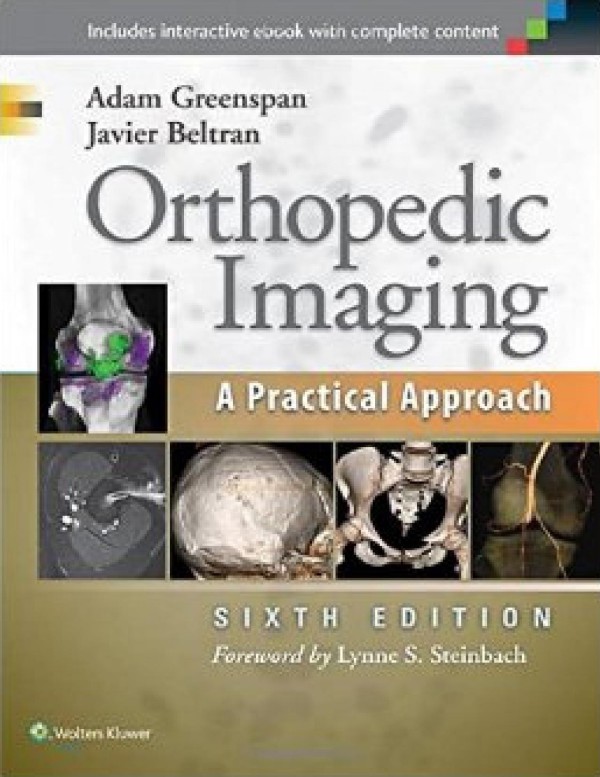
Orthopedic Imaging A Practical Approach 6th Edition by Adam Greenspan ISBN 1451191308 9781451191301
$50.00 Original price was: $50.00.$25.00Current price is: $25.00.
Authors:Adam Greenspan , Series:Surgery [36] , Tags:Medical / Orthopedics Medical / Radiology; Radiotherapy & Nuclear Medicine , Author sort:Greenspan, Adam , Languages:Languages:eng , Published:Published:Dec 2014 , Publisher:Wolters Kluwer , Comments:Comments:Interpret musculoskeletal images with confidence withOrthopedic Radiology: A Practical Approach!This trusted radiology reference has established itself as an ideal comprehensive source of guidance for radiologists and orthopedists at every level of training.
Orthopedic Imaging A Practical Approach 6th Edition by Adam Greenspan – Ebook PDF Instant Download/Delivery. 1451191308, 9781451191301
Full download Orthopedic Imaging A Practical Approach 6th Edition after payment
Product details:
ISBN 10: 1451191308
ISBN 13: 9781451191301
Author: Adam Greenspan
Interpret musculoskeletal images with confidence with Orthopedic Radiology: A Practical Approach! This trusted radiology reference has established itself as an ideal comprehensive source of guidance for radiologists and orthopedists at every level of training.Features
- Effectively interpret a full range of findings with the aid of more than 4,000 outstanding illustrations that encompass conventional radiography, ultrasound, CT, dual-energy CT, PET-CT, and all other diagnostic imaging modalities used to evaluate musculoskeletal disorders, including numerous examples of 3-D imaging.
- Master the latest trends in orthopedic radiology including the increasing emphasis on ultrasonography and MRI over other methods that expose patients to higher levels of radiation.
- Choose the best imaging approach for each patient with discussions of each technique’s accuracy, speed, and cost.
- Apply a state-of-the-art knowledge of magnetic resonance imaging interpretation with advanced guidance from renowned musculoskeletal MRI authorities.
- Find the information you need quickly and easily thanks to informative diagrams and schematics, and a quick-reference, high-yield format, including “Practical Points” summaries at the end of each chapter for quick review.
Now with the print edition, enjoy the bundled interactive eBook edition , offering tablet, smartphone, or online access to:
- Complete content with enhanced navigation
- A powerful search that pulls results from content in the book, your notes, and even the web
- Cross-linked pages , references, and more for easy navigation
- Highlighting tool for easier reference of key content throughout the text
- Ability to take and share notes with friends and colleagues
- Quick reference tabbing to save your favorite content for future use
Orthopedic Imaging A Practical Approach 6th Table of contents:
Chapter 1: Introduction to Orthopedic Imaging
- Role of Imaging in Orthopedic Diagnosis
- Basic Principles of Radiography, MRI, CT, and Ultrasound
- Imaging Modalities and Their Applications in Orthopedics
- Safety Considerations and Radiation Protection
- Image Quality and Optimization
Chapter 2: Basic Imaging Techniques and Interpretation
- X-ray Imaging: Principles and Techniques
- Computed Tomography (CT): Indications and Interpretation
- Magnetic Resonance Imaging (MRI): Techniques, Sequences, and Views
- Musculoskeletal Ultrasound: Basics and Advanced Techniques
- Nuclear Medicine and Bone Scintigraphy in Orthopedic Imaging
- Overview of Emerging Imaging Technologies
Chapter 3: Imaging of Bone and Joint Pathologies
- Normal Anatomy of Bones and Joints on Imaging
- Fractures: Classification, Imaging Signs, and Healing Stages
- Osteomyelitis: Imaging Characteristics and Diagnosis
- Bone Tumors: Benign vs. Malignant Lesions
- Bone Density and Osteoporosis: Imaging Techniques and Assessment
- Arthritis and Degenerative Joint Disease: Imaging Manifestations
Chapter 4: Imaging in Trauma and Emergency Orthopedics
- Fracture Imaging: Standard Views and Additional Projections
- Trauma to the Spine: Cervical, Thoracic, and Lumbar Imaging
- Pelvic and Acetabular Fractures: Imaging Approaches
- Upper Extremity Trauma: Shoulder, Elbow, and Wrist Imaging
- Lower Extremity Trauma: Hip, Knee, Ankle, and Foot Imaging
- Soft Tissue Injuries: MRI and Ultrasound in Ligament and Tendon Evaluation
Chapter 5: Spine Imaging and Disorders
- Normal Spine Anatomy on Imaging Modalities
- Degenerative Spine Conditions: Imaging Features of Disc Herniation and Spondylosis
- Spinal Trauma: Fractures, Dislocations, and Instability
- Spinal Tumors and Infections: MRI and CT Imaging
- Spinal Deformities: Scoliosis, Kyphosis, and Lordosis
- Imaging in Spinal Cord Pathologies: MRI Findings and Considerations
Chapter 6: Musculoskeletal Imaging in Children and Adolescents
- Pediatric Bone Growth and Development: Imaging of Growing Skeleton
- Common Pediatric Orthopedic Conditions: Imaging of Hip Dysplasia, Clubfoot, and Fractures
- Bone and Joint Infections in Children: Radiographic Features
- Pediatric Tumors: Benign vs. Malignant Lesions in Children
- Imaging for Congenital Skeletal Abnormalities
Chapter 7: Imaging of Soft Tissue and Tendon Pathologies
- Tendon Injuries: MRI, Ultrasound, and CT Imaging Techniques
- Ligament Injuries: Evaluation Using MRI and Ultrasound
- Bursitis and Soft Tissue Infections: Imaging Findings
- Muscle Injuries: MRI Evaluation of Strains and Tears
- Soft Tissue Tumors: Benign and Malignant Lesions on Imaging
Chapter 8: Joint Imaging in Orthopedics
- Imaging of the Shoulder Joint: Normal and Pathological Findings
- Elbow and Wrist Imaging: Common Pathologies and Imaging Approaches
- Hip Imaging: Evaluation of the Hip Joint, Femoroacetabular Impingement, and Labral Tears
- Knee Imaging: MRI and X-ray in Knee Pathologies
- Ankle and Foot Imaging: Fractures, Deformities, and Soft Tissue Pathologies
Chapter 9: Imaging in Orthopedic Infections and Inflammatory Diseases
- Radiographic Features of Bone and Joint Infections
- MRI and CT in Diagnosing Osteomyelitis and Septic Arthritis
- Imaging in Inflammatory Diseases: Rheumatoid Arthritis, Spondyloarthropathies
- Imaging of Soft Tissue Infections
- Osteonecrosis and Avascular Necrosis: Imaging Approaches and Diagnosis
Chapter 10: Imaging of Orthopedic Tumors and Bone Lesions
- Benign Bone Tumors: Radiographic Characteristics and Differential Diagnosis
- Malignant Bone Tumors: Imaging of Osteosarcoma, Ewing’s Sarcoma, and Metastases
- Soft Tissue Tumors: MRI and Ultrasound Features
- Imaging of Metastatic Bone Disease
- Bone Lesions in the Spine: Tumors, Infections, and Degenerative Changes
Chapter 11: Advanced Imaging Techniques in Orthopedics
- 3D Imaging and Virtual Reality in Orthopedic Imaging
- Functional Imaging in Orthopedic Disorders: PET and SPECT Scanning
- CT and MRI Arthrography in Joint Disorders
- Advanced MRI Sequences in Musculoskeletal Imaging
- Diffusion Tensor Imaging (DTI) and Other Emerging Techniques
- Molecular Imaging and Orthopedic Research Applications
Chapter 12: Imaging in Orthopedic Surgery and Interventional Procedures
- Preoperative Imaging Planning for Orthopedic Surgery
- Intraoperative Imaging: Use of Fluoroscopy, CT, and Ultrasound
- Imaging for Guidance in Joint Injection, Biopsy, and Arthroscopy
- Postoperative Imaging: Monitoring for Complications and Healing
- Image-Guided Orthopedic Interventions: Fracture Fixation and Arthroplasty
Chapter 13: Musculoskeletal Imaging in the Elderly
- Age-Related Changes in Bone and Joint Imaging
- Imaging of Osteoarthritis and Degenerative Conditions in Older Adults
- Fractures and Osteoporotic Changes in the Elderly: Evaluation and Management
- Imaging of Pathologies Unique to the Elderly: Avascular Necrosis, Sarcopenia
- Fall-Related Injuries and Imaging Challenges in Geriatric Patients
Chapter 14: Imaging in Orthopedic Research and Clinical Practice
- Radiologic Research in Orthopedic Disease and Treatment
- Imaging Biomarkers in Osteoarthritis and Cartilage Assessment
- The Role of Imaging in Orthopedic Drug Trials
- Radiologic Outcomes in Joint Replacement and Arthroplasty
- Imaging for Bone Regeneration and Tissue Engineering
Chapter 15: Case Studies and Clinical Applications
- Case Study 1: Trauma in the Upper Extremity
- Case Study 2: Degenerative Disc Disease and Spinal Stenosis
- Case Study 3: Bone Tumors and Differential Diagnosis
- Case Study 4: Imaging of Complex Fractures and Postoperative Evaluation
- Case Study 5: Pediatric Orthopedic Imaging: Fractures and Growth Disorders
People also search for Orthopedic Imaging A Practical Approach 6th:
orthopedic imaging a practical approach
orthopaedic imaging a practical approach orthopedic imaging a practical approach
orthopedic imaging a practical approach pdf
orthopedic imaging a practical approach 6th
what are orthopedic procedures
You may also like…
eBook PDF
Underwood Pathology A Clinical Approach 6th Edition by Simon Cross ISBN 0702046728 9780702046728
eBook PDF
Cosmetic Microbiology A Practical Approach 2nd edition by Philip Geis ISBN 0849314534 978-0849314537

