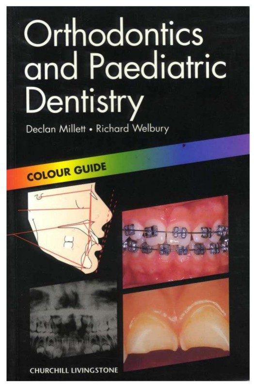
Orthodontics and Paediatric Dentistry 1st Edition by Declan T Millett ISBN 0443062870 9780443062872
$50.00 Original price was: $50.00.$25.00Current price is: $25.00.
Authors:Shayan Nemoodar Pub.Co. By : Hamed Forutanian , Author sort:Forutanian, Shayan Nemoodar Pub.Co. By : Hamed
Orthodontics and Paediatric Dentistry 1st Edition by Declan T Millett – Ebook PDF Instant Download/Delivery. 0443062870, 9780443062872
Full download Orthodontics and Paediatric Dentistry 1st Edition after payment
Product details:
ISBN 10: 0443062870
ISBN 13: 9780443062872
Author: Declan T Millett
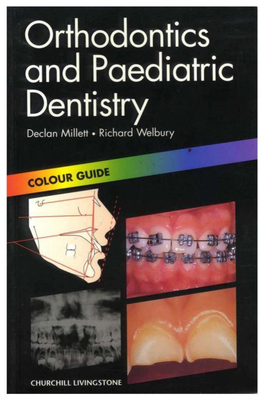
Orthodontics and paediatric dentistry are important areas of the undergraduate dental curriculum. No pocket colour atlas currently exists. All dentists have to know how to treat children, carry out basic orthodontic treatment and when to refer patients for specialist orthodontic treatment. Most orthodontic work is carried out on children, so the two subjects are conveniently integrated in this Colour Guide.
Orthodontics and Paediatric Dentistry 1st Table of contents:
1 / Normal development of the dentition and occlusion
Normal development
Primary dentition
Primary to mixed dentition
Dental arch development
Fig. 1
Fig. 2
Fig. 3
Normal permanent occlusion
Static occlusal relations (Andrews’ six keys)
Functional occlusal relations
Maturational changes in the occlusion
Fig. 4
Fig. 5
Fig. 6
2 / Malocclusion
Classification for diagnostic purposes
Angle’s classification based on molar relationship
British Standards Institute classification based on incisor relationship
Fig. 7
Fig. 8
Fig. 9
Fig. 10
Classification to assess treatment need
Fig. 11a
Fig. 11b
Classification to assess treatment outcome
Fig. 12
Fig. 13
3 / Cephalometric analysis
Aim and objective of cephalometric analysis
Fig. 14
Fig. 15
Fig. 16
Practice of cephalometric analysis
Cephalometric interpretation
Fig. 17
Fig. 18
4 / Mixed dentition I
Early loss of primary teeth
Fig. 19
Fig. 20
Fig. 21
Serial extraction
Fig. 22a
Fig. 22b
Fig. 23
5 / Mixed dentition II
Supernumerary teeth (see also p.101)
Mesiodens
Fig. 24
Fig. 25
Fig. 26
Hypodontia (see also pp. 23, 101)
Fig. 27a
Fig. 27b
Fig. 28
6 / Mixed dentition III
Buccal segment problems
Retained primary tooth
Infra-occluded primary molars
Impacted 6 (Fig. 30)
Posterior crossbite with mandibular displacement(Fig. 31)
Fig. 29
Fig. 30
Fig. 31
First permanent molars (FPM) with poor long-term prognosis
Fig. 32
Fig. 33
Fig. 34
7 / Mixed dentition IV
Labial segment problems
Upper median diastema
Dilaceration
Traumatic loss of 1
Fig. 35
Fig. 36
Fig. 37
Retained primary incisor
Incisors in crossbite
Finger/thumb-sucking habits
Early correction of increased overjet
Fig. 38
Fig. 39
Fig. 40
Ectopic maxillary canines
Fig. 41
Fig. 42a
Fig. 42b
Fig. 43
Fig. 44
Fig. 45
8 / Class I malocclusion
Fig. 46
Fig. 47
Fig. 48
Treatment
Fig. 49
Fig. 50a
Fig. 50b
9 / Class II Division 1 malocclusion
Fig. 51a
Fig. 51b
Fig. 51c
Treatment
Fig. 52a
Fig. 52b
Fig. 52c
Fig. 52d
10 / Class II Division 2 malocclusion
Fig. 53
Fig. 54
Fig. 55
Treatment
Fig. 56a
Fig. 56b
Fig. 57a
Fig. 57b
11 / Class III malocclusion
Fig. 58
Fig. 59
Fig. 60
Treatment
Fig. 61
Fig. 62a
Fig. 62b
12 / Crossbite
Fig. 63
Fig. 64
Fig. 65
Treatment
Fig. 66
Fig. 67
Fig. 68
13 / Anterior open bite (AOB)
Fig. 69
Fig. 70a
Fig. 70b
14 / Removable appliances
Active component
Fig. 71
Fig. 72
Fig. 73
Retention component
Baseplate
Fig. 74
Fig. 75
Fig. 76
15 / Anchorage
Fig. 77
Fig. 78
Fig. 79
16 / Fixed appliances
Fig. 80
Fig. 81
Fig. 82
Appliance types
Most common pre-adjusted appliances
Lingual appliance
Sectional appliance
Fixed – removable
Fig. 83
Fig. 84
Fig. 85
17 / Functional appliances
Fig. 86
Fig. 87
Fig. 88
Indications
Effects (Figs 89a-d)
Skeletal
Dental
Fig. 89a
Fig. 89b
Fig. 89c
Fig. 89d
18 / Adult orthodontics
Special considerations
Fig. 90
Fig. 91
Fig. 92
Treatment
Adjunctive
Comprehensive
Fig. 93a
Fig. 93b
Fig. 94
19 / Surgical orthodontic treatment
Fig. 95
Fig. 96
Fig. 97
Fig. 98
Orthodontic management
Surgical procedures
Fig. 99a
Fig. 99b
Fig. 99c
Fig. 99d
20 / Cleft lip and palate CL(P)
Common clinical features
Fig. 100
Fig. 101
Fig. 102
Management of care
Neonatal period to 18 months
Primary dentition
Mixed/permanent dentition
In late teenage years
Retention
Fig. 103a
Fig. 103b
Fig. 104
21 / Tooth eruption
Natal/neonatal teeth
Eruption cyst
Fig. 105
Fig. 106
22 / Nursing and rampant caries
Nursing caries
Fig. 107
Fig. 108
Fig. 109
Rampant caries
Fig. 110
Fig. 111
Fig. 112
23 / Tooth discoloration
Extrinsic
Fig. 113
Fig. 114
Fig. 115
Intrinsic
Fig. 116
Fig. 117
Fig. 118
24 / Enamel hypoplasia and fluorosis
Enamel hypoplasia
Fig. 119
Fig. 120
Fig. 121
Fluorosis
Fig. 122
Fig. 123
25 / Inherited anomalies
Amelogenesis imperfecta
Fig. 124
Fig. 125
Fig. 126
Dentinogenesis imperfecta
Fig. 127
Fig. 128
Fig. 129
26 / Anomalies of number and form
Hyperdontia
Hypodontia
Fig. 130
Fig. 131
Fig. 132
Talon cusp
Odontomes
Fig. 133
Fig. 134
Fig. 135
27 / Autotransplantation
Fig. 136
28 / Tooth surface loss
Erosion
Fig. 137
Fig. 138
Fig. 139
29 / Primary tooth trauma
Fig. 140
Fig. 141
Damage to permanent teeth after primary tooth trauma
Fig. 142
Fig. 143
Fig. 144
30 / Permanent tooth trauma I
Fig. 145
Fig. 146
Fig. 147
Permanent tooth trauma I: reattachment of crown fragments
Fig. 148
Fig. 149
Fig. 150
Permanent tooth trauma I: total or sub-total pulpotomy
Fig. 151
Fig. 152
Fig. 153
Permanent tooth trauma I: induced apical closure
Fig. 154
Fig. 155
Fig. 156
Fig. 157
31 / Permanent tooth trauma II
Root fractures
Fig. 158
Fig. 159
Permanent tooth trauma II: periodontal ligament injuries
Fig. 160
Fig. 161
Fig. 162
Permanent tooth trauma II: dentoalveolar fractures
Fig. 163
Fig. 164
Fig. 165
32 / Permanent tooth trauma III
Splinting
Fig. 166
Fig. 167
Permanent tooth trauma III: soft tissue injuries
Extraoral
Intraoral
Fig. 168
Fig. 169
Fig. 170
Permanent tooth trauma III: resorption
Internal resorption
External resorption
Replacement resorption
Fig. 171
Fig. 172
Fig. 179
Fig. 174
33 / Medical conditions
Down syndrome
Childhood cancer
Fig. 175
Fig. 176
Fig. 177
Congenital cardiac disease
Bleeding disorders
Fig. 178
Fig. 179
Fig. 180
34 / Viral infections
Primary herpetic gingivostomatitis
Secondary (recurrent) herpes labialis
Ocular herpes
Fig. 181
Fig. 182
Fig. 183
Herpes zoster
Hand-foot-and-mouth disease
Fig. 184
Fig. 185
35 / Aphthae
Recurrent aphthous stomatitis (RAS)
Fig. 186
Fig. 187
36 / Gingivitis
Acute necrotising ulcerative gingivitis (ANUG)
Chronic gingivitis
Fig. 188
Fig. 189
Fig. 190
37 / Periodontitis
Localised juvenile periodontitis
Neutrophil defects and neutropenias
Fig. 191
Fig. 192
Fig. 193
38 / Gingival recession
Gingivitis artefacta
Localised recession
Fig. 194
Fig. 195
39 / Gingival overgrowth
Localised gingival hyperplasia
Drug-induced gingival overgrowth
Hereditary gingival fibromatosis
Fig. 196
Fig. 197
Fig. 198
40 / Mucosal disease
Granulomas
Pyogenic granuloma
Giant cell granuloma (giant cell epulis)
Fig. 199
Fig. 200
Fig. 201
Traumatic lesions I
Fibroepithelial polyp
Mucocele
Ranula
Fig. 202
Fig. 203
Fig. 204
Traumatic lesions II
Burns
Sharp teeth and restorations
Local anaesthetic
Fig. 205
Fig. 206
Fig. 207
41 / Assorted mucosal lesions
Geographic tongue
Lichen planus
Orofacial granulomatosis
Fig. 208
Fig. 209
Fig. 210
Pericoronitis
Denture stomatitis
Infective papilloma
Fig. 211
Fig. 212
Fig. 213
Periapical infection
Fig. 214
Fig. 215
Fig. 216
Recommended reading
Orthodontics
Paediatric Dentistry
People also search for Orthodontics and Paediatric Dentistry 1st:
orthodontics and paediatric dentistry
clinical problem solving in orthodontics and paediatric dentistry
city orthodontics and paediatric dentistry
orthodontics and paediatric dentistry pdf
restorative dentistry paediatric dentistry and orthodontics pdf

