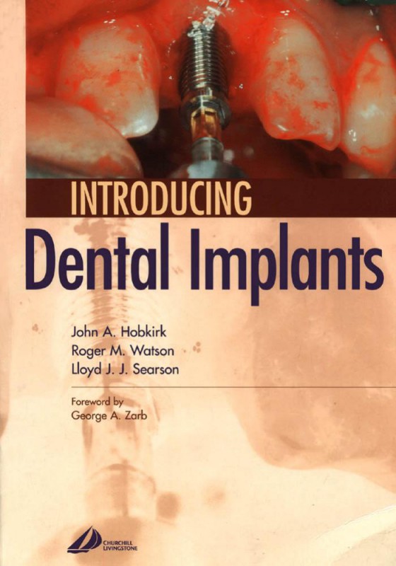
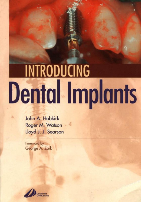
introducing dental implants 1st edition by John Hobkirk,Roger Watson,Lloyd Searson 9780702059339 0702059331
$50.00 Original price was: $50.00.$25.00Current price is: $25.00.
Authors:Churchill Livingstone; 1 edition (October 24, 2003) , Author sort:edition, Churchill Livingstone; 1 , Published:Published:Nov 2007
introducing dental implants 1st edition by John Hobkirk,Roger Watson,Lloyd Searson – Ebook PDF Instant Download/Delivery.9780702059339,0702059331
Full download introducing dental implants 1st edition after payment
Product details:
ISBN 10:0702059331
ISBN 13:9780702059339
Author:John Hobkirk,Roger Watson,Lloyd Searson
In recent years, dental implants have become a more common alternative to conventionally placed dentures, bridges and crowns. This accessible, well-illustrated introduction to the principles of implant dentistry provides readers coming into contact with implants for the first time with a working knowledge of the various implant systems available. Thorough discussions are also included on patient assessment and treatment planning, the use of implants in edentulous and partially edentulous patients and for single tooth restorations, as well as possible problems and maintenance procedures.
introducing dental implants 1st Table of contents:
1 Using this book
INTRODUCTION
IMPLANT TREATMENT
GENERAL TREATMENT DECISIONS
GATHERING INFORMATION AND TREATMENT PLANNING
IMPLANT SURGERY
THE EDENTULOUS CASE
THE PARTIALLY DENTATE CASE
THE SINGLE-TOOTH SCENARIO
OTHER APPLICATIONS
PROBLEMS
2 Implants: an introduction
MANAGEMENT OF MISSING TEETH
OSSEOINTEGRATION
3 General treatment decisions
INTRODUCTION
Treatment alternatives to dental implants for the partially dentate case
NO REPLACEMENT
4 Gathering information and treatment planning
WHAT ARE THE OBJECTIVES OF GATHERING INFORMATION AND PLANNING IMPLANT TREATMENT?
Box 4.1 Patient assessment leading to planned treatment
In which circumstances are dental implants most needed?
The edentulous patient
Box 4.2 Essential information
The partially dentate patient
What specific information should be sought in the medical, dental and social history of the patient?
5 Basic implant surgery
INTRODUCTION
INFORMATION REQUIRED AT THE SURGICAL STAGE
INFORMED CONSENT
MANAGEMENT
POST-OPERATIVE CARE
Wound management
Fig. 5.19 Using bone collected from the bone trap the implant site is augmented to provide better soft tissue support and cover any exposed implant threads.
Box 5.5 Grafting material
Postoperative haemorrhage
Postoperative analgesia
One-week review
SINUS LIFT/ELEVATION PROCEDURES
Contraindications
GRAFT MATERIALS
Problems seating the implant
Failure to place a cover screw or healing abutment over the implant
Failure to close the flap
Excess bleeding
Paraesthesia
Fig. 5.30 Wound breakdown around these three recently inserted implants has led to their exposure. They are associated with necrotic bone, and all three have failed.
Wound breakdown
Exposure of cover screws
Postoperative pain
FURTHER READING
6 The edentulous patient
INTRODUCTION: WHEN IS IMPLANT TREATMENT CONSIDERED APPROPRIATE?
IN WHICH CIRCUMSTANCES IS THIS TREATMENT LIKELY TO BE PROVIDED?
HOW MAY THE EDENTULOUS OR POTENTIALLY EDENTULOUS JAW BE TREATED?
Box 6.1 Complete dentures -influencing factors
Box 6.2 Complete implant-stabilized overdentures(s) -influencing factors
Box 6.3 Complete implant-stabilized fixed prosthesis(es) -influencing factors
Box 6.4 Problems associated with conventional complete dentures
HOW MAY THE EDENTULOUS JAW BE TREATED WITH IMPLANTS?
JAW RELATION
OCCLUSAL RELATION
POSITION OF RIDGE V. PROPOSED DENTAL ARCH
WELL-DESIGNED EXISTING COMPLETE DENTURES
Fig. 6.31 The OPT identifies the alignment of the bar with the condylar hinge axis.
Insertion of the prosthesis
Table 6.2 Complete over-denture anchorage to dental implants
Alternative procedure: relining/ adapting a denture
Preparation of fixed implant prostheses
Box 6.9 Choices in superstructure construction for fixed complete implant prostheses
Fig. 6.32 The wax pattern prepared for the cast alloy superstructure.
Fig. 6.33 A cast alloy superstructure showing the cantilever extensions and the enclosed gold alloy cylinders.
Fig. 6.34 A superstructure can be constructed by the ProceraTM technique of milling.
Fig. 6.35 Masking is applied to the titanium beam.
Fig. 6.36 Intra-oral radiographs of the ProceraTM prosthesis sited on the implants.
Fig. 6.37 Implants placed for the provision of fixed prostheses.
Fig. 6.38 Healing abutments will be removed and replaced by standard or multi-unit components.
Fig. 6.39 Healing caps have been placed on standard abutments and the patient’s denture modified to clear them.
Fig. 6.40 Access holes enable placement of the gold screws, which secure the prosthesis to the abutments.
Fig. 6.41 Radiograph showing a fixed mandibular prosthesis.
Fig. 6.42 A functioning maxillary prosthesis after 5 years.
Box 6.10 Patient complaints of new implant prostheses
LOOSENESS/INSTABILITY OF REMOVABLE PROSTHESIS
DIFFICULTY WITH ORAL HYGIENE
FOOD ACCUMULATION
SPEECH IMPAIRMENT
MONITORING AND MAINTENANCE
FURTHER READING
7 The partially dentate patient
INTRODUCTION
The partially dentate patient
Case assessment
SYSTEMIC FACTORS
Patients’ desires and needs
The patient’s expectations
Commitment
Resources
Residual life expectancy
Ability to cooperate
Ability to undergo surgery
LOCAL FACTORS
Treatment alternatives
Orthodontic treatment
Removable partial dentures
Resin retained bridges
Conventional bridges
Implant-based treatment
Missing posterior teeth
8 Single-tooth implants
INTRODUCTION
CAUSES OF THE MISSING SINGLE TOOTH
THE DECISION TO REPLACE A MISSING ANTERIOR TOOTH
Fig. 8.28 When designing an implant crown opposing a natural dentition, light contact in the intercuspal position is preferred while maintaining heavy contact between the adjacent natural dentition.
Fig. 8.29 If implant crowns oppose implant crowns, it is preferred that there is little or no contact of these crowns when the patient is in the intercuspal position.
Fig. 8.30 Initial blanching of the soft tissues upon insertion of the porcelain-fused-to-metal crown on 12.
Fig. 8.31 One-week review of 12 showing healthy soft tissue.
Two-week review
Fig. 8.32 Manufactured ‘CeraOne’ abutment replacing 46.
Fig. 8.33 Final crown cemented on ‘CeraOne’ abutment replacing 46.
Box 8.5 A final radiograph is taken to:
Danger signs at a review appointment
Fig. 8.34 Baseline radiograph after cementation of a crown on 11.
Fig. 8.35 Porcelain crown replacing 23 cemented on a single-tooth implant.
Fig. 8.36 Radiograph showing excess cement in the soft tissues.
Fig. 8.37 Radiograph showing loosening of an abutment screw due to failure to tighten it correctly.
Evaluation of marginal bone levels
Oral hygiene
FURTHER READING
9 Other applications
INTRODUCTION
INTRA-ORAL APPLICATIONS
Box 9.1 Other applications for osseointegrated implants
INTRA-ORAL
INTRA-ORAL AND FACIAL SKELETON
EXTRA-ORAL/FACIAL
Immediate loading
Fig. 9.1 Diagram showing a zygomatic implant engaging the palate and zygomatic bones of the skull lateral to the maxillary antrum and the orbit.
Orthodontic applications
How may implants assist restoration of the resected mandible?
Fig. 9.2 A resected mandible has been reconstructed with corticocancellous bone grafts.
Fig. 9.3 A computer image reconstructed from a CT scan, showing malocclusion arising from the graft of excessive length and inadequate height.
Box 9.2 Siting of dental implants in resected mandibles
Fig. 9.4 The mandible has been enhanced sufficiently by shortening and the use of cancellous iliac bone enclosed by a preformed titanium mesh tray. Dental implants now support a fixed prosthesis.
Box 9.3 Assessment of resected/reconstructed mandibles
Choice of prosthesis
Maxillary defects
Treatment stages
Problems
Treatment planning
Failure of osseointegration
Cleaning
Zygomatic implants
Introduction
Indications
Patient assessment
Surgical stage
Fig. 9.5 Partial restoration of the maxillary arch has been achieved with dental implants. The posterior sites in the premolar and first molar areas are restored with zygomatic implants.
Restoration
Fig. 9.6 The fixed maxillary prosthesis has been constructed with a cast alloy framework enclosing gold cylinders supporting a resin-based dental arch.
Box 9.4 Zygomatic implants
WHAT ARE THEY?
WHEN MIGHT THEY BE USED?
PATIENT ASSESSMENT
WHAT PROBLEMS MAY BE ENCOUNTERED WHEN USING THEM?
Problems
Fig. 9.7 A flanged implant body manufactured for use in the skull.
REHABILITATION OF EXTRA-ORAL DEFECTS
Facial prostheses
Fig. 9.8 Percutaneous abutments support cylinders linked by a bar.
The design and construction of facial prostheses
Fig. 9.9 The ear prosthesis is retained by clips on the bar.
Box 9.5 Treatment planning for facial prostheses
CLINICAL APPRAISAL OF DEFECTIVE TISSUE AREA
RADIOGRAPHIC EXAMINATION
LABORATORY ASSESSMENT
SURGICAL PREPARATION
PROSTHESIS DESIGN
Fig. 9.10 Reconstruction from the CT scan identifies, in an axial slice, the limited thickness of the mastoid bone on the defect side of the skull, where the ear is affected by hemifacial micosomia.
Surgical placement of skull implants
Fig. 9.11 Inserting a skull implant into the prepared site.
Prosthesis construction
Fig. 9.12 The patient has been born with hemifacial microsomia including a missing external ear (anotia).
Fig. 9.13 A computer image from an MRI scan may be manipulated to include a mirror image of the normal ear in a planned position on the defective side of the face.
Fig. 9.14 This image from the computer shows a slice file prepared for rapid proces modelling.
Fig. 9.15 Rapid process models can be prepared of the prosthesis fitted to the face and of the defect side for a precisely fitting wax replica.
Fig. 9.16 Rehabilitation achieved with the implant-retained silicone prosthesis.
Key issues
Fig. 9.17 Skull implants positioned in the orbital rim, support a bar carrying magnets for retention of a facial prosthesis.
Fig. 9.18 The orbital facial prosthesis is retained by keepers. The spectacles mask the perimeter.
Bone anchored hearing aids
Fig. 9.19 The skin has been penetrated by four abutments. The thick, hair-bearing skin has been replaced by a free graft to create thin, tightly bound tissue around the abutment, which is to be used for a bone-anchored hearing aid (BAHA).
Fig. 9.20 The ear prosthesis and BAHA are slightly separated.
12. Discuss the range of radiographic techniques which may be used in the planning of implant treatment.
People also search for introducing dental implants 1st :
introduction to implant dentistry a student guide
how to set up for dental implant surgery
how long after dental implant before crown
can one dental implant support 2 teeth
how long does it take for dental implants to integrate
You may also like…
eBook PDF
Mini Dental Implants Principles and Practice 1st edition by Victor Sendax 9781455744671 1455744670
eBook PDF
Osseointegration and Dental Implants 1st Edition by Asbjorn Jokstad ISBN 0813813417 9780813813417
eBook PDF
Osseointegration and Dental Implants 1st edition by Asbjorn Jokstad 9780813813417 0813813417
eBook PDF
Mini Dental Implants Principles and Practice 1st Edition by Victor Sendax 1455743860 9781455743865
eBook PDF
Mini Dental Implants Principles and Practices 1st edition by Victor Sendax 9781455744671 1455744670

