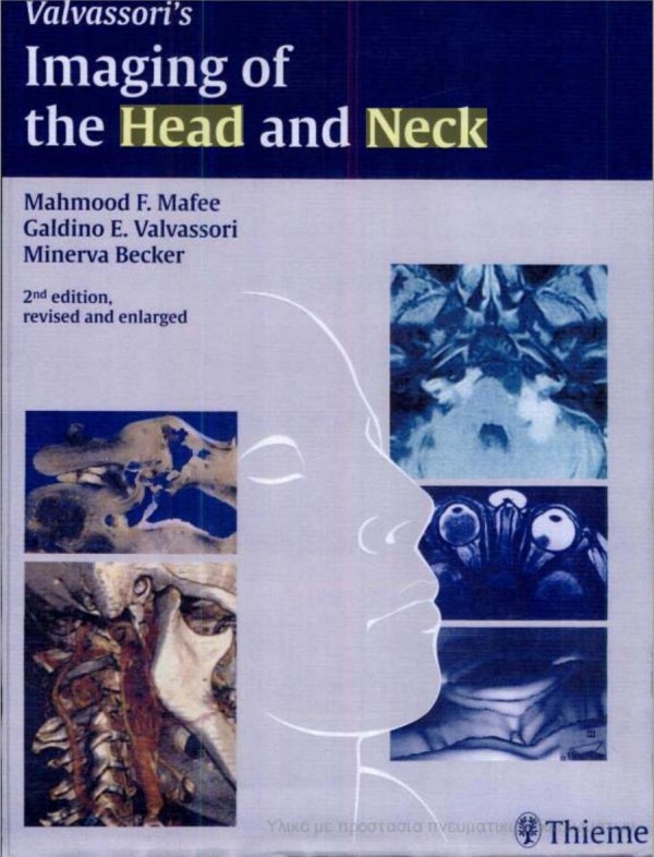
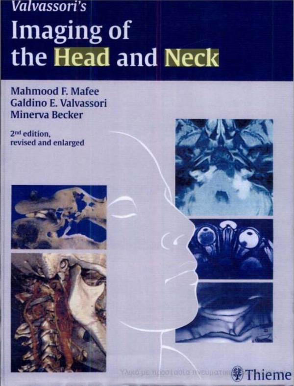
Imaging of the Head and Neck 2nd Edition by Mahmood Mafee, Galdino Valvassori, Minerva Becker ISBN 3131634820 9783131634825
$50.00 Original price was: $50.00.$25.00Current price is: $25.00.
Authors:Thieme; 2 Revised edition (November 10, 2004) , Author sort:edition, Thieme; 2 Revised , Published:Published:Mar 2008
Imaging of the Head and Neck 2nd Edition by Mahmood F. Mafee, Galdino E. Valvassori, Minerva Becker – Ebook PDF Instant Download/Delivery. 3131634820, 978-3131634825
Full download Imaging of the Head and Neck 2nd Edition after payment
Product details:
ISBN 10: 3131634820
ISBN 13: 978-3131634825
Author: Mahmood F. Mafee, Galdino E. Valvassori, Minerva Becker
More than 3,700 illustrations and systematic coverage of the latest technical developments make the new edition of Valvassori’s world-famous text your complete guide to head and neck imaging. Fully revised and updated to include a wider range findings in both adults and children, the book provides in-depth discussions of the eye and orbit, lacrimal drainage system, skull base, mandible and maxilla, temporomandibular joint, and suprahyroid and infrahyroid neck. CT and MRI scans acquired with the most advanced high-resolution equipment show all anatomic structures and pathological conditions, with actual cases clarifying every concept.
With thorough coverage of the newest imaging modalities, an abundance of high-quality graphics, and the expertise of worldwide leaders in the field, this is the reference of choice on head and neck imaging for experienced practitioners and residents-in-training.
Imaging of the Head and Neck 2nd Table of contents:
Section I: Temporal Bone
- Imaging of the Temporal Bone
- Sectional Anatomy of the Temporal Bone
- Anatomical Notes
- Sectional Anatomy
- Imaging Techniques:
- Conventional Radiography
- Computed Tomography (CT)
- Magnetic Resonance Imaging (MRI)
- Developmental Variations:
- Mastoid
- Lateral Sinus
- Tegmen
- Jugular Fossa
- Carotid Artery
- Arachnoid Granulations
- Petrous Apex
- Congenital Abnormalities of the Temporal Bone
- Imaging Assessment
- Embryology
- Anomalies of the Sound-Conducting System
- Facial Nerve Anomalies
- Anomalies of the Inner Ear
- Cerebrospinal Fluid Otorrhea
- Imaging Assessment for Cochlear Implantation
- Congenital Vascular Anomalies
- Syndromic Hearing Loss
- Temporal Bone Trauma
- Classification
- Clinical Findings
- Imaging Findings:
- Labyrinthine Concussion and Bleeding
- Longitudinal Fractures
- Transverse Fractures
- Comminuted Mastoid Fractures
- Ossicular Dislocation
- Projectile Injuries
- Traumatic Facial Nerve Lesions
- Iatrogenic Lesions
- Meningoencephalocele
- Cerebrospinal Fluid Otorrhea
- Acute Otitis Media, Mastoiditis, and Malignant Necrotizing External Otitis
- Imaging Findings
- Imaging Technique
- Differential Diagnosis
- Acute Labyrinthitis
- Chronic Labyrinthitis
- Facial Neuritis
- Chronic Otitis Media and Mastoiditis
- Clinical Findings
- Otoscopic Findings
- Imaging Techniques
- Imaging Findings
- Cholesteatoma of the Middle Ear
- Acquired Cholesteatoma:
- Clinical and Otoscopic Findings
- Imaging Techniques
- Diagnosis of Cholesteatoma
- Patterns of Imaging Findings:
- Pars Flaccida Cholesteatomas
- Epitympanic Retraction Pockets
- Pars Tensa Cholesteatomas
- Combined Pars Flaccida and Pars Tensa Cholesteatomas
- Evaluation of the Extent of Cholesteatoma
- Labyrinthine Fistulas
- Petrous Extension of Cholesteatoma
- Facial Nerve
- Congenital Cholesteatoma
- Postoperative and Postirradiation Imaging:
- Simple and Radical Mastoidectomies
- Tympanoplasty
- Acquired Cholesteatoma:
- Tumors
- Benign Tumors
- Malignant Tumors
- Internal Auditory Canal and Acoustic Schwannomas
- Pathology of the Internal Auditory Canal
- Otosclerosis and Bone Dystrophies:
- Otosclerosis
- Paget Disease
- Osteogenesis Imperfecta
- Osteopetrosis
- Fibrous Dysplasia
- Craniometaphyseal Dysplasia
Section II: Eye and Orbit, Base of the Skull
- Eye and Orbit
- Part 1: The Eye
- Embryology
- Lens
- The Ciliary Body, Iris, and Suspensory Ligaments of the Lens
- The Vitreous
- The Choroid
- The Sclera
- The Cornea
- Vascular System
- Extraocular Muscles
- The Eyelids
- The Lacrimal Gland
- The Lacrimal Sac and Nasolacrimal Duct
- Anatomy of the Orbit
- Imaging Techniques:
- CT Technique
- MRI Technique
- Pathology:
- Congenital Anomalies
- Ocular Detachments
- Ocular Inflammatory Disorders
- Benign Reactive Lymphoid Hyperplasia
- Papilledema
- Intraocular Calcifications
- Retinoblastoma and Masslike Lesions
- Retinal Dysplasia, Retinopathy of Prematurity, and Other Retinal Conditions
- Malignant Uveal Melanoma and Metastases
- Ocular Trauma
- Part 2: The Orbit
- Embryology
- Anatomy
- Bony Orbit
- Orbital Septum and Eyelids
- Orbicularis Oculi
- Extraocular Muscle Sheaths
- Orbital Fatty Reticulum
- Muscles of the Eye
- Innervation and Vascular Anatomy
- Lacrimal Apparatus
- Imaging Techniques:
- CT
- MRI
- Pathology:
- Hypertelorism, Hypotelorism, Exophthalmos
- Acquired Orbital Cysts and Inflammatory Diseases
- Tumors and Vascular Conditions
- Neural Lesions
- Orbital Trauma
- Part 1: The Eye
Section III: Nasal Cavity and Paranasal Sinuses
- Imaging of the Nasal Cavity and Paranasal Sinuses
- Embryology and Development of the Nasal Cavity and Paranasal Sinuses:
- Maxillary Sinuses
- Ethmoid Sinuses
- Frontal Sinuses
- Sphenoid Sinuses
- Anatomy:
- Nasal Cavity and Mucous Membrane
- Paranasal Sinuses (Maxillary, Frontal, Ethmoidal, Sphenoidal)
- Functional Endoscopic Sinus Surgery
- Imaging Techniques:
- CT
- MRI
- Pathology:
- Congenital Anomalies
- Inflammatory Diseases: Cysts, Polyps, Retention Cysts
- Tumors and Tumorlike Lesions
- Trauma and Postoperative Findings
- Embryology and Development of the Nasal Cavity and Paranasal Sinuses:
Section IV: Masticatory System
-
Temporomandibular Joint
- Embryology and Anatomy
- Diagnostic Imaging Techniques:
- CT, MRI, Nuclear Medicine
- Pathology:
- Inflammatory Conditions
- Internal Derangements
- Osteoarthritis and Other Arthritic Conditions
- Traumatic Injuries and Neoplasms
- Postoperative Conditions
-
Mandible and Maxilla
- Embryology and Development of Dental Anatomy
- Imaging Techniques:
- Intraoral Radiography
- CT, MRI
- Pathology:
- Congenital and Developmental Anomalies
- Inflammation and Osteoradionecrosis
- Benign and Malignant Tumors
Section V: Suprahyoid Neck
-
Nasopharynx
- Embryology and Anatomy
- Imaging Techniques
- Pathology:
- Congenital Anomalies
- Inflammatory Conditions
- Tumors (e.g., Nasopharyngeal Carcinoma)
-
Parapharyngeal and Masticator Spaces
- Anatomy and Pathology
- Imaging Techniques
-
Salivary Glands
- Embryology, Anatomy, and Imaging Techniques:
- Conventional Radiography
- CT, MRI, MR Sialography, Ultrasound, Nuclear Medicine
- Pathological Conditions:
- Neoplasms, Inflammatory Diseases, and Congenital Lesions
- Embryology, Anatomy, and Imaging Techniques:
-
Oral Cavity and Oropharynx
- Normal Anatomy and Imaging Techniques
- Pathology:
- Tumors (e.g., Squamous Cell Carcinoma)
- Inflammatory Lesions
Section VI: Infrahyoid Neck
-
Larynx and Hypopharynx
- Anatomy and Imaging Techniques:
- CT, MRI
- Pathology:
- Tumors (e.g., Squamous Cell Carcinoma, Non-Squamous Tumors)
- Infectious and Inflammatory Conditions
- Congenital Lesions and Trauma
- Anatomy and Imaging Techniques:
-
Other Infrahyoid Neck Lesions
- Anatomy and Imaging Techniques
- Pathology:
- Neoplastic Lesions (Thyroid, Parathyroid)
- Inflammatory and Infectious Conditions
- Vascular Malformations
People also search for Imaging of the Head and Neck 2nd:
ultrasonography of the head and neck an imaging atlas
multimodality imaging of paragangliomas of the head and neck
does an mri of the head include the neck
imaging for head and neck cancer
spaces of the head and neck radiology assistant
You may also like…
eBook PDF
Head and Neck Imaging Case Review Series 4th Edition by David Yousem ISBN 1455776297 9781455776290

