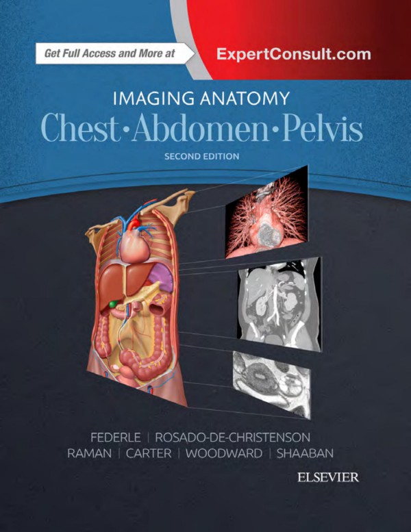
Imaging Anatomy Chest Abdomen Pelvis 2nd Edition by Michael Federle, Melissa Rosado de Christenson, Siva Raman, Brett Carter, Paula Woodward, Akram Shaaban ISBN 0323509460 9780323509466
Original price was: $50.00.$25.00Current price is: $25.00.
Authors:Michael P Federle; Melissa L. Rosado-De-christenson; Siva P. Raman; Brett W. Carter; Paula J. Woodward; Akram M. Shaaban , Series:Anatomy [86] , Tags:Medical; Radiology; Radiotherapy & Nuclear Medicine; Anatomy; Allied Health Services; Imaging Technologies; (Imaging Anatomy) , Author sort:Federle, Michael P & Rosado-De-christenson, Melissa L. & Raman, Siva P. & Carter, Brett W. & Woodward, Paula J. & Shaaban, Akram M. , Ids:9780323509442 , Languages:Languages:eng , Published:Published:Dec 2016 , Publisher:Elsevier Health Sciences , Comments:Comments:Designed to help you quickly learn or review normal anatomy and confirm variants, Imaging Anatomy: Chest, Abdomen, Pelvis provides detailed views of anatomic structures in successive imaging slices in each standard plane of imaging. Axial, coronal, sagittal, and 3D reconstructions accompany highly accurate and detailed medical drawings, assisting you in making an accurate diagnosis. Comprehensive coverage of the chest, abdomen, and pelvis, combined with an orderly, easy-to-follow structure, make this unique title unmatched in its field. Includes all relevant imaging modalities, 3D reconstructions, and highly accurate and detailed medical drawings that illustrate the fine points of the imaging anatomy Depicts common anatomic variants and covers common pathological processes as a part of its comprehensive coverage Provides a detailed overview of airway and interstitial network anatomy—the basis for understanding and diagnosing interstitial lung disease Features representative pathologic examples to highlight the effect of disease on human anatomy Includes plain radiography, the latest generation of multi-planar advanced cross-sectional MR and CT, ultrasound for pelvis/renal/liver/gallbladder, barium for GI tract, and much more Offers state of the art, detailed pelvic floor imaging and perianal/perirectal fistula imaging using high-resolution CT and MR, including 3T MR

