
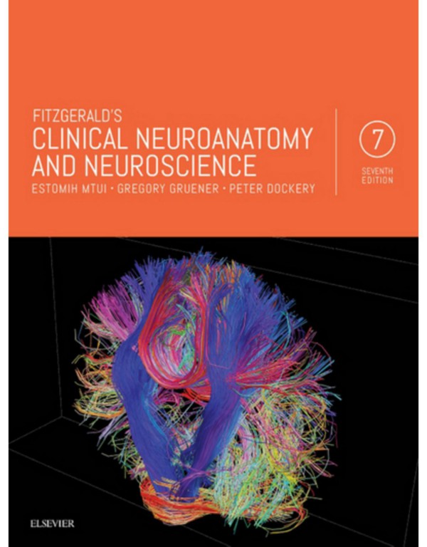
Fitzgerald Clinical neuroanatomy and neuroscience 7th Edition by Estomih Mtui, Gregory Gruener, Peter Dockery ISBN 9780702068140 0702068144

Original price was: $50.00.$25.00Current price is: $25.00.
Authors:Estomih Mtui , Series:Anatomy [241] , Tags:Clinical Neuroanatomy and Neuroscience , Author sort:Mtui, Estomih , Languages:Languages:eng , Published:Published:Dec 2015 , Publisher:Elsevier , Comments:« less;
Fitzgerald Clinical neuroanatomy and neuroscience 7th Edition by Estomih Mtui, Gregory Gruener, Peter Dockery – Ebook PDF Instant Download/Delivery. 9780702068140 ,0702068144
Full download Fitzgerald Clinical neuroanatomy and neuroscience 7th Edition after payment

Product details:
ISBN 10: 0702068144
ISBN 13: 9780702068140
Author: Estomih Mtui, Gregory Gruener, Peter Docker
Utilizing clear text and explanatory artwork to make clinical neuroanatomy and neuroscience as accessible as possible, this newly updated edition expertly integrates clinical neuroanatomy with the clinical application of neuroscience. It’s widely regarded as the most richly illustrated book available for guidance through this complex subject, making it an ideal reference for both medical students and those in non-medical courses.
- Complex concepts and subjects are broken down into easily digestible content with clear images and concise, straightforward explanations.
- Boxes within each chapter contain clinical information assist in distilling key information and applying it to likely real-life clinical scenarios.
- Chapters are organized by anatomical area with integrated analyses of sensory, motor and cognitive systems, and are designed to integrate clinical neuroanatomy with the basic practices and clinical application of neuroscience.
- Opening summaries at the beginning of each chapter feature accompanying study guidelines to show how the chapter contents apply in a larger context.
- Core information boxes at the conclusion of each chapter reinforce the most important facts and concepts covered.
- Bulleted points help expedite study and retention.
- Explanatory illustrations are drawn by the same meticulous artists who illustrated Gray’s Anatomy.
- Thoroughly updated content reflects the latest knowledge in the field.
Fitzgerald Clinical neuroanatomy and neuroscience 7th Edition Table of contents:
1: Embryology
Spinal cord
Brain
2: Cerebral Topography
Surface features
Internal anatomy of the cerebrum
3: Midbrain, Hindbrain, Spinal Cord
Brainstem
Spinal cord
Cerebellum
4: Meninges
Cranial meninges
Spinal meninges (Figure 4.10)
Circulation of the cerebrospinal fluid (Figure 4.12)
5: Blood Supply of the Brain
Arterial supply of the forebrain
Arterial supply to hindbrain
Venous drainage of the brain
Regulation of blood flow
The blood–brain barrier
6: Neurons and Neuroglia
Neurons
Synapses
Neuroglial cells of the central nervous system
7: Electrical Events
Structure of the plasma membrane
Response to stimulation: action potentials
8: Transmitters and Receptors
Electrical synapses
Chemical synapses
Transmitters and modulators
9: Peripheral Nerves
General features
Microscopic structure of peripheral nerves
Degeneration and regeneration
10: Innervation of Muscles and Joints
Motor innervation of skeletal muscle
Sensory innervation of skeletal muscle
Innervation of joints
11: Innervation of Skin
Sensory units
Nerve endings
12: Electrodiagnostic Examination
Nerve conduction studies
Electromyography
13: Autonomic Nervous System
Components of the autonomic nervous system
Sympathetic nervous system
Parasympathetic nervous system
Neurotransmission in the autonomic system
Regional autonomic innervation
Interaction of the autonomic and immune systems
Visceral afferents
14: Nerve Roots
Development of the spinal cord
Adult anatomy
Distribution of spinal nerves
15: Spinal Cord: Ascending Pathways
General features
Ascending sensory pathways
Somatic sensory pathways
Other ascending pathways
16: Spinal Cord: Descending Pathways
Anatomy of the ventral grey horn
Descending motor pathways
Blood supply of the spinal cord
17: Brainstem
General arrangement of cranial nerve nuclei
Background information
C1 segment of the spinal cord (Figure 17.10)
Spinomedullary junction (Figure 17.11)
Middle of the medulla oblongata (Figure 17.12)
Upper part of the medulla oblongata (Figure 17.13)
Pontomedullary junction (Figure 17.14)
Mid-pons (Figure 17.15)
Upper pons (Figure 17.16)
Lower midbrain (Figure 17.17)
Upper midbrain (Figure 17.18)
Midbrain–thalamic junction (Figure 17.19)
Orientation of brainstem slices in magnetic resonance images (Figure 17.20)
18: The Lowest Four Cranial Nerves
Hypoglossal nerve
Spinal accessory nerve
Glossopharyngeal, vagus, and cranial accessory nerves
19: Vestibular Nerve
Introduction
Vestibular system
20: Cochlear Nerve
Auditory system
21: Trigeminal Nerve
Trigeminal nerve
22: Facial Nerve
Facial nerve
Nervus intermedius
23: Ocular Motor Nerves
The nerves
Nerve endings
Pupillary light reflex (Figure 23.4)
Accommodation
Notes on the sympathetic pathway to the eye
Ocular palsies
Control of eye movements
24: Reticular Formation
Organisation
Functional anatomy
25: Cerebellum
Functional anatomy
Microscopic anatomy
Representation of body parts
Afferent pathways
Efferent pathways (Figure 25.10)
Anticipatory function of the cerebellum
Clinical disorders of the cerebellum
The cerebellum and higher brain functions
26: Hypothalamus
Gross anatomy
Functions
27: Thalamus, Epithalamus
Thalamus
Epithalamus
28: Visual Pathways
Retina
Central visual pathways
29: Cerebral Cortex
Structure
Cortical areas
Sensory areas
Motor areas
30: Electroencephalography
Neurophysiologic basis of the electroencephalogram
Technique
Types of patterns
31: Evoked Potentials
Sensory evoked potentials
Motor evoked potentials
32: Hemispheric Asymmetries
Handedness
Parietal lobe (Figure 32.6)
Prefrontal cortex
33: Basal Ganglia
Basic circuits
34: Olfactory and Limbic Systems
Olfactory system
Limbic system
35: Cerebrovascular Disease
Anterior circulation of the brain
Posterior circulation of the brain
Transient ischaemic attacks
Clinical anatomy of vascular occlusions
Glossary
Index
People also search for Fitzgerald Clinical neuroanatomy and neuroscience 7th Edition:
fitzgerald’s clinical neuroanatomy and neuroscience 8th edition
fitzgerald’s clinical neuroanatomy and neuroscience 2021
fitzgerald’s clinical neuroanatomy and neuroscience 7th edition
can you do neuroscience after psychology
You may also like…


eBook PDF
Biometrics for Dummies 1st Edition by Peter Gregory, Michael Simon ISBN 0470292881 9780470292884
eBook PDF
Snell Clinical Neuroanatomy 8th Edition by Ryan Splittgerber ISBN 1496346750 9781496346759

eBook PDF
Introduction To Personal Computers Windows 7th Edition by Peter Norton ISBN 1426019335 9781426019333