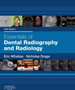
Essentials of Dental Radiography and Radiology 5th Edition by Eric Whaites, Nicholas Drage ISBN 0702051683 9780702051685


Original price was: $50.00.$25.00Current price is: $25.00.
Authors:Eric Whaites; Nicholas Drage , Series:Dentistry [245] , Author sort:Whaites, Eric & Drage, Nicholas , Ids:9780702051685 , Languages:Languages:eng , Published:Published:Jul 2013 , Publisher:Elsevier Health Sciences , Comments:Comments:Medical, Dentistry, General, Allied Health Services, Imaging TechnologiesThis is a new edition of a classic text that presents all of the information that a dental student needs to know in order to safely capture high-quality clinical images and accurately interpret their findings.In this latest edition, both traditional methods of imaging and new modalities are included, such as cone beam CT, and the author team has been expanded to bring a fresh approach to the subject area.Written in an accessible manner which avoids unnecessary detail, each page spread has been carefully designed to ensure clarity of understanding by the reader to ensure both exam success and confidence and safety in the clinical situation. Topics address the whole curriculum and range from the physics of imaging to radiation protection and image interpretation.Suitable for undergraduate students and post-graduates alike, this book has become essential reading for all readers who intend to practice clinical dentistry.Provides a comprehensive account of the radiology and radiography topics usually examined at undergraduate and postgraduate levelClear and accessible approach to the subject makes learning especially easyMore than 1100 illustrations present clinical, diagnostic and practical information in an accessible mannerWritten by a world authority on the subject areaContains recent classifications and advanced imaging modalities including cone beam CT imaging techniquesIncludes a new chapter on cone beam technology which includes the latest RCS (Eng) Guidelines for patient selectionContains an on-line self-assessment bank to aid exam preparationChapter on legislation now on-line to ensure constant currency of information
Essentials of Dental Radiography and Radiology 5th Edition by Eric Whaites, Nicholas Drage – Ebook PDF Instant Download/Delivery. 0702051683, 978-0702051685
Full download Essentials of Dental Radiography and Radiology 5th Edition after payment

Product details:
ISBN 10: 0702051683
ISBN 13: 978-0702051685
Author: Eric Whaites, Nicholas Drage
This is a new edition of a classic text that presents all of the information that a dental student needs to know in order to safely capture high-quality clinical images and accurately interpret their findings.
In this latest edition, both traditional methods of imaging and new modalities are included, such as cone beam CT, and the author team has been expanded to bring a fresh approach to the subject area.
Written in an accessible manner which avoids unnecessary detail, each page spread has been carefully designed to ensure clarity of understanding by the reader to ensure both exam success and confidence and safety in the clinical situation. Topics address the whole curriculum and range from the physics of imaging to radiation protection and image interpretation.
Suitable for undergraduate students and post-graduates alike, this book has become essential reading for all readers who intend to practice clinical dentistry.
- Provides a comprehensive account of the radiology and radiography topics usually examined at undergraduate and postgraduate level
- Clear and accessible approach to the subject makes learning especially easy
- More than 1100 illustrations present clinical, diagnostic and practical information in an accessible manner
- Written by a world authority on the subject area
- Contains recent classifications and advanced imaging modalities including cone beam CT imaging techniques
- Includes a new chapter on cone beam technology which includes the latest RCS (Eng) Guidelines for patient selection
- Contains an on-line self-assessment bank to aid exam preparation
- Chapter on legislation now on-line to ensure constant currency of information
Essentials of Dental Radiography and Radiology 5th Table of contents:
Part 1: Introduction
- The Radiographic Image
- Introduction
- Nature of the Radiographic Image
- Quality of the Radiographic Image
- Perception of the Radiographic Image
- Common Types of Dental Radiographs
Part 2: Radiation Physics, Equipment, and Radiation Protection
-
The Production, Properties, and Interactions of X-rays
- Introduction
- Atomic Structure
- X-ray Production
- Interaction of X-rays with Matter
-
Dental X-ray Generating Equipment
- Ideal Requirements
- Main Components of the Tubehead
- Main Components of the Control Panel
- Circuitry and Tube Voltage
- Other X-ray Generating Apparatus
-
Image Receptors
- Radiographic Film
- Digital Receptors
-
Image Processing
- Chemical Processing
- Computer Digital Processing
-
Radiation Dose, Dosimetry, and Dose Limitation
- Dose Units
- Dose Limits
- Dose Rate
- Dose Limitation
-
Biological Effects Associated with X-rays, Risk, and Practical Radiation Protection
- Radiation-induced Tissue Damage
- Classification of Biological Effects
- Practical Radiation Protection
Part 3: Radiography
-
Dental Radiography – General Patient Considerations, Including Control of Infection
- General Guidelines on Patient Care
- Specific Requirements When X-raying Children and Patients with Disabilities
- Control of Infection
-
Periapical Radiography
- Main Indications
- Ideal Positioning Requirements
- Radiographic Techniques
- Positioning Difficulties in Periapical Radiography
-
Bitewing Radiography
- Main Indications
- Ideal Technique Requirements
- Positioning Techniques
- Resultant Radiographs
-
Occlusal Radiography
- Terminology and Classification
-
Oblique Lateral Radiography
- Introduction
- Terminology
- Main Indications
- Equipment Required
- Basic Technique Principles
- Positioning Examples for Various Oblique Lateral Radiographs
- Bimolar Technique
-
Skull and Maxillofacial Radiography
- Equipment, Patient Positioning, and Projections
-
Cephalometric Radiography
- Main Indications
- Equipment
- Main Radiographic Projections
- Cephalometric Posteroanterior of the Jaws (PA Jaws)
-
Tomography and Panoramic Radiography
- Introduction
- Tomographic Theory
- Panoramic Tomography
- Selection Criteria
- Equipment
- Technique and Positioning
-
Cone Beam Computed Tomography (CBCT)
- Main Indications
- Equipment and Theory
- Technique and Positioning
-
The Quality of Radiographic Images and Quality Assurance
- Introduction
- Film-based Image Quality
- Practical Factors Influencing Film-based Image Quality
- Typical Film Faults
- Patient Preparation and Positioning Errors
- Quality Assurance in Dental Radiology
- Digital Image Quality
- Practical Factors Influencing Digital Image Quality
- Typical Digital Image Faults
- Quality Control Procedures for Digital Radiography
-
Alternative and Specialized Imaging Modalities
- Introduction
- Contrast Studies
- Radioisotope Imaging
- Computed Tomography (CT)
- Ultrasound
- Magnetic Resonance (MR)
Part 4: Radiology
-
Introduction to Radiological Interpretation
- Essential Requirements for Interpretation
- Conclusion
-
Dental Caries and the Assessment of Restorations
- Introduction
- Classification of Caries
- Diagnosis and Detection of Caries
- Other Important Radiographic Appearances
- Limitations of Radiographic Detection of Caries
- Radiographic Assessment of Restorations
- Limitations of the Radiographic Image
- Suggested Guidelines for Interpreting Bitewing Images
-
The Periapical Tissues
- Introduction
- Normal Radiographic Appearances
- Radiographic Appearances of Periapical Inflammatory Changes
- Other Important Causes of Periapical Radiolucency
- Suggested Guidelines for Interpreting Periapical Images
-
The Periodontal Tissues and Periodontal Disease
- Introduction
- Selection Criteria
- Radiographic Features of Healthy Periodontium
- Classification of Periodontal Disease
- Radiographic Features of Periodontal Disease
- Assessment of Bone Loss and Furcation Involvement
- Evaluation of Treatment Measures
- Limitations of Radiographic Diagnosis
-
Implant Assessment
- Introduction
- Main Indications
- Treatment Planning Considerations
- Radiographic Examination
- Postoperative Evaluation and Follow-up
-
Developmental Abnormalities
- Introduction
- Classification of Developmental Abnormalities
- Typical Radiographic Appearances of the More Common and Important Developmental Abnormalities
- Radiographic Assessment of Mandibular Third Molars
- Radiographic Assessment of Unerupted Maxillary Canines
-
Radiological Differential Diagnosis – Describing a Lesion
- Introduction
- Detailed Description of a Lesion
-
Differential Diagnosis of Radiolucent Lesions of the Jaws
- Introduction
- Step-by-Step Guide
- Typical Radiographic Features of Cysts
- Typical Radiographic Features of Tumors and Tumor-like Lesions
- Typical Radiographic Features of Bone-related Lesions
-
Differential Diagnosis of Lesions of Variable Radiopacity in the Jaws
- Typical Radiographic Features of Abnormalities of the Teeth
- Typical Radiographic Features of Conditions of Variable Opacity Affecting Bone
- Typical Radiographic Features of Soft Tissue Calcifications
- Typical Radiographic Features of Foreign Bodies
-
Bone Diseases of Radiological Importance
- Introduction
- Developmental or Genetic Disorders
- Infective or Inflammatory Conditions
- Hormone-related Diseases
- Blood Dyscrasias
- Diseases of Unknown Cause
-
Trauma to the Teeth and Facial Skeleton
- Introduction
- Injuries to the Teeth and Their Supporting Structures
- Skeletal Fractures
- Fractures of the Mandible
- Fractures of the Middle Third of the Facial Skeleton
- Other Fractures and Injuries
-
The Temporomandibular Joint
- Introduction
- Normal Anatomy
- Investigations
- Main Pathological Conditions Affecting the TMJ
-
The Maxillary Antra
- Introduction
- Normal Anatomy
- Normal Appearance of the Antra on Conventional Radiographs
- Antral Disease
- Investigation and Appearance of Disease within the Antra
- Other Paranasal Air Sinuses
-
The Salivary Glands
- Salivary Gland Disorders
- Investigations
People also search for Essentials of Dental Radiography and Radiology 5th:
essentials of dental radiography and radiology 6th edition pdf
whaites and drage essentials of dental radiography and radiology 2020
what are the five steps in processing dental radiographs
dental radiography techniques
importance of quality assurance in dental radiography
You may also like…