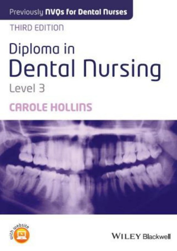
Diploma in Dental Nursing Level 3 3rd Edition by Carole Hollins ISBN 1118905059 9781118905050
$50.00 Original price was: $50.00.$25.00Current price is: $25.00.
Authors:Hollins, Carole , Tags:EBC Converted , Author sort:Hollins, Carole , Published:Published:Jul 2014
Diploma in Dental Nursing Level 3 3rd Edition by Carole Hollins – Ebook PDF Instant Download/Delivery. 1118905059, 9781118905050
Full download Diploma in Dental Nursing Level 3 3rd Edition after payment
Product details:
ISBN 10: 1118905059
ISBN 13: 9781118905050
Author: Carole Hollins
Diploma in Dental Nursing, Level 3 is the new edition of the must-have study companion for trainee dental nurses preparing for the City & Guilds Level 3 Diploma in Dental Nursing (formerly NVQ). The book offers comprehensive support on the units assessed by portfolio – from first aid and health and safety to specific chairside support procedures – as well as the four areas of the course tested by multiple choice questions: infection control, oral health assessment, dental radiography and oral health management.
Diploma in Dental Nursing Level 3 3rd Table of contents:
1 Unit 301: Ensure Your Own Actions Reduce Risks to Health and Safety
Learning outcomes
Outcome 1 assessment criteria
Outcome 2 assessment criteria
Outcome 3 assessment criteria
Overview of responsibilities
Employers’ responsibilities
Figure 1.1 A health and safety poster.
Risk assessment to identify the hazards
Employees’ responsibilities – role of the dental nurse
Figure 1.2 Hazard sign indicating a wet floor.
Personal presentation
Personal hygiene
Use of PPE
Figure 1.3 A hand washing poster.
Suitable clothing and accessories
Summary of health and safety requirements
Fire Precaution (Workplace) Regulations
Fire detection
Figure 1.4 A smoke alarm.
Figure 1.5 A fire extinguisher.
Firefighting
Evacuation and escape routes
Figure 1.6 A fire exit pictogram.
Figure 1.7 A fire action poster.
Smoking in the workplace
First aid regulations
Knowledge and use of emergency equipment
Figure 1.8 A first aid box.
Figure 1.9 A first aid box location poster.
Control of Substances Hazardous to Health (COSHH)
Figure 1.10 Symbols of Control of Substances Hazardous to Health (COSHH) risk categories.
Figure 1.11 Example of a Control of Substances Hazardous to Health (COSHH) assessment sheet.
Storage
Ventilation and temperature control
Occupational hazardous chemicals
Mercury
Inhalation
Absorption
Figure 1.12 A pre-loaded amalgam capsule.
Figure 1.13 A waste amalgam tub.
Figure 1.14 A tub for waste amalgam capsules.
Ingestion
Handling of mercury spillages
Figure 1.15 A page of an accident book.
Figure 1.16 Collection of mercury droplets.
Figure 1.17 Mercury droplets collected in a syringe.
Figure 1.18 An amalgamator with a lid.
Figure 1.19 A mercury spillage kit.
Figure 1.20 An acid etchant gel.
Acid etchant
Bleach (and other disinfectants)
Figure 1.21 Examples of disinfectants.
Figure 1.22 Bottle label displaying hazard symbol.
Reporting of Injuries, Diseases, and Dangerous Occurrences Regulations (RIDDOR)
Waste disposal
Figure 1.23 Current waste classifications relevant to the dental workplace. PPE, personal protective equipment; LA, local anaesthetic.
Figure 1.24 Blue-lidded sharps box.
Figure 1.25 An orange hazardous waste sack.
Figure 1.26 A sharps box.
Figure 1.27 Waste processing chemical storage drums.
Waste handling training
Ionising radiation
Figure 1.28 Controlled area and safety zone.
Occupational hazards
Inoculation injury
Figure 1.29 A re-sheathing device. (a) Needle guard in place. (b) Syringe re-sheathed in the device.
General safety measures
Manual handling
Weight and dimensions
Awkward movements and frequency
Excessive movements
Distance and stairs
Physical ability and medical conditions
Training
2 Unit 302: Reflect On and Develop Your Practice
Learning outcomes
Outcome 1 assessment criteria
Outcome 2 assessment criteria
Outcome 3 assessment criteria
Outcome 4 assessment criteria
Outcome 5 assessment criteria
Reflect on performance
Use of diaries or portfolios
Use of personal development plans
Use of staff appraisal
Figure 2.1 SWOT analysis.
Figure 2.2 A sample staff appraisal sheet.
Implement a performance improvement plan
Actions needed
Prioritising
SMART objectives
Development opportunities
Continuing professional development
Figure 2.3 Continuing professional development (CPD) certificate.
Figure 2.4 An example of a continuing professional development (CPD) record sheet.
Post-registration qualifications
Extended duties
Direct access
Evaluate the effectiveness of the plan
Compliance with registration requirements
Figure 2.5 General Dental Council’s Standards for the Dental Team document.
1 Put patients’ interests first
1.1 Listen to your patients
1.2 Treat every patient with dignity and respect at all times
1.3 Be honest and act with integrity
1.4 Take a holistic and preventative approach to patient care which is appropriate to the individual patient
1.5 Treat patients in a hygienic and safe environment
1.6 Treat patients fairly, as individuals and without discrimination
1.7 Put patients’ interests before your own or those of any colleague, business or organisation
1.8 Have appropriate arrangements in place for patients to seek compensation if they have suffered harm
1.9 Find out about laws and regulations that affect your work and follow them
2 Communicate effectively with patients
2.1 Listen to them and give them time to consider information, taking their views and individual needs into consideration
2.2 Recognise and promote patients’ rights and responsibilities for making decisions about their health priorities and care
2.3 Give patients the information they need, in a way they can understand, so that they can make informed decisions
2.4 Give patients clear information about costs
3 Obtain valid consent
3.1 Obtain valid consent before starting treatment, explaining all the relevant options and the possible costs
3.2 Make sure that patients (or their representatives) understand the decisions they are being asked to make
3.3 Make sure that the patient’s consent remains valid at each stage of investigation or treatment
4 Maintain and protect patients’ information
4.1 Make and keep contemporaneous, complete and accurate patient records
4.2 Protect the confidentiality of patients’ information and only use it for the purpose for which it was given
4.3 Only release a patient’s information without their permission in exceptional circumstances
4.4 Ensure that patients can have access to their records
4.5 Keep patients’ information secure at all times, whether your records are held on paper or electronically
5 Have a clear and effective complaints procedure
5.1 Make sure that there is an effective complaints procedure readily available for patients to use, and follow that procedure at all times
5.2 Respect a patient’s right to complain
5.3 Give patients who complain a prompt and constructive response
6 Work with colleagues in a way that is in patients’ best interests
6.1 Work effectively with your colleagues and contribute to good teamwork
6.2 Be appropriately supported when treating patients
6.3 Delegate and refer appropriately and effectively
6.4 Only accept a referral or delegation if you are trained and competent to carry out the treatment and you believe that what you are being asked to do is appropriate for the patient
6.5 Communicate clearly and effectively with other team members and colleagues in the interests of patients
6.6 Demonstrate effective management and leadership skills if you manage a team
7 Maintain, develop and work within your professional knowledge and skills
7.1 Provide good quality care based on current evidence and authoritative guidance
7.2 Work within your knowledge, skills, professional competence and abilities
7.3 Update and develop your professional knowledge and skills throughout your working life
8 Raise concerns if patients are at risk
8.1 Always put patients’ safety first
8.2 Act promptly if patients or colleagues are at risk and take measures to protect them
8.3 Make sure if you employ, manage or lead a team that you encourage and support a culture where staff can raise concerns openly and without fear of reprisal
8.4 Make sure if you employ, manage or lead a team, that there is an effective procedure in place for raising concerns, that the procedure is readily available to all staff and that it is followed at all times
8.5 Take appropriate action if you have concerns about the possible abuse of children or vulnerable adults (including the elderly)
9 Make sure your personal behaviour maintains patients’ confidence in you and the dental profession
9.1 Ensure that your conduct, both at work and in your personal life, justifies patients’ trust in you and the public’s trust in the dental profession
9.2 Protect patients and colleagues from risks posed by your health, conduct or performance
9.3 Inform the GDC if you are subject to criminal proceedings or a regulatory finding is made against you, anywhere in the world
9.4 Co-operate with any relevant formal or informal inquiry and give full and truthful information
3 Unit 268: First Aid Essentials
Learning outcomes
Outcome 1 assessment criteria
Outcome 2 assessment criteria
Outcome 3 assessment criteria
Outcome 4 assessment criteria
Outcome 5 assessment criteria
Outcome 6 assessment criteria
Outcome 7 assessment criteria
Outcome 8 assessment criteria
Roles and responsibilities of the first aider
Health and safety requirements
Infection control and minimising cross-infection
Correct reporting of incidents
Knowledge and use of emergency equipment
Figure 3.1 An automatic external defibrillator.
Figure 3.2 An emergency drugs kit.
Emergency equipment/drugs
Figure 3.3 A Geudel airway.
Communication and care of the casualty
Incident assessment
Primary survey
Secondary survey
Management of an unresponsive casualty who is breathing normally
Danger
Response
Shout
Airway
Breathing
Figure 3.4 A manual suction device.
Figure 3.5 Head tilt to open airway.
Figure 3.6 Jaw thrust to open airway.
Figure 3.7 Look, listen and feel for signs of breathing.
Figure 3.8 The recovery position.
Recovery position
Modified recovery position
Casualty having a seizure
Management of an unresponsive casualty with breathing problems
Circulation
Figure 3.9 Carrying out external cardiac compressions.
Figure 3.10 Hand lock for chest compressions.
Figure 3.11 Arm lock for chest compressions.
Rescue breathing
Figure 3.12 Use of the ventilation bag.
Figure 3.13 Rescue breathing mouth to mouth.
Cardiopulmonary modifications
Babies and young children
Pregnant women
Management of a choking casualty
Figure 3.14 Back slaps between shoulder blades.
Figure 3.15 Abdominal thrusts.
Choking in young children
Figure 3.16 Baby back slaps using fingers.
Choking in babies
Management of a wounded and bleeding casualty
Figure 3.17 Faint recovery position.
Management of a casualty in shock
Management of minor injuries
Small cuts, grazes, and bruises
Minor burns and scalds
Small splinters
4 Unit 304: Prepare and Maintain Environment, Instruments and Equipment for Clinical Dental Procedures
Learning outcomes
Outcome 1 assessment criteria
Outcome 2 assessment criteria
Outcome 3 assessment criteria
Outcome 4 assessment criteria
Application of standard precautions of infection control
Standard precautions
Personal protective equipment
Figure 4.1 Items of personal protective equipment.
Figure 4.2 Personal protective equipment items in use.
Maintenance of a clean and tidy work environment
Correct use of cleaning equipment
Maintenance of suitable environmental factors
Application of health and safety measures
Personal hygiene and hand washing
Hand washing techniques
Figure 4.3 Sink-side dispensers.
Figure 4.4 Elbow-operated tap in use.
Equipment maintenance
Figure 4.5 Illuminated X-ray machine controls.
Figure 4.6 Velopex machine warning lights.
Figure 4.7 “TST” strip for checking autoclave function.
Figure 4.8 An autoclave log printout.
Figure 4.9 Helix device for checking vacuum autoclave function.
Storage of equipment and materials
Actions to take in the event of a spillage
Water spillages
Chemical spillages
Body fluid spillages
Reporting of hazards
Inoculation injury
Application of suitable sterilisation methods
Transportation to the decontamination room
Decontamination room layout
Preparation of items for sterilisation
Handpieces
Dental instruments
Figure 4.10 Use of the illuminated magnifier for instrument inspection.
Figure 4.11 An ultrasonic bath.
Figure 4.12 A washer-disinfector machine.
Sterilisation procedures
Figure 4.13 Downward displacement autoclave.
Figure 4.14 A vacuum autoclave.
Storage of sterilised items
Figure 4.15 A date-stamped sterilisation pouch.
Safe disposal of hazardous and non-hazardous waste
Figure 4.16 A waste ‘tooth pot’.
Waste handling training
5 Unit 305: Offer Information and Support to Individuals on the Protection of Their Oral Health
Learning outcomes
Outcome 1 assessment criteria
Outcome 2 assessment criteria
Correct identification of the patient
Gaining valid consent
Communication skills
Provide appropriate oral health information
Dental caries
Non-carious tooth surface loss
Gingivitis and periodontal disease
Diet
Figure 5.1 Collection of unhealthy snacks.
Figure 5.2 Collection of “hidden sugar” products.
Specific issues
Figure 5.3 An example of a “quit smoking” leaflet.
Demonstrate and advise on appropriate oral hygiene techniques
Figure 5.4 Plaque visible on teeth after using a disclosing liquid.
Figure 5.5 Supervised child tooth brushing.
Tooth brushing demonstration and advice
Figure 5.6 A Sonicare electric toothbrush.
Figure 5.7 Gingival crevice brushing.
Figure 5.8 Extensive abrasion cavities.
Figure 5.9 New and worn toothbrushes.
Figure 5.10 Examples of different toothpastes.
Toothpastes
Figure 5.11 High-concentration fluoride toothpaste.
Figure 5.12 Desensitising toothpaste.
Figure 5.13 Enamel repair toothpaste.
Interdental cleaning
Figure 5.14 Dental flosses and tapes.
Figure 5.15 Interdental flossettes.
Figure 5.16 Interdental brush detail.
Figure 5.17 Use of an interdental brush for posterior cleaning.
Figure 5.18 Flossing technique.
Figure 5.19 Types of mouthwash.
Use of mouthwashes
Figure 5.20 A desensitising mouthwash.
Figure 5.21 A peroxyl mouthwash.
Figure 5.22 A corsodyl daily mouthwash.
Figure 5.23 A fluoride mouthwash.
Oral hygiene advice for denture wearers
Oral hygiene advice for orthodontic patients
Fixed appliances
Removable appliances
6 Unit 306: Provide Chairside Support during the Assessment of Patients’ Oral Health
Learning outcomes
Outcome 1 assessment criteria
Outcome 2 assessment criteria
Types of patient records
Figure 6.1 A typical NHS FP25 record card.
Personal details
Medical history
Dental charts
Radiographs
Figure 6.2 An example of completed charting grid.
Figure 6.3 Example of a completed basic periodontal examination (BPE) chart.
Figure 6.4 A detailed periodontal assessment sheet.
Figure 6.5 A soft tissue assessment sheet.
Figure 6.6 A horizontal bitewing radiograph.
Figure 6.7 A periapical radiograph.
Figure 6.8 A dental pantomograph.
Figure 6.9 A lateral skull view.
Photographs
Study models
Figure 6.10 A set of study models.
Orthodontic measurements
Figure 6.11 An orthodontic assessment sheet.
Retrieve and make available the patient records
Setting up for the assessment
Observing hard and soft tissues
Figure 6.12 Mouth mirror, angled probe and tweezers.
Figure 6.13 Cotton wool roll and cotton wool pledget.
Figure 6.14 Briault probe.
Measuring and making records
Orthodontic assessment
Figure 6.15 Periodontal probes. (a) Calculus probe, (b) BPE/CPITN probe and (c) pocket measuring probe.
Figure 6.16 A measuring ruler to record the overjet and help record the overbite.
Figure 6.17 Alginate mixing equipment.
Recording the verbal assessment information
Observation of hard and soft tissues
Tooth charting
Palmer notation
Figure 6.18 A manual charting grid.
Two-digit FDI World Dental Federation notation
Figure 6.19 Surfaces of the teeth – mesial aspect.
Figure 6.20 Surfaces of the teeth – occlusal aspect.
BPE and periodontal charting
Figure 6.21 Basic periodontal examination (BPE) probe.
Orthodontic assessment
Figure 6.22 Class I occlusion.
Figure 6.23 Class II division 1 malocclusion with lip trap.
Figure 6.24 Class II division 2 malocclusion with increased overbite.
Figure 6.25 Class III malocclusion with reverse overjet.
Figure 6.26 Overjet and overbite.
Recording relevant medical conditions
Figure 6.27 An example of a medical history form.
Maintaining confidentiality of the information
7 Unit 307: Contribute to the Production of Dental Images
Learning outcomes
Outcome 1 assessment criteria
Outcome 2 assessment criteria
Outcome 3 assessment criteria
Maintain the health and safety of all persons
Correct identification of the patient
Other health and safety actions
Set up for the taking of dental images
Figure 7.1 Intra-oral films: (a) child size; (b) adult size.
Figure 7.2 A vertical bitewing radiograph.
Figure 7.3 Maxillary anterior occlusal.
Figure 7.4 Extra-oral dental panoramic tomograph (DPT) cassette.
Knowledge of the correct setting up of the area for the procedure
Figure 7.5 Digital sensor plate and cable.
Figure 7.6 Bitewing holders: horizontal (left) and vertical (right).
Figure 7.7 Periapical holders: anterior (left) and posterior (right).
Figure 7.8 Close-up of holder showing beam direction arrow.
Process the dental films and mount them correctly
Automatic processing
Figure 7.9 Velopex processing machine – internal detail.
Manual processing
Figure 7.10 Darkroom layout.
Figure 7.11 Viewing screen.
Figure 7.12 Correctly mounted upper periapical radiograph.
Figure 7.13 Correctly mounted lower periapical radiograph.
Store or save the images correctly
Contribute to quality assurance checks
Figure 7.14 Radiograph scoring 1 on the quality assurance (QA) scale, showing a periapical abscess.
Figure 7.15 Radiograph of UR5 scoring 2 on the quality assurance (QA) scale.
Figure 7.16 Radiograph scoring 3 on the quality assurance (QA) scale.
8 Unit 308: Provide Chairside Support during the Prevention and Control of Periodontal Disease and Caries, and the Restoration of Cavities
Learning outcomes
Outcome 1 assessment criteria
Outcome 2 assessment criteria
Before the procedure
Correct identification of the patient
Figure 8.1 Correctly mounted dental pantomograph.
Correct identification of the procedure
Knowledge of the correct setting up of the area
Figure 8.2 Use of a soap dispenser.
Figure 8.3 Barrier protection on handles.
Knowledge of the instruments, materials and equipment required
Prevention and control of periodontal disease by scaling and debridement
Figure 8.4 An ultrasonic scaler.
Figure 8.5 Curettes.
Figure 8.6 Polishing cup and brush.
Prevention and control of caries using fissure sealants
Prevention and control of caries by fluoride application
Figure 8.7 Duraphat varnish.
Restoration of cavities
Material differences
Figure 8.8 Handpieces.
Figure 8.9 A bur block stand.
Amalgam
Figure 8.10 The Siqveland matrix system.
Figure 8.11 A plastic amalgam carrier.
Composite
Figure 8.12 A transparent matrix strip.
Figure 8.13 A curing light.
Figure 8.14 A finishing strip.
Figure 8.15 A glass ionomer cervical matrix system.
Glass ionomer
Knowledge of the actions to take if preparation cannot be completed
During the procedure
Preparation of the patient
Assistance during the administration of local anaesthesia
Figure 8.16 Handing over the loaded syringe.
Identification and correct handling of the instruments
Prevention and control of periodontal disease
Prevention and control of caries
Restoration of cavities
Figure 8.17 Composite compoule in gun device.
Figure 8.18 Zinc oxide–eugenol cement – to mix.
Zinc oxide and eugenol
Zinc phosphate
Figure 8.19 Zinc phosphate cement – to mix.
Figure 8.20 Zinc polycarboxylate cement – to mix.
Zinc polycarboxylate
Adequate provision of moisture control and tissue retraction
Figure 8.21 Aspirator and ejector tips.
Monitoring and support of the patient
After the procedure
Complete the patient records
Correct handling and disposal of waste amalgam
Precautions to be followed by all staff
Mercury spillage
Decontaminate the surgery and instruments
9 Unit 309: Provide Chairside Support during the Provision of Fixed and Removable Prostheses
Learning outcomes
Outcome 1 assessment criteria
Outcome 2 assessment criteria
Outcome 3 assessment criteria
Outcome 4 assessment criteria
Before the procedure
Correct identification of the patient
Correct identification of the procedure
Knowledge of the correct setting up of the area
Knowledge of the instruments, materials and equipment required
Fixed prosthesis preparation
Figure 9.1 Crown preparation diamond burs.
Figure 9.2 Retraction cord and solution.
Figure 9.3 Examples of suitable impression trays.
Figure 9.4 A crown former.
Figure 9.5 Beebee crown shears.
Figure 9.6 A shade guide.
Figure 9.7 Post systems.
Figure 9.8 A conservation tray.
Figure 9.9 Rubber dam equipment.
Fixed prosthesis fitting
Figure 9.10 Miller forceps and articulating paper.
Figure 9.11 Polishing burs.
Removable prosthesis appointment
First impressions
Second impressions
Bite registration
Figure 9.12 Stock impression trays – edentulous and boxed.
Figure 9.13 A denture shade guide.
Figure 9.14 A work ticket (docket).
Figure 9.15 Special trays and initial models.
Figure 9.16 Wax bite rims with models.
Figure 9.17 Pink wax, wax knife and burner.
Figure 9.18 Willis bite gauge in position.
Try-in
Fitting
Figure 9.19 Le Cron carver.
Figure 9.20 Acrylic burs (a) and stones (b).
Orthodontic appliances
Removable appliance fitting and adjustment
Fixed appliance bonding and adjustment
Figure 9.21 Adams universal pliers.
Figure 9.22 Straight handpiece and acrylic trimming bur.
Figure 9.23 Archwire, bracket and molar band with tube.
Figure 9.24 Bracket and band removers.
Retainer fit
Knowledge of the actions to take if preparation cannot be completed
During the procedure
Preparation of the patient
Assistance during the administration of local anaesthesia
Assistance during the placement of rubber dam
Identification and correct handling of the instruments, etc
Figure 9.25 Rubber dam in place on lower molar tooth.
Adequate provision of moisture control and tissue retraction
Mixing impression materials and loading trays
Alginate impression material
Figure 9.26 Alginate mixing stages (a–d) and tray loading (e, f).
Silicone impression material
Figure 9.27 A graduated elastomer mixing pad.
Figure 9.28 Expressed material before applying mixing tube.
Polyether impression material
Figure 9.29 Polyether collection technique.
Correct handling of impressions and other items
Mixing luting cements and temporary cements
Zinc phosphate
Zinc polycarboxylate
Figure 9.30 Glass ionomer powder, scoop and liquid.
Glass ionomers
Light-cure and dual-cure materials
Monitoring and support of the patient
After the procedure
Provide oral hygiene instructions
Fixed prostheses
Removable prostheses
Figure 9.31 Superfloss.
Orthodontic appliances
Patient advice for fixed appliances
Patient advice for removable appliances
Complete the patient records
Decontaminate the surgery and instruments
10 Unit 310: Provide Chairside Support during Non-surgical Endodontic Treatment
Learning outcomes
Outcome 1 assessment criteria
Outcome 2 assessment criteria
Before the procedure
Correct identification of the patient
Correct identification of the procedure
Knowledge of the correct setting up of the area
Knowledge of the instruments, materials and equipment required
Pulpectomy
Figure 10.1 Endodontic instruments: (a) barbed broach; (b) reamer; (c) file; (d) spiral paste filler.
Figure 10.2 A Gates Glidden drill.
Figure 10.3 An endodontic irrigation syringe.
Figure 10.4 An apex locator.
Figure 10.5 A finger spreader.
Figure 10.6 An endodontic instrument tray.
Figure 10.7 Ledermix paste.
Figure 10.8 Paper points.
Figure 10.9 Cresophene antiseptic solution.
Figure 10.10 Glyde lubricant.
Figure 10.11 Gutta-percha points.
Pulpotomy
Pulp capping
Knowledge of the actions to take if preparation cannot be completed
During the procedure
Preparation of the patient
Assistance during the administration of local anaesthesia
Assistance during the placement of rubber dam
Identification and correct handling of the instrument
Adequate provision of moisture control and tissue retraction
Monitoring and support of the patient
After the procedure
Complete the patient records
Decontaminate the surgery and instruments
11 Unit 311: Provide Chairside Support during the Extraction of Teeth and Minor Oral Surgery
Learning outcomes
Outcome 1 assessment criteria
Outcome 2 assessment criteria
Outcome 3 assessment criteria
Before the procedure
Correct identification of the patient
Correct identification of the procedure
Extractions
Knowledge of the correct setting up of the area
Figure 11.1 Bite pack.
Figure 11.2 Haemostatic sponges.
Knowledge of the instruments, materials and equipment required
Figure 11.3 Extraction forceps: (a) upper straight, root, premolar; (b) upper left and right molar, bayonets; (c) lower anterior, root, molar, cowhorns.
Figure 11.4 A luxator.
Figure 11.5 Elevators.
Figure 11.6 A disposable fine-bore aspirator.
Figure 11.7 Coupland chisels – sizes 1, 2, and 3.
Figure 11.8 Scalpel blade and handle.
Figure 11.9 An osteotrimmer.
Figure 11.10 A periosteal elevator.
Figure 11.11 Tissue dissecting forceps.
Figure 11.12 A pair of needle holders.
Figure 11.13 A suture pack.
Figure 11.14 Suture scissors.
Figure 11.15 An implant kit.
Knowledge of the actions to take if preparation cannot be completed
During the procedure
Preparation of the patient
Assistance during the administration of local anaesthesia
Identification and correct handling of the instruments
Figure 11.16 No-touch technique of passing bagged items.
Adequate provision of moisture control, irrigation and tissue retraction
Figure 11.17 Retractors: (a) Austin; (b) Kilner; (c) tissue rake.
Correct handling of body tissues
Assistance during the placement of haemostats/sutures
Monitoring and support of the patient
Figure 11.18 Cutting the suture.
Recognition of complications
Actions to take in the event of a complication
After the procedure
Provide postoperative instructions
Figure 11.19 Postoperative instruction leaflet.
Complete the patient records
Discharge the patient
Decontaminate the surgery and instruments
Figure 11.20 Handwritten procedure entry.
12 Unit 312: Principles of Infection Control in the Dental Environment
Learning outcomes
Outcome 1 assessment criteria
Outcome 2 assessment criteria
Outcome 3 assessment criteria
Outcome 4 assessment criteria
Outcome 5 assessment criteria
Background knowledge of microorganisms
Figure 12.1 Shapes of bacteria.
Figure 12.2 Structure of a virus.
Infection
Response of the body to infection
Inflammatory response
Immune response
Occupational hazards – cross-infection and inoculation injury
Hepatitis B
High-risk groups
Prevention of cross-infection
Treatment of known carriers
Hepatitis C
AIDS
Infectivity
High-risk groups
Prevention
Treatment of known carriers
New variant Creutzfeld–Jakob disease (vCJD)
Inoculation injury
Infection control
Basic principles of infection control
Cleaning of the hands
Use of PPE
Cleaning of the clinical environment
Figure 12.3 Sealed pouch and lidded tray.
Figure 12.4 A non-foaming aspirator disinfectant.
Cleaning of equipment, handpieces and instruments
Details of cleaning techniques for reusable items
Manual cleaning
Ultrasonic bath
Maintenance and testing of ultrasonic bath
Washer-disinfector
Maintenance and testing of washer-disinfector
Autoclave
Handling and storage of sterilised items
Maintenance and testing of autoclave
Other methods of sterilisation
Decontamination room
Figure 12.5 Layout and operation of the decontamination room.
Hazardous waste disposal
Waste classification
Figure 12.6 Hazardous waste designations. LA, local anaesthetic; PPE, personal protective equipment.
Relevant health and safety legislation, policies and guidelines
Health and Safety at Work Act
Care Quality Commission
Decontamination in primary dental care practices – HTM 01-05
Control of Substances Hazardous to Health (COSHH)
Reporting of Injuries, Diseases, and Dangerous Occurrences Regulations (RIDDOR)
Special Waste and Hazardous Waste Regulations
Environmental Protection Act
13 Unit 313: Assessment of Oral Health and Treatment Planning
Learning outcomes
Outcome 1 assessment criteria
Outcome 2 assessment criteria
Outcome 3 assessment criteria
Outcome 4 assessment criteria
Outcome 5 assessment criteria
Oral health assessments
Methods used to carry out assessment
Extra-oral soft tissue assessment
Figure 13.1 A “cold sore” lesion on upper lip.
Intra-oral soft tissue assessment
Figure 13.2 A white patch on buccal mucosa.
Periodontal assessment
Figure 13.3 Division of mouth into sextants for recording of the basic periodontal examination (BPE).
Figure 13.4 Periodontal pocket probing procedure.
Tooth charting
Palmer notation
Two-digit FDI notation
Use of radiographs
Figure 13.5 Radiograph showing dental caries.
Figure 13.6 Radiograph showing periodontal disease.
Figure 13.7 Radiograph showing filling overhangs UL6 and UL7.
Figure 13.8 Radiograph showing unerupted teeth.
Types of views used in dental radiography
Use of photographs
Use of study models
Vitality tests
Figure 13.9 Ethyl chloride liquid in container.
Figure 13.10 Greenstick compound for vitality testing.
Materials used in oral assessments
Legislation and guidelines in relation to patient records and confidentiality
Importance of records
Clinical records
Dental history
Present oral health status
Treatment
National Health Service records
Confidentiality of patient records
Disclosure without patient consent
Access to health records
General Dental Council (GDC) Standards Guidance and confidentiality
Information governance
Consent to treatment
Key definitions
Who can give consent?
GDC Standards Guidance and patient consent
Patient complaints
“In house” complaints procedure
GDC Standards Guidance and complaints handling
Summary of oral assessment
Orthodontic clinical assessment
Figure 13.11 Class I molar and canine relationship.
Types of malocclusion
Crowding
Protruding upper incisors
Prominent lower jaw
Orthodontic appliances
Figure 13.12 Class II division 1 malocclusion.
Figure 13.13 Class II division 2 malocclusion.
Figure 13.14 Class III malocclusion.
Fixed orthodontic appliances
Patient advice for fixed appliances
Removable orthodontic appliances
Figure 13.15 Removable upper orthodontic appliance.
Figure 13.16 Types of spring: (a) palatal finger spring; (b) “Z” spring.
Figure 13.17 Types of retractor: (a) buccal canine retractor; (b) Roberts retractor.
Patient advice for removable appliances
Functional appliances
Figure 13.18 Functional appliance.
Role of the dental nurse during orthodontic assessment and treatment
Disease-related changes of the oral tissues
Diseases of the oral soft tissues
Oral white and red patches
Inflammatory disorders
Effects of ageing on the soft tissues
Medical conditions that affect the oral tissues
Oral cancer
Herpes
Human immunodeficiency virus (HIV)
Hepatitis
Diabetes
Epilepsy
Eating disorders
Digestive disorders
Medical emergencies
Causes of collapse
Faint
Asthma attack
Anaphylaxis
Figure 13.19 Inhaler administration.
Figure 13.20 Giving oxygen using mask.
Epileptic fit
Hypoglycaemia and diabetic coma
Angina
Figure 13.21 Glucogel tube from an emergency kit.
Figure 13.22 Sublingual glyceryl trinitrate (GTN) spray administration.
Myocardial infarction
Choking in adults
Choking in young children
Choking in babies
Cardiac arrest and respiratory arrest
Immediate assessment of the casualty
DRSABC in detail
Danger
Response
Shout
Airway
Breathing
Circulation
Rescue breathing
BLS modifications
Babies and young children
Pregnant women
Basic structure and function of oral and dental anatomy
Teeth
Figure 13.23 The structure of a tooth.
Microscopic structure of the teeth
Enamel
Dentine
Cementum
Pulp
Tooth morphology
Primary dentition (deciduous teeth)
Figure 13.24 Primary dentition.
Figure 13.25 Secondary dentition.
Secondary dentition (permanent teeth)
Anatomy of individual teeth
Figure 13.26 Illustrated tooth anatomy.
Figure 13.27 Features of individual teeth: (a) upper right lateral incisor; (b) upper right central incisor; (c) lower left central incisor; (d) lower left lateral incisor; (e) upper right canine; (f) lower left canine; (g) upper right second premolar; (h) upper right first premolar; (i) lower left second premolar; (j) lower left first premolar; (k) upper right third molar; (l) upper right second molar; (m) upper right first molar; (n) lower right first molar; (o) lower left second molar; (p) lower left third molar.
Normal occlusion
Figure 13.28 Normal occlusion.
Figure 13.29 Supporting structures of a tooth.
Supporting structures
Alveolar bone
Gingiva
Figure 13.30 The three gingival areas.
Periodontal ligament
Figure 13.31 Fibre groups of the periodontal ligament.
Salivary glands
Parotid gland
Figure 13.32 Salivary glands.
Submandibular gland
Sublingual gland
Functions of saliva
Figure 13.33 Floor of the mouth in cross-section.
Figure 13.34 Lingual supragingival calculus.
Disorders of the salivary glands
Xerostomia
Ptyalism
Muscles of mastication
Figure 13.35 The skull in detail.
Other relevant muscle groups
Suprahyoid muscles
Figure 13.36 Oral musculature.
Muscles of facial expression
Anatomy of the skull
Figure 13.37 The three regions of the skull.
Figure 13.38 The face in detail.
Jaws – maxilla and mandible
Maxilla and palatine bones
Figure 13.39 Facial bones and air spaces.
Mandible
Figure 13.40 Palate and maxillary teeth.
Figure 13.41 The mandible: (a) outer side; (b) inner side.
Figure 13.42 Temporo-mandibular joint.
Temporo-mandibular joint and chewing movements
Disorders of the temporo-mandibular joint
Figure 13.43 Effects of cheek biting.
Nerve supply to the teeth and supporting structures
Trigeminal nerve
Maxillary division
Figure 13.44 Nerve supply of the upper teeth.
Figure 13.45 Nerve supply of the lower teeth.
Mandibular nerve
Trigeminal neuralgia
Facial nerve
Glossopharyngeal nerve
Hypoglossal nerve
Blood supply to the head and neck
Common oral diseases
14 Unit 314: Dental Radiography
Learning outcomes
Outcome 1 assessment criteria
Outcome 2 assessment criteria
Outcome 3 assessment criteria
Outcome 4 assessment criteria
Use of ionising radiation in dentistry
Figure 14.1 Dentigerous cyst surrounding unerupted lower right third molar tooth – margins indicated by probes.
Figure 14.2 Passage of X-rays in human tissue.
Nature of ionising radiation
Effect of ionising radiation on the body cells – radiation hazards
Ionising radiation regulations
Figure 14.3 X-ray machine rectangular collimator tube.
Compliance with IRR99
Figure 14.4 A radiation warning sign.
Compliance with IR(ME)R 2000
Roles and responsibilities
Patient protection
Radiation protection file
Principles of dental radiography
Views used in dental radiography
Figure 14.5 Contents of an intra-oral film packet.
Radiographic techniques
Figure 14.6 Bitewing holders – loaded.
Figure 14.7 A posterior periapical holder – loaded.
Figure 14.8 A posterior periapical radiograph.
Figure 14.9 Paralleling technique.
Figure 14.10 Bisecting angle technique.
Figure 14.11 Maxillary anterior occlusal positioned.
Figure 14.12 Lateral oblique position.
Figure 14.13 Dental panoramic tomograph machine with the patient in position.
Digital radiography
Figure 14.14 Digital image on computer screen.
Formation of the conventional image
Film processing
Automatic processing
Manual processing
Mounting and viewing films
Care of processing equipment and film packets
Radiograph exposure, handling and processing faults
Exposure faults
Figure 14.15 Radiograph exposure faults: (a) elongation; (b) foreshortening; (c) coning; (d) reversed film.
Handling faults
Figure 14.16 Radiograph handling faults: (a) scratched; (b) splashed; (c) crazed; (d) insufficient washing.
Figure 14.17 Radiograph processing faults: (a) dark film; (b) blank film; (c) fogged film; (d) faint film.
Processing faults
Quality assurance of films
Handling of chemicals
Figure 14.18 Developer solution bottle.
Figure 14.19 Fixer solution bottle.
Storage and waste collection
Spillages
15 Unit 315: Scientific Principles in the Management of Oral Health Diseases and Dental Procedures
Learning outcomes
Outcome 1 assessment criteria
Outcome 2 assessment criteria
Outcome 3 assessment criteria
Outcome 4 assessment criteria
Common oral diseases
Dental caries
Bacterial plaque
Figure 15.1 Gingival plaque.
Carbohydrates and sugars
Acid formation
Sites of caries
Caries and cavity formation
Figure 15.2 Cavity formation (ADJ, amelodentinal junction).
Figure 15.3 Brown spot lesion.
Figure 15.4 Tooth cavity.
Irreversible pulpitis
Alveolar abscess
Figure 15.5 Chronic abscess with sinus.
Role of saliva in oral health
Reduced salivary flow
Diagnosis of caries
Figure 15.6 Enamel undermined by caries.
Figure 15.7 Transillumination technique.
Periodontal disease
Causes of periodontal disease
Periodontal tissues in health
Figure 15.8 Periodontium in health.
Chronic gingivitis
Figure 15.9 Formation of chronic gingivitis.
Chronic periodontitis
Figure 15.10 Development of chronic periodontitis.
Diagnosis of periodontal disease
Medical history
Signs
Summary
Prevention and management of dental disease
Control of bacterial plaque
Tooth brushing
Toothpastes
Interdental cleaning
Mouthwashes
Other methods of plaque removal
Increase tooth resistance to acid attack
Topical fluorides
Systemic fluorides
Fissure sealing
Enamel fluorosis
Modification of the diet
Figure 15.11 Examples of good snacks.
Modify the contributory factors
Control the host response
Non-surgical periodontal treatment
Figure 15.12 Periodontal pocket treatments.
Supragingival plaque control
Figure 15.13 Flossing technique.
Figure 15.14 Interdental and interspace brushes.
Subgingival plaque control
Non-carious tooth surface loss
Figure 15.15 Erosion of the enamel.
Figure 15.16 Tooth attrition.
Other periodontal conditions
Sub-acute pericoronitis
Figure 15.17 Abfraction with caries present.
Figure 15.18 Pericoronitis around lower right third molar.
Acute herpetic gingivitis
Acute necrotising ulcerative gingivitis
Acute lateral periodontal abscess
Patient evaluation and motivation
Communication skills
Communicating with patients whose first language is not English
Communicating with different age groups
Review of patient progress
Figure 15.19 National Institute for Health and Care Excellence (NICE) guidelines for dental recall.
Effect of general health on oral health
Patients with disabilities
Elderly patients
Summary
Manage and handle materials and instruments, and understand the purpose and stages of different dental procedures
Administration of local anaesthesia
Local anaesthesia
Local anaesthetic drugs
Figure 15.20 Local anaesthetic cartridges.
Local anaesthetic equipment
Figure 15.21 Local anaesthetic syringes: (a) side-loading; (b) breech-loading.
Figure 15.22 Self-aspirating syringe plungers.
Figure 15.23 Local anaesthetic needles.
Figure 15.24 Topical anaesthetic gel.
Local anaesthetic administration techniques
Nerve block
Figure 15.25 Types of local anaesthetic injection.
Figure 15.26 Inferior dental nerve block technique.
Figure 15.27 Mental nerve block technique.
Local infiltration
Intra-ligamentary injection
Figure 15.28 Ligmaject syringe.
Intra-osseous injection
Local anaesthesia for extractions
Upper teeth
Figure 15.29 Intra-osseous system.
Lower teeth
Local anaesthesia for restorative treatments
Figure 15.30 Injections for upper teeth.
Figure 15.31 Injections for lower teeth.
Upper teeth
Lower teeth
Preparation for local anaesthesia
Patient advice following local anaesthesia
Cavity restoration with fillings
Classification of cavities
Cavity preparation
Retention of fillings
Figure 15.32 Undercutting of cavities to achieve retention.
Cavity lining
Moisture control
High-speed and low-speed suction
Absorbent materials
Rubber dam equipment
Handpieces
Burs
Figure 15.33 Slow-speed burs.
Figure 15.34 High-speed burs.
Figure 15.35 Burs: (a) steel for straight handpiece; (b) steel, latch grip, for low-speed contra-angle handpiece; (c) steel, latch grip, for miniature contra-angle handpiece; (d) tungsten carbide, friction grip, for air turbine handpiece; (e) diamond, friction grip, for air turbine handpiece.
Figure 15.36 Bur shapes: (a) round; (b) pear; (c) flat fissure; (d) tapered fissure; (e) end-cutting.
Figure 15.37 Amalgam squirting out of a class II cavity.
Matrix systems
Figure 15.38 Mandrels and brushes: (a) Huey mandrel; (b) Moore mandrel; (c) pinhead mandrel; (d) polishing brushes; (e) abrasive rubber cups and discs for Huey mandrel; (f) mounted fine abrasives.
Polishing instruments
Care of instruments
Temporary restorations
Linings
Permanent restorations
Amalgam
Mercury poisoning
Precautions to be followed by all staff
Surgery hygiene
Safe disposal of waste amalgam
Figure 15.39 Waste amalgam separator trap.
Mercury spillage
Composite
Safe handling and usage of composite
Glass ionomer cements
Safe use of glass ionomers
Periodontal therapy
Flap surgery
Figure 15.40 Periodontal flap incision.
Gingivectomy and gingivoplasty
Figure 15.41 Gingivectomy procedure and Blake knife.
Figure 15.42 An electrosurgery unit.
Non-surgical endodontics
Diagnosis of irreversible pulpitis
Figure 15.43 Radiograph showing periapical areas.
Treatment option considerations
Complications
Pulpectomy – conventional root canal therapy
Instrument details
Pulpotomy
Open apex root filling
Pulp capping
Prosthodontics
Impression materials used in prosthodontics
Alginate impression material
Figure 15.44 An alginate impression.
Addition silicone impression material
Figure 15.45 Express putty.
Figure 15.46 Xantopren paste.
Figure 15.47 Express soft body material in delivery gun.
Polyethers
Figure 15.48 Impregum paste.
Impression handling
Impression trays
Figure 15.49 Edentulous impression trays.
Figure 15.50 A boxed impression tray.
Figure 15.51 Triple trays.
Fixed prosthodontics
Figure 15.52 A full gold crown on a model.
Figure 15.53 A permanent bridge on a model.
Figure 15.54 Porcelain veneers.
Figure 15.55 A cemented gold inlay.
Crowns
Instruments and materials required
Post crowns
Figure 15.56 Porcelain bonded crowns.
Figure 15.57 A post crown.
Temporary crowns
Bridges
Figure 15.58 Odus Pella crown forms.
Figure 15.59 Metal temporary crowns.
Figure 15.60 Bridge components.
Figure 15.61 A fixed–fixed bridge.
Figure 15.62 A fixed–moveable bridge.
Figure 15.63 A simple cantilever bridge.
Figure 15.64 A spring cantilever bridge.
Figure 15.65 An adhesive bridge.
Adhesive bridges
Temporary bridges
Oral hygiene instruction for crowns and bridges
Veneers
Figure 15.66 Veneer tooth preparations.
Inlays
Figure 15.67 An inlay preparation.
Removable prosthodontics
Full and partial acrylic dentures
Figure 15.68 A full acrylic denture.
Figure 15.69 A partial denture on a model.
Figure 15.70 An example of a denture clasp.
Denture construction
Figure 15.71 Try-in stage of a lower full denture.
Aftercare instructions and advice
Full and partial chrome-cobalt dentures
Figure 15.72 A denture adhesive.
Figure 15.73 A chrome skeleton denture design.
Denture construction
Aftercare instructions and advice
Immediate replacement dentures
Aftercare instructions and advice
Figure 15.74 A spoon denture.
Other removable prosthetic procedures
Relines and rebases
Additions
Tissue conditioners
Obturators
Overdentures
Orthodontics
Fixed orthodontic appliances
Figure 15.75 A fixed appliance – bonded arch with metal brackets.
Patient advice for fixed appliances
Removable orthodontic appliances
Patient advice for removable appliances
Functional appliances
Dental implants
Figure 15.76 Dental implant components: (a) ball abutment for overdenture; (b) implant fixture.
Implant and prosthesis placement procedures
Figure 15.77 Radiograph of implant placement.
Figure 15.78 An implant procedure: (a) stage 1 – implant fixture in bone; (b) stage 2 – crown or bridge abutment fitted.
Extractions and minor oral surgery
Extractions
Reasons for tooth extraction
Simple extractions
Figure 15.79 Upper and lower patterns of forceps.
Figure 15.80 Blade details of forceps.
Preoperative and postoperative instructions
Surgical extractions
Extraction involving tooth sectioning
Extractions involving mucoperiosteal flaps
Surgical instruments for flap procedures
Tooth impaction
Figure 15.81 Mesioangular impaction.
Complications of extractions
Tooth fracture
Oro-antral fistula
Loss of the tooth
Bleeding
Infection
Accidental extraction
Figure 15.82 Position of deciduous second molar and premolar.
Use of antibiotics with MOS
Other MOS procedures
Operculectomy
Alveolectomy and alveoplasty
Figure 15.83 Bone rongeurs.
Gingivectomy and gingivoplasty
Periodontal flap surgery
Soft tissue biopsies
Cyst removal
Frenectomy
People also search for Diploma in Dental Nursing Level 3 3rd:
diploma in dental nursing
nebdn diploma in dental nursing
level 3 diploma in dental nursing
level 3 diploma in dental nursing equivalent
nebdn diploma in dental nursing ucas points
You may also like…
eBook PDF
Basic Guide to Dental Procedures 1st Edition by Carole Hollins ISBN 1405153970 9781405153973
eBook PDF
Levison Textbook for Dental Nurses 10th Edition by Carole Hollins ISBN 1405175575 9781405175579
eBook PDF
Questions and Answers for Dental Nurses 3rd Edition by Carole Hollins ISBN 1118341643 9781118341643
eBook PDF
Levison Textbook for Dental Nurses 10th edition by Carole Hollins 1405175575 9781405175579
eBook PDF
Diploma in Dental Nursing Level 3 3rd Edition by Carole Hollins ISBN 1118905059 9781118905050

