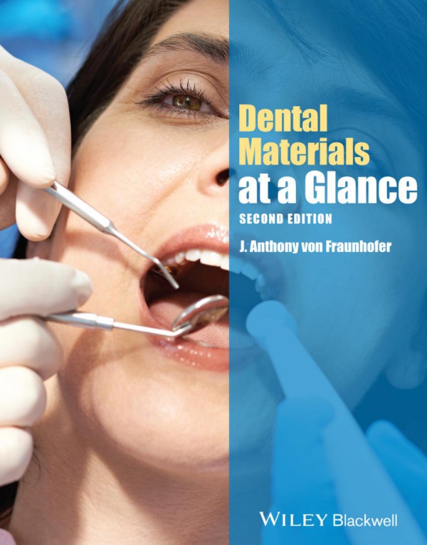
Dental Materials at a Glance 2nd Edition by J Anthony von Fraunhofer ISBN 1118684583 9781118684580
$50.00 Original price was: $50.00.$25.00Current price is: $25.00.
Authors:J. Anthony von Fraunhofer , Author sort:Fraunhofer, J. Anthony von
Dental Materials at a Glance 2nd Edition by J Anthony von Fraunhofer – Ebook PDF Instant Download/Delivery. 1118684583, 9781118684580
Full download Dental Materials at a Glance 2nd Edition after payment
Product details:
ISBN 10: 1118684583
ISBN 13: 9781118684580
Author: J Anthony von Fraunhofer
Dental Materials at a Glance 2nd Table of contents:
Part I Fundamentals
1 Properties of materials—tensile properties
Box 1.1 Desirable properties of dental materials
Table 1.1 Typical mechanical properties of dental biomaterials
Figure 1.1 Applied forces and specimen deformations.
Figure 1.2 Load versus stress for feet.
Figure 1.4 Stress–strain curves for brittle, elastic, and ductile materials.
Figure 1.3 The stress–strain curve of a nonferrous metal.
Figure 1.5 Elastic and plastic regions of a stress–strain curve.
2 Toughness, elastic/plastic behavior, and hardness
2.1 Elastic and plastic behavior
Figure 2.1 Optimal loading (stress and strain) region for a resilient material.
2.2 Viscoelasticity
2.3 Stress relaxation
Figure 2.2 Stress relaxation: decrease in induced stress as a result of creep.
2.4 Fracture toughness
2.5 Determining mechanical properties
Figure 2.3 Diametral disc test for determining the tensile strength of brittle materials.
Figure 2.4a Transverse testing of a specimen.
Figure 2.4b Loads and resultant stresses in a specimen under transverse testing.
2.6 Abrasion and wear resistance
3 Physical properties of materials
3.1 Thermal and electrical properties
Table 3.1 Thermal properties of various dental materials
Table 3.2 Coefficients of thermal expansion
Figure 3.1 Effect of temperature rise on a restoration and tooth with different coefficients of thermal expansion.
Table 3.3 Electrical constants for dental materials and teeth
3.2 Optical properties (color and appearance)
Table 3.4 Wavelengths of visible light
4 Adhesion and cohesion
Box 4.1 Molecular forces determining cohesive strength of an adhesive
Figure 4.1 Adhesion and cohesion.
Figure 4.2 Adhesive and cohesive failures of cemented restorations.
4.1 Forces in cohesion
4.2 Forces in adhesion
Table 4.1 Basic types of adhesion
4.3 Mechanisms of adhesion
4.3.1 Chemical adhesion
Table 4.2 Bond energies and bond lengths in adhesive forces
4.3.2 Dispersive adhesion
4.3.3 Diffusive adhesion
4.4 The adhesion zone
Figure 4.3 Adhesion zone between adhesive and substrate (schematic).
5 Mechanical adhesion
5.1 Contact angle and surface tension
Figure 5.1 Adhesive–substrate contact angles for clean, slightly contaminated, and contaminated surfaces.
Table 5.1 Relationships among surface wetting, substrate surface energy, contact angles, and adhesive/cohesive forces
Figure 5.2 Interfacial tensions for a drop of liquid on a surface: θ, contact angle between liquid and substrate; γSA, solid–air interfacial tension (surface energy of substrate); γLA, liquid–air interfacial tension (surface tension of liquid); γSL, solid–liquid interfacial tension (adhesion between liquid and solid).
5.2 Micromechanical adhesion
6 Dental hard tissues
Box 6.1 Changes in dental enamel with patient age
Box 6.2 Proteinaceous components of dentin
Table 6.1 Characteristics of dental hard tissues
Table 6.2 Average inorganic constituents content (wt.%) of mineralized tissue
6.1 Dental enamel
Table 6.3 Inorganic and organic content (wt.%) of enamel and dentin
Figure 6.1 Scanning electron micrograph (×1000) of etched enamel.
6.2 Dentin
Table 6.4 Characteristics of dentinal tubules
Figure 6.2 Scanning electron micrograph (×1000) of dentin tubules with smear layer present.
6.3 Cementum
6.4 Fluoridation of enamel
6.5 Strontium
7 Bone
Table 7.1 Composition of bone
Table 7.2 Inorganic constituents of bone (wt.%)
Table 7.3 Average composition of bone mineral
Table 7.4 Cells responsible for formation, maintenance, and resorption of bone
7.1 Dental implants and bone
Table 7.5 Classification of bone types
Part II Laboratory materials
8 Gypsum materials
8.1 Dental gypsum materials
Table 8.1 Dental gypsum products
8.2 Setting reaction
8.3 Water-to-powder (W/P) ratio
Table 8.2 Water-to-powder (W/P) ratios for gypsum materials
Table 8.3 Effect of W/P ratio on characteristics of mixed gypsum materials
8.4 Factors in setting
8.4.1 Expansion
8.4.2 Spatulation
8.4.3 Water temperature
8.4.4 Humidity
8.4.5 Colloids
8.5 Physical properties
8.5.1 Mixing
8.5.2 Setting time
8.5.3 Compressive strength
Figure 8.1 Effect of drying time on compressive strength of dental plaster.
Figure 8.2 Effect of the W/P ratio (shown above the columns) on the strength of gypsum materials.
8.5.4 Tensile strength
8.5.5 Surface hardness
8.5.6 Setting expansion
8.6 Comparative properties
Table 8.4 Comparative properties of dental gypsum products
Figure 8.3 Wet and dry compressive strengths of gypsum materials.
9 Die materials
9.1 Gypsum products
Table 9.1 W/P ratios, setting times, and setting expansions of dental and die stones
Figure 9.1 Compressive strengths of dental and die stones.
9.2 Handling of gypsum materials
9.3 Polymeric die materials
9.4 Pouring the impression
Figure 9.2 A cast prepared for construction of a crown.
10 Dental waxes
Table 10.1 Dental waxes
10.1 Composition
Table 10.2 Natural waxes
Table 10.3 Effect of wax additions on the properties of paraffin wax
10.1.1 Mineral waxes
10.1.2 Plant waxes
10.1.3 Animal waxes
10.1.4 Synthetic waxes
10.1.5 Gums, fats, and resins
10.2 Wax properties
10.2.1 Melting and thermal expansion
Table 10.4 Wax melting ranges and thermal expansions
10.2.2 Mechanical properties
10.2.3 Flow
10.3 Dental waxes
10.3.1 Pattern waxes
10.3.2 Baseplate wax
10.3.3 Casting waxes
10.3.4 Inlay waxes
10.3.5 Sticky wax
10.3.6 Impression waxes
11 Investments and casting
Box 11.1 Requirements of investment materials
11.1 Casting and investments
11.1.1 Composition
11.2 Gypsum-bonded investment
11.2.1 Composition
11.2.2 Properties
Table 11.1 Expansion requirements for gypsum-bonded investments
11.2.3 Effect of temperature
Table 11.2 Thermal expansions of crystallographic forms of SiO2
Figure 11.1 Thermal transformations of silica.
11.2.4 Setting and hygroscopic expansion
Table 11.3 Effect of manipulation variables on investment expansion
11.2.5 Other factors in expansion
11.3 High-temperature investments
11.3.1 Phosphate-bonded investments
11.3.2 Silica-bonded investments
Figure 11.2 Polymerized silica.
11.4 Considerations in casting
Table 11.4 Effect of casting variables on porosity
Part III Dental biomaterials
12 Inelastic impression materials
12.1 Factors in impression-making
12.2 Inelastic impression materials
12.2.1 Impression plaster
12.2.2 Impression compound
Table 12.1 Impression compound
Table 12.2 Flow properties of impression and tray compound
12.2.3 Zinc oxide—eugenol
Table 12.3 Zinc oxide–eugenol impression material
Figure 12.1 Structure of eugenol.
Figure 12.2 Structure of zinc eugenolate.
12.2.4 Noneugenol pastes
12.2.5 Surgical pastes
13 Elastic impression materials
Figure 13.1 Compression of impression material at the bulbosity of the tooth as tray is withdrawn from the mouth.
Figure 13.2 Approximate permissible distortions for 100% recovery of elastomerics.
13.1 Alginate (irreversible) hydrocolloids
Table 13.1 Approximate composition of alginate impression material
Table 13.2 Problems with alginate impressions
13.2 Agar–agar (reversible) hydrocolloid
13.3 Polysulfide rubber
Table 13.3 Polysulfide elastomeric impression material
13.4 Condensation-cured silicones
13.5 Addition-cured silicones (polysiloxanes)
13.6 Polyether impression materials
13.7 Properties of elastic impression materials
13.7.1 Setting properties
Table 13.4 Setting behavior of principal medium-body elastomeric impression materials
13.7.2 Mechanical properties
Table 13.5 Mechanical properties of medium-body elastomeric impression materials
Figure 13.3 Contact angle of water and castability of dental gypsum into impression materials.
13.8 Tray adhesives
14 Occlusal (bite) registration materials
Box 14.1 Requirements of an occlusal registrationmaterial
14.1 Traditional registration materials
14.2 Modern registration materials
Figure 14.1 Clinical photograph showing wax block-outs of missing maxillary posterior teeth and a lower RPD in place.
Figure 14.2 Occlusal registration taken with Regisil® vinyl polysiloxane
Figure 14.3 Wax maxillary denture base fabricated on VPS registration.
Figure 14.4 Casts of maxilla and mandible with VPS occlusal registration in place.
14.2.1 Pattern resins
15 Precious metal alloys
Box 15.1 Requirements of casting alloys
Box 15.2 Noble metals
15.1 Gold and noble metals
15.2 Precious metals
15.3 Gold alloys
15.3.1 Carat and fineness of gold
15.3.2 Gold–copper alloys
Figure 15.1 Face-centered tetragonal superlattice of AuCu.
Table 15.1 Effect of hardening on gold alloys
15.3.3 Dental gold casting alloys
Table 15.2 Dental gold casting alloys
Table 15.3 Average properties of dental casting alloys
15.3.4 Low-carat gold alloys
Table 15.4 Average properties of low-gold casting alloys
Table 15.5 Casting conditions for low-gold alloys
15.4 Heat treatment of gold
Table 15.6 Heat treatments for gold alloys
15.5 Problems with castings
Table 15.7 Casting defects
Table 15.8 Effect of casting variables on porosity
16 Base metal alloys
Box 16.1 Characteristics of titanium and titanium alloys
Table 16.1 Dental applications of base metal alloys
16.1 Cast chromium alloys
16.1.1 Cast chromium alloys
Table 16.2 Compositions of Cobalt–chromium and nickel–chromium casting alloys
Table 16.3 Average properties of Co–Cr and Ni–Cr alloys
16.1.2 Alloying elements
16.2 Investments and casting
16.3 Microstructure
16.4 Heat treatment
16.5 Physical and mechanical properties
16.6 Titanium and titanium alloys
16.6.1 Pure titanium
16.6.2 Titanium alloys
17 Porcelain bonding alloys
Box 17.1 Requirements for PFM restoration materials
Box 17.2 Requirements for metalloceramic alloys
Box 17.3 Problems with PFM restorations
17.1 PFM restorations
17.2 Ceramic–noble metal systems
Table 17.1 Average compositions of noble metal ceramic-bonding alloys
Table 17.2 Average properties of noble metal ceramic-bonding alloys
17.3 Ceramic–base metal systems
Table 17.3 Compositions of base metal alloys for metalloceramic restorations*
Table 17.4 Properties of base metal metalloceramic alloys
18 Implant metals
Figure 18.1a Modern dental implant.
Figure 18.1b Modern tapered dental implant.
Figure 18.2 Radiograph showing osseointegration of implants in bone.
Figure 18.3 (a,b) Site preparation and placement of a “skinny” implant.
18.1 Implant alloys
Figure 18.4 Hydroxyapatite (HA) coating on the surface of a dental implant (scanning electron micrograph, ×1000).
19 Partial denture base materials
19.1 Metallic RPDs
Figure 19.1 Cast Vitallium removable partial denture.
Figure 19.2 Removable partial denture with a metal base mounted on a cast, showing the acrylic gumwork, prosthetic teeth, and the denture clasps positioned on the abutment tooth.
Table 19.1 Base metal casting alloys
Table 19.2 Mechanical properties of partial denture alloys
Figure 19.3 Corrosion and surface tarnishing of a chromium-cobalt removable partial denture framework due to the action of a hypochlo-rite-containing denture cleanser.
19.2 Polymeric RPDs
Figure 19.4 Flexible resin partial denture.
Table 19.3 Physical properties of flexible partial denture materials
20 Complete denture bases-acrylic resin
Box 20.1 Required properties of denture base materials (in alphabetical order)
Figure 20.1 Maxillary complete denture.
20.1 Polymerization reactions
Figure 20.2 Reaction initiation—decomposition of benzoyl peroxide molecule into two free radicals.
Figure 20.3 Reaction propagation—interaction between free radicals and monomer.
Figure 20.4 Reaction termination by annihilation, disproportionation, or transfer reaction.
Table 20.1 Stages during monomer–polymer reaction
20.2 Polymer properties
Table 20.2 Glass transition temperatures (Tg) of methacrylate resins
Table 20.3 Residual monomer levels
20.3 Problems with denture bases
Table 20.4 Problems with denture bases and their causes
20.3.1 Palatal vault
20.4 Denture teeth
20.5 Soft dentures
21 Modified acrylics and other denture base resins
Table 21.1 Properties of denture base resins
21.1 High-impact resins
Figure 21.1 PalaXpress Ultra overdenture.
21.2 Pour-type resins
Figure 21.2 Pouring mixed pour resin and liquid into a mold.
Figure 21.3 As-molded poured resin denture.
Figure 21.4 Processed and polished poured resin Lucitone® Fas-Por™ denture.
21.3 Rapid-cure resins
21.4 Light-cure resins
Figure 21.5 Triad® light-cured denture base with clear palate.
22 Denture fracture and repair
Figure 22.1 Modern cold-cure denture repair material.
Box 22.1 Criteria for a satisfactory denture repair
22.1 Denture repair
Figure 22.2 Arrangement of a fractured denture for repair.
Figure 22.3 Fractured denture edges prepared with round profiles and a gap of 3 mm between broken edges for optimal repair.
Figure 22.4 Triad® custom curing unit for urethane dimethacrylate repair material.
23 Denture liners
23.1 Resilient (soft) liners
Figure 23.1 Placement of PermaSoft within a denture.
23.1.1 Resilient liner materials—modified acrylic resins
23.1.2 Resilient liner materials—silicone elastomers
Figure 23.2 Schematic representation of shock-absorbing capability of silicone and polyphosphazene elastomeric resilient liners.
23.2 Tissue conditioners
Figure 23.3 Elastic, anelastic, and permanent deformation of a viscoelastic gel under loading and after load release.
Figure 23.4 Schematic representation of the deformation of a tissue conditioner under rapid (dynamic) loading and slow (quasi-static) loading.
23.3 Problems with denture liners
23.3.1 Useful life
23.3.2 Liner thickness
23.3.3 Liner separation
23.3.4 Liner sanitization
24 Denture cleansing
Box 24.1 Bacteria and fungi identified from dentures
Figure 24.1 Cameo surface of a dirty denture.
24.1 Denture plaque
24.2 Cleansing methods
24.2.1 Brushing of dentures
Figure 24.2 Numbers of colony-forming units (CFUs) recovered from dentures after cleaning.
24.2.2 Effervescent cleansers
25 Dental luting
Box 25.1 Cement selection criteria
Figure 25.1 Failure patterns for thin and thick cement films beneath crowns.
25.1 Dental cements—general considerations
25.2 Cement properties
25.3 Luting agent selection
Figure 25.2 Bond strength of adhesive to etched and silanated porcelain exceeds the cohesive strength of porcelain.
25.4 Cement film thickness
Figure 25.3 Cement film thicknesses in µm beneath vented and unvented full-coverage crowns for different taper angles.
26 Cavity varnishes, liners, and bases
Box 26.1 Requirements of bases and liners
Box 26.2 Factors affecting integrity and efficacy of liners and base
Box 26.3 Requirements of high-strength bases
26.1 Cavity varnishes
26.1.1 Composition
26.1.2 Varnish film characteristics
26.1.3 Applications
26.2 Cavity liners
Figure 26.1 Cavity liner reducing the adverse effects of heat, chemicals, and galvanic effects on the dental pulp.
26.2.1 Composition
26.2.2 Characteristics
26.3 Low-strength bases
26.3.1 Ca(OH)2 bases
26.3.2 ZOE bases
26.3.3 Visible light–cured bases
26.3.4 Properties of low-strength bases
26.4 High-strength bases
Table 26.1 Average mechanical properties of low- and high-strength bases
26.4.1 Characteristics of high-strength bases
27 Provisional (temporary) dental cements
Figure 27.3 Setting reaction of zinc oxide–eugenol.
Box 27.1 Desirable properties of provisional cements
Figure 27.1 Provisional restoration in single quadrant impression.
Figure 27.2 Loading Integrity® provisional restoration with provisional dental cement, prior to luting onto prepared tooth.
27.1 Zinc oxide–eugenol cements
27.2 Noneugenol systems
Figure 27.4 Dispensed paste-paste provisional cement.
Table 27.1 Properties of provisional dental cements
27.3 Cement removal
28 Inorganic (acid–base reaction) cements
Box 28.1 Types of permanent dental cement
28.1 Zinc phosphate
Table 28.1 Approximate composition of zinc phosphate (ZNP) cement
28.1.1 Manipulation
28.2 Zinc polyacrylate (polycarboxylate) cement
Table 28.2 Average properties of inorganic cements
Table 28.3 Zinc polycarboxylate and GIC characteristics compared with those of zinc phosphate (ZNP) cement
28.3 Glass ionomer cement (GIC)
Figure 28.1 Setting reaction of glass ionomer cement (GIC).
28.3.1 Manipulation and properties
28.4 New luting agent technology
29 Resin-modified and resin cements
Box 29.1 Requirements of resin-modified and resin cements
29.1 Hybrid ionomer cements
Table 29.1 Types of RMGI and their applications
Table 29.2 Components of liquid portion of RMGI cements
Table 29.3 Components of several RMGI cements
29.1.1 Setting reaction
29.1.2 Properties
29.1.3 Applications
29.2 Resin cements
Table 29.4 Components of resin cements
29.2.1 Properties
Figure 29.1 Bond strengths for conventional and adhesive resin cements.
Table 29.5 Average properties of GICs, RMGIs, and resin cements
29.2.2 Applications
Table 29.6 Types of resin cements and their applications
29.3 Adhesive resin cements
30 Denture adhesives
30.1 Denture adhesives
Figure 30.1 Schematic representation of hydration of a denture adhesive to form a gel that swells and fills voids between the CD and its supporting tissues.
Figure 30.2 Cohesive and adhesive forces in a denture adhesive between a CD base and its supporting structures.
30.1.1 Dental adhesive materials
Figure 30.3 Gel denture adhesive applied to the intaglio surface of a lower complete denture.
Figure 30.4 Adhesive strips applied to intaglio surface of a complete upper denture.
30.1.2 Mechanism of retention
30.1.3 Clinical use of denture adhesives
30.1.4 Adverse reactions to denture adhesives
31 Dental amalgam
31.1 Dental amalgam alloy
Table 31.1 Specification composition limits (wt.%) of amalgam alloys
Table 31.2 Compositions (wt.%) of amalgam alloys
31.2 Setting reaction
Figure 31.1 Setting reaction of conventional amalgam alloy.
Figure 31.2 Setting reaction of dispersion-modified (admixed) amalgam alloy.
Figure 31.3 Setting reaction of single-phase amalgam alloy.
Figure 31.4 Increase in amalgam compressive strength with time.
31.3 Physical properties
Table 31.3 Average properties of dental amalgams
31.4 Manipulation and handling properties
31.5 Corrosion
Figure 31.5 Corrosion process with a conventional dental amalgam.
32 Adhesive dentistry
Box 32.1 Factors in dental adhesion
Box 32.2 Causes of problems in enamel bonding
32.1 Dental adhesion
Figure 32.1 (a) Adhesive surface tension exceeds critical surface energy. (b) Critical surface energy greater than surface tension of adhesive.
32.1.1 Composition of enamel and dentin
Figure 32.2 Compositions of enamel and dentin.
32.1.2 Etching of enamel
Figure 32.3 Schematic profiles of smooth, abraded, and etched surfaces.
Table 32.1 Common etch patterns of dental enamel
32.1.3 Adhesion to enamel
32.1.4 Bonding to enamel
33 Bonding to dentin
Box 33.1 Common dentin conditioning agents
33.1 Dentin bonding
Table 33.1 Developments (generations) in dentin-bonding systems
33.1.1 Conditioning
33.1.2 Priming
Figure 33.1 Schematic representation of the coupling action of a dentin-bonding agent providing adhesion between the resin matrix and tooth surface.
Figure 33.2 Bifunctional HEMA molecule: methacrylate group can bond to resin, whereas the active end can react with dentin substrate.
Figure 33.3 Schematic diagram of 4-META and its bond to calcium in hydroxyapatite of conditioned dentin.
Table 33.2 Dentin-bonding agents
33.1.3 Bonding
33.1.4 Combination systems
Figure 33.4 Schematic diagram of formation of a hybrid layer on dentin.
33.2 Bonded dentin
34 Composite restorative resins
Box 34.1 Clinical requirements of esthetic filling materials
Box 34.2 Factors determining properties of composites
34.1 Dental composites
34.1.1 Resin matrix
Figure 34.1 Molecular structure of bis-GMA (Bowen’s resin) and urethane dimethacrylate (UDMA).
34.1.2 Curing systems
34.1.3 Filler particles
Table 34.1 Particle size of fillers in composite resins
34.1.4 Filler loading
Table 34.2 Physical properties of composite restorative materials
34.1.5 Composite strengthening
34.2 Compomers
35 Endodontic filling materials
Box 35.1 Factors in successful root canal therapy
Box 35.2 Ideal characteristics of endodontic irrigants
Box 35.3 Requirements of endodontic sealer cements
35.1 Canal instrumentation
35.2 Canal irrigation
35.3 Sealer cements
Table 35.1 Components of traditional endodontic sealer cements
35.4 Canal obturation
Table 35.2 Potential problems with traditional obturation techniques
35.5 Endodontic leakage
Figure 35.1 Relative leakage behavior of endodontic obturation techniques.
36 Provisional filling materials and restorations
Box 36.1 Ideal properties of a provisional restorative material
Box 36.2 Advantages of light-curing provisional restorative materials
36.1 Traditional materials
36.2 Resin systems
36.3 Coronal restorations
36.4 Temporization
Table 36.1 Average properties of temporization resins
Figure 36.1 (a) Clinical photograph of patient’s mouth before treatment. (Courtesy of Heraeus Kulzer US.) (b) Temporized patient with Venus Temp 2 provisional restorations. (Courtesy of Heraeus Kulzer US.)
Figure 36.2 Triad® light-cured provisional restorations.
37 Materials in periodontics
Box 37.1 Properties of an ideal periodontal dressing material
37.1 Zinc oxide materials
Table 37.1 Components of modern periodontal dressing materials
37.2 Polymeric periodontal dressings
37.3 Dressing properties
Figure 37.1 Water solubilities of periodontal dressing materials at room and mouth temperature.
Figure 37.2 Water sorption behavior at 37°C.
Figure 37.3 Adhesion
38 Dental porcelain
Box 38.1 Clinical applications of dental porcelains
38.1 Manufacture
Figure 38.1 Schematic diagram of the two-dimensional structure of sodium silicate glass.
Table 38.1 Compositions (wt.%) of dental and decorative porcelains*
38.2 Composition
38.2.1 Feldspar
38.2.2 Silica
38.2.3 Kaolin
38.2.4 Metallic oxides
Table 38.2 Effects of metal oxides on appearance of porcelain
38.3 Dental porcelains
Table 38.3 Classification of dental porcelains by fusion temperature
Table 38.4 Compositional differences between decorative, high-fusing, and low-fusing porcelains
38.3.1 High-fusing porcelains
38.3.2 Medium-fusing porcelains
38.3.3 Low-fusing porcelains
38.3.4 Very low fusing porcelains
38.3.5 Glazes
38.3.6 Stains
38.4 Structure
39 Manipulation and properties of porcelain
Box 39.1 Condensation methods for porcelain slurries
39.1 Manipulation
39.1.1 Paste
39.1.2 Condensation
39.1.3 Dies
39.1.4 Drying
39.1.5 Firing (Sintering)
Table 39.1 Factors in sintering
Table 39.2 Stages in firing of porcelain
Figure 39.1 Effect of surface condition and firing regimen on porcelain strength.
39.2 Physical properties
39.2.1 Strength
39.2.2 Porosity
39.2.3 Hardness
40 Strengthening porcelain
40.1 Porcelain strengthening
40.1.1 Aluminous porcelain
Figure 40.1 Reinforcing filler particle acting as a “crack stopper” for a propagating crack.
40.1.2 Alumina inserts and cores
40.1.3 Spinel-based cores
40.1.4 Leucite-reinforced porcelain
Table 40.1 Strengths of dental hard tissues and dental ceramics
40.1.5 Metal-bonded porcelain
Figure 40.2 Schematic representation of a PFM (metallo-ceramic) crown.
Figure 40.3 Schematic representation of a crown constructed with a glass-infiltrated (In-Ceram) core.
41 Advanced ceramic systems
Table 41.1 Dental ceramics and methods used for restoration fabrication
41.1 Hot-pressed ceramics
41.1.1 High-leucite molded glass
41.1.2 Lithium ceramics
41.1.3 Slip-cast ceramics
41.1.4 Castable glass–ceramic
Figure 41.1 Schematic representation of a crown constructed with a glass-ceramic (Dicor) core.
Figure 41.2 Flexural strengths (MPa) of porcelain and castable glasses.
41.2 Machinable ceramics
Figure 41.3 Average flexural strengths (MPa) of porcelains and advanced ceramic systems.
41.3 Veneers
42 CAD-CAM restorations
Figure 42.1 Dental laboratory CAD-CAM systems.
Figure 42.2 E4D CAD-CAM system for the dental office.
42.1 Digital imaging
Figure 42.3a Patient before restoration.
Figure 42.3b Patient after restoration.
42.2 Restorative materials
Figure 42.4 Finishing and characterization of a CAD-CAM ceramic restoration.
42.3 CAD-CAM dentistry
42.3.1 Restoration luting
43 Orthodontic materials
43.1 Intraoral appliances
43.2 Extraoral appliances and jaw orthopedics
Table 43.1 Appliances for jaw orthopedics
43.3 Removable orthodontic appliances
43.4 Fixed orthodontic appliances
Table 43.2 Characteristics of common orthodontic arch wires
Table 43.3 Yield strength, elastic modulus, and YS/E ratio for orthodontic arch wires
Figure 43.1 Schematic diagram of orthodontic elastomeric chains (A, closed loop; B, short open loop; C, long open loop).
Figure 43.2 Force required to distract three- and four-loop segments of elastomeric chain in the dry (as-received) condition and after immersion in water and a citrate-containing power drink.
44 Grinding, polishing, and finishing
Box 44.1 Effects of cutting, grinding, and polishing on surfaces
44.1 Material removal
44.1.1 Abrasives
Table 44.1 Common abrasives
Table 44.2 Abrasive particle and grit sizes
44.1.2 Dental burs
Table 44.3 Cutting action of carbide and diamond burs and bur selection for different substrates
44.2 Coolant and lubricant effects
Figure 44.1 Effect of external temperature on internal (pulpal) temperature of teeth.
Figure 44.2 Effect of coolant flow rate on dental cutting rates.
Figure 44.3 Chemomechanically enhanced dental cutting.
44.3 Finishing and polishing regimens
45 Adverse effects of dental biomaterials
Figure 45.1 Prevalence of adverse reactions in dental specialties.
Figure 45.2 Potential adverse reactions.
45.1 Toxicity and carcinogenicity
Table 45.1 Carcinogenic and toxic substances used in dentistry
45.2 Hypersensitivity and allergic reactions
45.2.1 Hypersensitivity
45.2.2 Allergic reactions
Table 45.2 Principal allergic reactions
Table 45.3 Type I and Type IV allergic reactions
Table 45.4 Common allergens in dentistry
45.2.3 Metal allergies
Figure 45.3 Corrosion of metal by reaction with sweat or saliva.
Table 45.5 Prevalence of metal allergies
45.2.4 Reaction to mercury
Figure 45.4 Mechanism of mercury release from amalgam restorations.
45.3 Conclusion
46 Dental erosion
Figure 46.1 Enamel dissolution caused by excessive consumption of citrus soft drinks.
Figure 46.2 Enamel dissolution caused by consumption of a sports drink.
46.1 Soft drink consumption
46.2 Beverage acidity
Figure 46.3 Enamel dissolution in soft drinks (14 days).
46.3 Acids in beverages
46.4 Factors in erosion
46.5 Other causes of erosion
People also search for Dental Materials at a Glance 2nd:
dental materials at a glance
dental materials at a glance pdf
what are dental materials
dental material companies
dental materials important questions
You may also like…
eBook PDF
Research Writing in Dentistry 1st edition by Anthony von Fraunhofer 081380762X 9780813807621
eBook PDF
Pre Clinical Dental Skills at a Glance 1st edition by James Field 9781118766620 1118766628
eBook PDF
Dental Materials at a Glance 2nd edition by Anthony von Fraunhofer 9781118684580 111868458
eBook PDF
Research Writing in Dentistry 1st Edition by Anthony von Fraunhofer ISBN 081380762X 9780813807621

