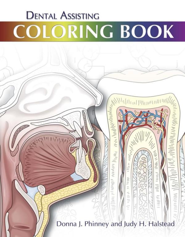
Dental Assisting Coloring Book 1st Edition by Donna Phinney, Judy Halstead ISBN 1133170269 9781133170266
$50.00 Original price was: $50.00.$25.00Current price is: $25.00.
Authors:premedia , Author sort:premedia , Published:Published:Jul 2010
Dental Assisting Coloring Book 1st Edition by Donna Phinney, Judy Halstead – Ebook PDF Instant Download/Delivery. 1133170269, 9781133170266
Full download Dental Assisting Coloring Book 1st Edition after payment
Product details:
ISBN 10: 1133170269
ISBN 13: 9781133170266
Author: Donna Phinney, Judy Halstead
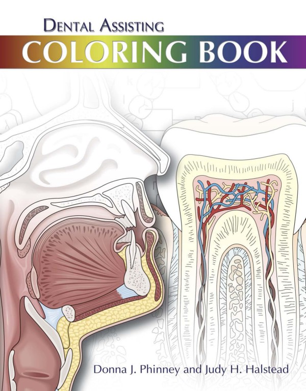
Dental Assisting Coloring Book is an interactive tool designed as both a study aid and a review guide to enhance learning in the field of dentistry. The format makes review and learning creative and fun. Unlike other coloring books, Delmar’s Dental Assisting Coloring Book contains questions presented in a variety of formats, in addition to coloring and labeling, to test recall and overall comprehension of key concepts. It covers a wide variety of topics encountered in lectures, clinics, and labs, including general anatomy, tooth anatomy and dental charting, equipment and dental instruments, procedures, radiology equipment, and x-ray landmarks. Dental Assisting Coloring Book is an effective way to enhance learning and improve retention of concepts critical to success in the field of dental assisting.
Dental Assisting Coloring Book 1st Table of contents:
Ch 1: General Anatomy
Basic Cell Structures
Body Planes
Body Directions
Body Cavities
Axial and Appendicular Skeleton
Anatomic Features of the Bone
Skeletal Joints
Types of Muscle Tissue
Tendons and Ligaments
Structure of a Neuron
Simple Refl ex Arc
Structures of the Endocrine System
System and Pulmonary Circulation
Structures of the Heart
Structures of the Digestive System
Salivary Glands and Ducts
Structures of the Respiratory System
Structures of the Bronchi
The Lungs
Tonsils
The Immune System
Ch 2: Head and Neck Anatomy
Landmarks of the Face
Structures of the Oral Cavity—Maxillary View
Structures of the Oral Cavity—Mandibular View
Landmark on the Buccal Mucosa
Landmarks of the Oral Pharynx Area
Landmarks of the Palate
Landmarks on the Dorsal Surface of the Tongue
Landmarks on the Ventral Surface of the Tongue
Basic Taste Buds of the Tongue
Salivary Glands and Ducts
Lateral Aspect of the Cranium and Face
Frontal View of the Bones of the Cranium and Face
Landmarks of the Palate
Lateral View of the External Surface of the Mandible
Internal Lingual View of the Mandible
Temporomandibular Joint (TMJ)
Movement of the Temporomandibular Joint (TMJ)–Hinge Joint
Movement of the Temporalmandibular Joint (TMJ)–Gliding Joint
Muscles of Mastication–Lateral View
Muscles of Facial Expression
Extrinsic Muscles of the Tongue
Muscles of the Floor of the Mouth
The Hyoid Bone
Muscles of the Soft Palate
Muscles of the Neck
Nerves of the Maxillary Arch
Medial View of the Branches of the Pterygopalatine Nerve
Mandibular Nerves
Arteries of the Face and Oral Cavity
Veins of the Face and Oral Cavity
Ch 3: Tooth and Tissue Structures
The Three Primary Embryonic Layers
Embryology
Developing Embryo with Primary Layers Identifi ed
Facial Processes Shown on an Embryo (Child and Adult)
Development of the Palate
Bilateral Cleft of the Lip (Alveolar Process and Primary Palate)
Life Cycle of the Tooth
Enamel Rods
Tissues of the Tooth
Enamel
Dentin
Pulp
Cementum
Tooth and Surrounding Tissues
Sharpey’s Fibers and Cementum
Periodontal Ligaments and Alveolar Crests
Cross Section of Mandibular Molar Tissues of the Tooth Identified
Gingival Fiber Groups
Periodontium
Alveolar Mucosa
Ch 4: Tooth Anatomy
Adult Dentition
Deciduous Dentition
Primary Dentition
Permanent Dentition
Primary Teeth
Permanent Dentition
Permanent Dentition
Permanent Dentition
Permanent Dentition
Anatomical Structures
Anatomical Landmarks
Anatomical Landmarks
Maxillary Central Incisors
Maxillary Lateral Incisors
Maxillary Canine
Maxillary First Bicuspid (Premolar)
Maxillary Second Bicuspid (Premolar)
Maxillary First Molar
Maxillary Second Molar
Maxillary Third Molar
Mandibular Central Incisors
Mandibular Lateral Incisors
Mandibular Cuspids
Mandibular First Bicuspids (Premolars)
Mandibular Second Bicuspids (Premolars)
Mandibular First Molar
Mandibular Second Molar
Mandibular Third Molar
Mixed Dentition of a Seven- or Eight-Year-Old
Contact and Embrasure
Deciduous Maxillary Teeth
Deciduous Mandibular Teeth
Identification of Teeth
Eruption Dates for Primary Teeth
Exfoliation Dates for Primary Teeth
Ch 5: Dental Charting
Universal Numbering System
International Standards Organization Numbering System
Palmer Numbering System
Charting Example #1
Charting Example #2
Charting Example #3
Charting Example #4
Charting Example #5
Charting Example #6
Charting Example #7
Charting Example #8
Charting Example #9
Charting Example #10
Ch 6: Introduction to the Dental Office and Basic Chairside Assisting
Small Dental Offi ce Blueprint
Sterilizing Area
Laboratory Area
Dental Treatment Room
Operator’s Mobile Cart
Air-Water Syringe
Activity Zones
Ch 7: Basic Chairside Instruments and Tray Systems
Parts of an Instrument and Different Shanks
Instruments with Black’s Three-Number Formula
Instruments with Black’s Four-Number Formula
Chisels, Hatchets, and Hoes
Gingival Margin Trimmers and Angle Formers
Excavators
Explorers, Periodontal Probe, and Cotton Pliers
Cement Spatulas and Burnishers
Condensors, Carvers, and Plastic Filling Instruments
Parts of a Bur and Shanks
Cutting Bur Shapes and Number Ranges
Diamond Burs, Finishing Burs, Surgical Burs, and Laboratory Burs
High- and Low-speed Handpieces and Attachments
Double Color coding
Triple Color coding
Color-coding for Procedure Sequence
Ch 8: Anesthesia and Sedation
Types of Anesthetic Injections
Maxillary Arch Injections and Site Locations
Mandibular Arch Injections and Site Locations
Aspirating Syringe
Needle Parts
Parts of an Anesthetic Cartridge
Information on the Anesthetic Cartridge
Equipment and Supplies Needed to Prepare an Anesthetic Syringe
Ch 9: Dental X-ray Film and Holding Devices
Electromagnetic Energy Spectrum and Applications
Primary, Secondary, and Leakage Radiation
Parts of Dental Arm Assembly
Tube Head, PID, and Vertical Indicator Scale
Tube Head and X-ray Tube
X-ray Tube
Composition of Dental X-ray Film
Sizes of Dental X-ray Film
Film Packet
Film Holding Devices
Rinn XCP
Processing Room
Manual Processing Tank
Ch 10: Radiology Landmarks
Landmark Planes for Exposing Radiographs of the Face
Landmarks for the Tooth and Surrounding Tissues
Landmarks for the Surrounding Tissues Continued
Landmarks for the Maxillary Arch
Landmarks for the Mandibular Arch
Ch 11: Miscellaneous
Maslow’s Hierarchy of Needs
Food Guide Pyramid
People also search for Dental Assisting Coloring Book 1st:
dental assisting coloring book
dental assisting coloring book answer key
dental hygienist coloring book
dental hygiene coloring sheet
dental hygiene coloring pages
You may also like…
eBook PDF
Delmar Dental Assisting Exam Review 1st Edition by Melissa Thibodeaux ISBN 0827390718 9780827390713
eBook PDF
Modern Dental Assisting 1st Edition by Doni Bird, Debbie Robinson ISBN 1437717292 9781437717297
eBook PDF
Comprehensive Dental Assisting 2nd Edition by Lippincott Williams ISBN 1284564533 9781284564532
eBook PDF
Comprehensive Dental Assisting 1st Edition by Jones Bartlett Learning ISBN 1284564533 9781284564532

