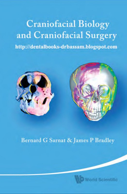
Craniofacial Biology and Craniofacial Surgery 1st Edition by Bernard Sarnat, James Breadley ISBN 9789812839299 9812839291
$50.00 Original price was: $50.00.$25.00Current price is: $25.00.
Authors:Bernard George Sarnat; James P. Bradley , Series:Surgery [16] , Tags:Medical; Surgery; Oral & Maxillofacial; Orthopedics; Plastic & Cosmetic; World Scientific Publishing Company , Author sort:Sarnat, Bernard George & Bradley, James P. , Ids:Google; 9789812839282 , Languages:Languages:eng , Published:Published:Oct 2010 , Publisher:World Scientific , Comments:Comments:This book is unique. It deals primarily with and brings together a wide-ranging group of essays spanning more than half a century’s worth of research done by Bernard G Sarnat. Much of this historical review remains significant and germane today. Some material antedates the emergence of the specialties of craniofacial biology, craniofacial surgery, and bone biology, while many of the reports preceded the period of molecular biology. This book thus represents a fundamental pioneering contribution to a representative portion of the specialties. Building on past data reported by Sarnat, James P Bradley contributes significantly to the present by including recent works which cover issues dealing with stem cell, tissue regeneration and tissue engineering research. In addition, appropriately selected clinical work is included a result of the further development and maturity of the specialties. And what does the future hold? No doubt unpredictable gigantic advances. The purpose of this selective, organized, and limited review, analysis, and summary of personally conducted experiments is to relate certain aspects of differential growth and change and nonchange to age, sites, rates, factors, and mechanisms. In many instances, correlations are made between research findings and clinical practice, and this retrospective study brings all of them together.
Craniofacial Biology and Craniofacial Surgery 1st Edition by Bernard Sarnat, James Breadley – Ebook PDF Instant Download/Delivery. 9789812839299 ,9812839291
Full download Craniofacial Biology and Craniofacial Surgery 1st Edition after payment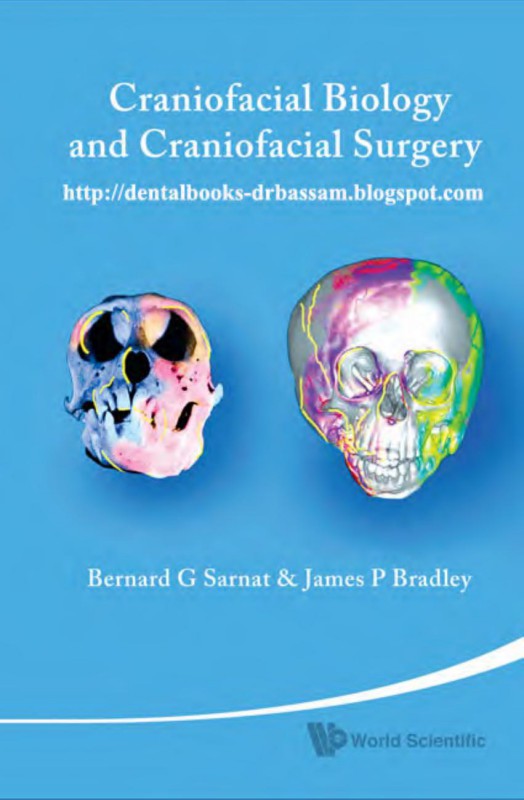
Product details:
ISBN 10: 9812839291
ISBN 13: 9789812839299
Author: Bernard Sarnat, James Breadley
Craniofacial Biology and Craniofacial Surgery 1st Edition Table of contents:
Chapter 1 Introduction
BONE GROWTH AND CHANGE
NORMAL CRANIOFACIAL GROWTH
Environment and Growth
REFERENCES
PART I THE LOWER FACE
Chapter 2 Growth Pattern of the Pig Mandible
INTRODUCTION
REVIEW OF THE LITERATURE
MATERIAL AND METHODS
Surgical Procedures
Lateral Cephalometric Roentgenographs
Tracings of the Roentgenographs
Preparation of Gross Material
FINDINGS
Gross
Serial Lateral Cephalometric Roentgenographs and Tracings
Increase in Size
Ramus height
Ramus width
Body height
Total mandibular length
Eruption of the Permanent Mandibular First and Second Molars Correlated with Mandibular Growth
DISCUSSION
Method of Study
Sites of Growth
Condyle and posterior border of the ramus
Anterior border
Alveolar border
Inferior border
Relationship between the ramus and body growth
Correlation of mandibular growth and development of the permanent molars
SUMMARY AND CONCLUSIONS
REFERENCES
Chapter 3 Mandibular Condylectomy in Young Monkeys
INTRODUCTION
MATERIALS AND METHODS
FINDINGS
Anterior Aspect of the Skull
Lateral Aspect of the Skull
Basal Aspect of the Skull
COMMENTS
Facial Growth
SUMMARY AND CONCLUSIONS
REFERENCES
Chapter 4 Mandibular Condylectomy in Adult Monkeys
INTRODUCTION
MATERIALS AND METHODS
Animals
Anesthesia
Surgical Procedure
Photographs and Roentgenographs
Histological Sections
RESULTS
Antemortem Observations
Postmortem Observations
Region of the temporomandibular joint of the operated-on side
Lateral aspect of the skull
Anterior aspect of the skull
Ventral aspect of the skull
Roentgenographic and Photographic Observations
Histological Observations
DISCUSSION
Comparison of Findings after Condylectomy in Adult and Growing Monkeys
Dental Occlusion
Roentgenographic Changes
Controls
SUMMARY
REFERENCES
Chapter 5 Temporalis Muscle and Coronoid Process
REFERENCES
Chapter 6 Fractured Mandible and Incisor
INTRODUCTION
Dental Changes
Healing of Bone
REFERENCES
Chapter 7 The Temporomandibular Joint
FOREWORD TO THE FOURTH EDITION
FOREWORD TO THE FOURTH EDITION
PREFACE TO THE FOURTH EDITION
FOREWORD TO THE THIRD EDITION
PREFACE TO THE THIRD EDITION
PREFACE TO THE SECOND EDITION
PREFACE TO THE FIRST EDITION
Chapter 8 Condylar Tumors
UNILATERAL HYPERPLASIA OF THE MANDIBULAR CONDYLE
Chapter 9 Overgrowth of Coronoid Processes
INTRODUCTION
REPORT OF CASE
Examination
Operation
Postoperative Course
REFERENCES
Chapter 10A Surgery of the Mandible: Some Clinical and Experimental Considerations
INTRODUCTION
ABNORMAL GROWTH: SOME SURGICAL CONSIDERATIONS
UNDERDEVELOPMENT OF THE MANDIBLE
Local Causes
Trauma
Inflammation
ANKYLOSIS AND CONDYLAR RESECTION
Systemic Causes
Hereditary and congenital conditions
Inflammatory lesions
Dietary deficiencies
Endocrine disturbances: Cretinism
OVERDEVELOPMENT OF THE MANDIBLE
Local Causes
Intrinsic factors: Tumors
Extrinsic factors: Enlarged tongue
Idiopathic
Unilateral hyperplasia of the mandibular condyle
Prenatal unilateral hypertrophy of the face
Prognathic mandible
Endocrine Disturbances: Giantism and Acromegaly
Giantism
Acromegaly
DISTORTION OF THE MANDIBLE
OTHER SURGICAL CONSIDERATIONS
Chronic Anterior Dislocation of the Mandible
REFERENCES
Chapter 10B The Mandible: Clinical Considerations
PEDIATRIC MANDIBULAR TRAUMA
MANDIBULAR ASYMMETRY
MANDIBULAR DEVIATION WITH UNILATERAL CORONAL SYNOSTOSIS
PIERRE ROBIN SEQUENCE
DISTRACTION OSTEOGENESIS
History
Biology
Mandibular Distraction
Devices and Vectors
Neonatal Distraction
CRANIOFACIAL MICROSOMIA
TREACHER–COLLINS SYNDROME
GENIOPLASTY DISTRACTION
TEMPOROMANDIBULAR JOINT ANKYLOSIS
SUMMARY
REFERENCES
PART II THE MIDFACE
Chapter 11 Osteology of the Rabbit Face
REFERENCES
Chapter 12A Normal Growth of the Suture
INTRODUCTION
MATERIALS AND METHODS
Animals
Anesthesia
Metallic Implants (Direct Measurements)
Serial Cephalometric Roentgenography (Indirect Measurements)
RESULTS
Skulls
Increased separation of paired implants at the frontonasal suture
Increased separation of paired implants at the internasal and interfrontal sutures
Serial Roentgenographs
DISCUSSION
Implants
SUMMARY AND CONCLUSIONS
REFERENCES
Chapter 12B Rabbit Snout After Extirpation of the Frontonasal Suture
INTRODUCTION
MATERIAL AND METHODS
Animals
Anesthesia
Extirpation of the Frontonasal Suture
Metallic Implants (Direct Measurements)
Serial Cephalometric Roentgenography (Indirect Measurements)
Gross Observations
Direct Measurement
Total Longitudinal Growth Between Implants
Components of Longitudinal Growth Between Implants
Indirect Measurements: Serial Roentgenographs
DISCUSSION
Implants
Extirpation Site
Some Considerations of Sutural Growth
SUMMARY AND CONCLUSIONS
REFERENCES
Chapter 13 Growth Pattern of the Nasal Bone Region
INTRODUCTION AND PURPOSE
MATERIALS AND METHODS
Animals
Anesthesia
Metallic Implants
Serial Cephalometric Roentgenography
RESULTS
Gross
Serial Cephalometric Roentgenographs and Tracings
DISCUSSION
Clinical Comment
REFERENCES
Chapter 14 Rabbit Nasal Septum
INTRODUCTION AND PURPOSE
REVIEW OF THE LITERATURE
MATERIALS AND METHODS
Animals
Anesthesia
Surgical Procedure
Photographs and Roentgenographs
RESULTS
Growing Rabbits
Resected septovomeral region and/or cartilaginous nasal septum (large amounts)
Antemortem observations
Postmortem observations
Roentgenographic observations
Adult Rabbits
DISCUSSION
The Face and Jaws After Surgical Experimentation with the Septovomeral Region
Dental Changes
Clinical Considerations
SUMMARY AND CONCLUSIONS
REFERENCES
Chapter 15 Growth of Multiple Facial Sutures
INTRODUCTION
MATERIAL
METHODS
Anesthesia
Metallic Implants (Direct Measurements)
Cephalometric Roentgenographs and Tracings (Indirect Measurements)
Preparation of Material
FINDINGS
Examination of Skulls
Tissue reaction to individual implants
Separation of paired implants
Examination of Roentgenographs
Individual implants
Paired implants
Rate of separation
Examination of Superposed Tracings of Serial Roentgenographs
DISCUSSION
Appositional Growth
Sutural Growth
Total Facial Growth
Comparison between the Roentgenographic and Direct Methods of Measuring Growth
SUMMARY AND CONCLUSIONS
Chapter 16 Maxillary Sinus
INTRODUCTION
BRIEF DESCRIPTION OF THE GROSS ANATOMY OF THE MAXILLARY SINUS OF THE DOG
BRIEF GROSS DESCRIPTION OF THE MAXILLARY DENTITION OF THE DOG
MATERIAL AND METHODS
FINDINGS
DISCUSSION
CLINICAL APPLICATION
SUMMARY AND CONCLUSIONS
REFERENCES
Chapter 17 The Palate
INTRODUCTION
MATERIAL AND METHODS
FINDINGS
Postoperative Findings
Postmortem Findings
Soft tissues of the hard palate
Bony palate
Other structures
DISCUSSION
Healing After Palatal Surgery
Palatal and Facial Growth After Palatal Surgery
Maxillary Underdevelopment: Some Possible Causes
Genetic and prenatal
Surgical interference
Scar tissue
Need for Further Study
SUMMARY AND CONCLUSIONS
REFERENCES
Chapter 18 The Midface: Clinical Considerations
SARNAT’S GROWTH STUDIES IN CLINICAL PRACTICE
MIDFACE HYPOPLASIA
Monobloc Distraction
Le Fort I Distraction
Septal Surgery
ALVEOLAR CLEFT
Boneborne Rapid Expansion
Bone Morphogenetic Protein
CLEFT PALATE
Fibroblast Growth Factor
Delayed Cleft Palate Repair
SUMMARY
REFERENCES
PART III THE ORBIT AND EYE
Chapter 19 Osteology of the Orbit
Chapter 20 Deceleration of Growth of the Orbit
INTRODUCTION AND PURPOSE
MATERIALS AND METHODS
The Orbit
Volumetric Determination
Comparison of Direct and Indirect Orbital Volumetric Determinations
Surgical Experimentation
Orbital volume after reduction of orbital contents
Young rabbit orbital volume after evisceration, enucleation, or exenteration
Orbital volume after evisceration or enucleation with an implant52
Adult rabbit orbital volume after enucleation
RESULTS
Orbital Volume in Young and Adult Rabbits
Comparison of Direct and Indirect Orbital Volumetric Determinations
Surgical Experimentation
Orbital volume after reduction of orbital contents
Young rabbit orbital volume after evisceration, enucleation, or exenteration
Orbital volume after evisceration or enucleation with an implant
Adult rabbit orbital volume after enucleation
DISCUSSION
Methods of Determining Orbital Volume
Orbital Volume of Young and Adult Rabbits
Comparison of Linear Measurements and Estimated Volumes of Orbital, Imprint, and Roentgenographic Im
Surgical Experimentation
Orbital volume after reduction of orbital contents
Young rabbit orbital volume after evisceration, enucleation, or exenteration
Orbital volume after evisceration or enucleation with an implant
Adult rabbit orbital volume after enucleation
SUMMARY AND CONCLUSIONS
REFERENCES
Chapter 21 Orbital Volume After Increase of Orbital Contents
YOUNG RABBIT ORBITAL VOLUME AFTER PERIODIC INTRABULBAR INJECTIONS OF SILICONE
ADULT RABBIT ORBITAL VOLUME AFTER PERIODIC INTRABULBAR INJECTIONS OF SILICONE
RESULTS
Orbital Volume After Increase of Orbital Contents
Young rabbit orbital volume after periodic intrabulbar injections of silicone
Adult rabbit orbital volume after periodic intrabulbar injections of silicone
DISCUSSION
Orbital Volume After Increase of Orbital Contents
Young rabbit orbital volume after periodic intrabulbar injections of silicone
Adult rabbit orbital volume after periodic intrabulbar injection of silicone
Chapter 22 The Eye
INTRODUCTION
VOLUMETRIC DETERMINATION
COMPARISON OF LINEAR AND VOLUMETRIC EYE DETERMINATIONS
RESULTS
Eye Volume in Young and Adult Rabbits
Comparison of Linear and Volumetric Eye Determinations
Relationship of Eye and Orbital Volume to Each Other and to Age, Weight, and Sex
DISCUSSION
Methods of Determining Eye Volume
Comparison of the linear and gravimetric methods of determining eye volume
Eye Volume of Young and Adult Rabbits
Relationship of Eye and Orbital Volume to Each Other and to Age, Weight, and Sex
SUMMARY AND CONCLUSIONS
Chapter 23 The Upper Face and Orbit: Clinical Considerations
SARNAT’S GROWTH STUDIES OF THE ORBIT AND UPPER FACE
MICROPHTHALMIA
CONGENITAL ORBITAL DYSTOPIA
Horizontal Dystopia
Correction of Hypertelobitism; K Stitch Reconstruction
Vertical Dystopia
Craniofrontonasal Dysplasia
Craniofacial Cleft
ORBITAL TRAUMA
SUMMARY
REFERENCES
PART IV TOOTH DEVELOPMENT AND ASSOCIATED CONDITIONS
Chapter 24 Tooth Development
EXPERIMENTS OF NATURE: DENTAL AND FACIAL DEVELOPMENT
Chapter 25 Effects of Hibernation on Tooth Development
INTRODUCTION
Chapter 26 Yellow Phosphorus and Teeth
INTRODUCTION
Chapter 27 Anodontia
INTRODUCTION
GROWING TOOTH
REPORT OF A CASE
Period from 2 to 5 Years of Age
Period from 6 to 16 Years of Age
Gross Study of Increase in the Size of the Jaws
Serial Cephalometric-Roentgenographic Study
SUMMARY AND CONCLUSIONS
Chapter 28 Ameloblastoma
INTRODUCTION
DIAGNOSIS
Clinical Findings
Roentgenographic Findings
Gross Findings
Microscopic Findings
SURGICAL TREATMENT
Intraoral Approach
Extraoral Approach
Maintenance of Jaw Fragments
Reconstruction of the Defect
SUMMARY AND CONCLUSIONS
REFERENCES
Chapter 29 Congenital Syphilis
Chapter 30 Enamel Hypoplasia
PART V THE CRANIUM
Chapter 31A The Skull Base, Sutures, and Long Bones
INTRODUCTION
METHODS AND MATERIALS
RESULTS
COMMENT
Growing Bone
SUMMARY
REFERENCES
Chapter 31B Cranial Sutures: Clinical Considerations
CRANIOSYNOSTOSIS
Normal Suture Fusion: Rodent Model
Dura mater influence
Silastic barrier experiment
Suture rotation experiment
Murine cranial suture fusion
Murine in vitro study
Murine in vitro suture rotation
Cytokine signaling
Insulin-like growth factors
Transforming growth factor beta, fibroblast growth factors, and other candidate genes
Pathologic Suture Fusion: Rabbit Model
Correction of unilateral coronal synostosis in rabbits leads to resolution of mandibular asymmetry
Intracranial pressure changes in craniosynostotic rabbits
Noggin and Runx2 in craniosynostotic rabbits
Rescue of coronal suture fusion using transforming growth factor beta 3 (TGF- β3)
Stress-induced Suture Fusion
Stress-induced in vitro cranial suture fusion
CLINICAL CONSIDERATIONS
REFERENCES
PART VI SOME LESSONS LEARNED
Chapter 32 Differential Growth and Healing of Bones and Teeth
INTRODUCTION
EXPERIMENTAL CONSIDERATIONS
Bone Growth
Sutural growth
Several facial sutures
Shell
Cartilaginous growth
Long bone; base of the skull
Mandibular condyle
Nasal septum
Appositional and resorptive (remodeling) growth
Nasal bone
Mandible
Growth of Cavities
Orbit
Normal orbital volume
Maxillary sinus
Growth of Teeth
Hibernation
Sutural Growth
Cartilaginous Growth
Appositional and Resorptive (Remodeling) Growth
Cavities
Teeth
Healing of Mineralized Tissues
Chapter 33 Sutural Growth
REGROWTH OF SUTURES
HISTOLOGICAL FINDINGS
THE SNOUT AFTER EXTIRPATION OF THE FRONTONASAL SUTURE REGION IN YOUNG RABBITS
SUTURAL GROWTH
GROWTH AT THE FRONTONASAL SUTURE IN YOUNG RABBITS
GROWTH AT SEVERAL SUTURES IN YOUNG TURTLE SHELLS
PATTERN OF SUTURAL GROWTH OF BONES
COMMENT
REFERENCES
Chapter 34 Effects and Noneffects of Personal Environmental Experimentation on Postnatal Craniofacia
EDITOR’S NOTE
INTRODUCTION
LOCAL SURGICAL INTERVENTION
Changes After Mandibular Condylectomy
Effects of Extirpation of the Nasal Septum
Experimental Changes After Decrease and Increase in Orbital Contents Volume
SYSTEMIC FACTORS
Yellow Phosphorus and Its Effect on Long Bones, the Base of the Skull, and Teeth
Effect of Decreased Temperature on Growth
Chapter 35 Interstitial Growth of Bones
INTRODUCTION
MATERIALS AND METHODS
Turtle Hyoplastron and Hypoplastron
RESULTS
Pig Mandible
Rabbit Frontal and Nasal Bones
DISCUSSION
REFERENCES
Chapter 36 Some Methods of Assessing Growth of Bones
INTRODUCTION
METHODS USED
Anthropometry
Impressions and Casts
Orbit
Orbital imprints
Eye
Maxillary sinus
Dental arches
Serial photographs
Vital markers
Alizarin red S
Hibernation
Yellow phosphorus
Implant markers (and serial roentgenographs)
Single bones (apposition and resorption)
Mandible: Surgical procedure
Nasal bone
Rib (endochondral)
Multiple bones — sutural (apposition)
Frontonasal suture and multiple facial sutures
Multiple turtle shell sutures
Histology
Serial cephalometric roentgenographs
Serial cephalometric roentgenographs and implant markers (see “Implant markers”)
Roentgenographs and metaphysial bands
Autoroentgenographs (cartilaginous nasal septum) (see Chap. 11)
REFERENCES
Chapter 37 Cartilage and Cartilage Implants
INTRODUCTION
BASIC CONSIDERATIONS OF CARTILAGE
HYALINE CARTILAGE
Gross Morphology
Distribution and Function
Histologic Appearance
Histogenesis
Physiology
Nutrition
Metabolism
Growth
Repair
Ectopic cartilage formation
Age changes
Pathology
Degeneration
Inflammation
Abnormal growth and development
Neoplasms
Effects of irradiation
ELASTIC CARTILAGE AND FIBROCARTILAGE
CARTILAGE IMPLANTS
Brief Historical Review
Classification
Donor source
Autogenous cartilage grafts
Homogenous cartilage implants
Heterogenous cartilage implants
Donor sites
Physical state
Clinical Uses of Cartilage Implants
Definition of a Successful Implant
Factors Influencing Successful Implantation of Cartilage
Fate of Implanted Cartilage
Gross findings
Shape
Variations in size
Histologic findings
Histochemical findings
Nutrition and metabolism
SUMMARY
REFERENCES
PART VII PUBLIC HEALTH ASPECTS
Chapter 38A The Teeth as Recorders of Systemic Disease
Chapter 38B Rickets
Chapter 38C Congenital Syphilis
Chapter 38D Sickle Cell Anemia
Chapter 38E Oral and Facial Cancer
FOREWORD
PREFACE TO THE FIRST EDITION
PREFACE
Chapter 38F Oral Occupational Disease
REFERENCE
Appendix A Becoming a Plastic Surgeon — Yesterday (BGS)
INTRODUCTION
VILRAY P. BLAIR (1871–1955)
VILRAY P. BLAIR AND THE AMERICAN BOARD OF PLASTIC SURGERY
JAMES B. BROWN (1899–1971)
LOUIS T. (BILL) BYARS (1906–1969)
FRANK MCDOWELL (1911–1981)
BERNARD G. SARNAT (1912– )
BARNES HOSPITAL
REFERENCES
Appendix B Becoming a Plastic Surgeon — Today (JPB)
REFERENCES
Index
People also search for Craniofacial Biology and Craniofacial Surgery 1st Edition:
craniofacial biology in orthodontics
craniofacial biology
craniofacial anatomy
craniofacial development

