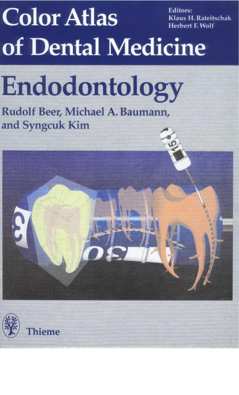
Color Atlas of Endodontology 1st Edition by Rudolf Beer, Michael A Baumann, Syngcuk Kim, Richard Jacobi ISBN 0865778566 9780865778566
$50.00 Original price was: $50.00.$25.00Current price is: $25.00.
Authors:Thieme; 1 edition (December 15, 1999) , Author sort:edition, Thieme; 1 , Published:Published:Aug 2002
Color Atlas of Endodontology 1st Edition by Rudolf Beer, Michael A Baumann, Syngcuk Kim, Richard Jacobi – Ebook PDF Instant Download/Delivery. 0865778566, 978-0865778566
Full download Color Atlas of Endodontology 1st Edition after payment
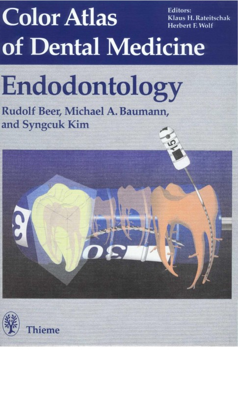
Product details:
ISBN 10: 0865778566
ISBN 13: 978-0865778566
Author: Rudolf Beer, Michael A Baumann, Syngcuk Kim, Richard Jacobi
You can buy this product at Gangaram Jinnah Medical Books Shop for home delivery and Cash on delivery to all over Pakistan. All kind of medical books are available.
Color Atlas of Endodontology 1st Table of contents:
1 Incipient enamel caries
2 Extent of caries
3 Extent of caries
4 Clinical appearance and bitewing radiograph
Diagnosis of Fissure Caries
5 Fissure anatomy
6 ECM caries meter
7 Extent of fissure caries
8 Extent of fissure caries
9 Extent of fissure caries
10 Summary
11 Breakdown of the enamel surface
12 Fiberoptic transillumination (FOTI)
13 Fiberoptic transillumination (FOTI)
14 Summary
Smooth Surface Caries
15 Incipient lesion (chalky spot)
16 Incipient lesion (10 years later)
17 Summary
Reversible Pulpitis
18 Dentinal caries and pulpitis
19 Reversible pulpitis
20 Nerve fibers in the pulp
21 Pain mechanism
Acute Irreversible Pulpitis
22 Caries and pulpitis
23 Accumulation of inflammatory cells
24 Inflammation-free root canal pulp tissue
25 Periapical inflammation
Presumptive Diagnosis
26 Inadequate restorations
27 Caries excavation
28 Acute reaction
29 Access preparations and root canal treatment
30 Root canal preparation
31 Root canal filling
Carious Pulp Exposure
32 Pulpitis aperta granulomatosa, “pulp polyp”
33 Necrosis of the pulp tissue in the root canal
34 Open pulpitis
35 Tissue removal
36 Treatment of the open pulpitis
Necrosis of the Pulp Tissue
37 Carious exposure of the pulp with necrosis
38 Tissue necrosis
39 The boundaries of necrosis
40 Periapical inflammation
Intentional Devitalization
41 Emergency treatment
42 Condition following intentional devitalization of the pulp
43 Uncovering the entrances to the root canals
44 Root canal preparation
45 Root canal filling
46 Two-year recall
Filling Materials and Pulp Necrosis
47 Pulp response to acid etching
48 Necrosis and aspiration
49 Acute inflammation
50 Root canal preparation
51 Root canal filling
Bacterial Infection in the Root Canal
52 Penetration of caries into the pulp tissue
53 Necrosis in the root canal with apical periodontitis
54 Bacteria within dentin
55 Bacteria in the root canal
Treatment of Bacterial Infection
56 Bacterial infection
57 Apical periodontitis
58 Emergency treatment
59 Removal of bacterially infected material from the canal
60 Interim dressing and coronal seal
61 Root canal filling and coronal restoration
Acute Apical Periodontitis
62 Caries progression and apical periodontitis
63 Necrosis and lysis of the radicular pulp
64 Inflammation in the apical root canal pulp
65 Acute apical periodontitis
Periapical Abscess
66 Palatal abscess
67 Opening and drainage through the root canal
68 Root canal preparation
69 Interim dressing
70 Antibacterial effect
71 Root canal filling
Chronic Apical Periodontitis
72 Necrosis and chronic periapical lesion
73 Necrosis of the coronal pulp
74 Periapical granulation tissue
75 Periapical microcysts
Chronic Apical Periodontitis and Radicular Cysts
76 Fistula and radiographic periapical lesion
77 Root canal preparation
78 Interim dressing
79 Clinical monitoring
80 Root canal obturation
81 Radiographic monitoring of the results of conservative treatment
Radicular Cysts
82 Formation and components of a radicular cyst
83 Contents of the cystic lumen
Examination and Diagnosis
Extraoral Examination
84 Extraoral ulcer
85 Extraoral swelling
86 Extraoral fistula
Intraoral Examination
87 Caries and filling
88 Fistulae and perforations
89 Tooth discoloration
Sensitivity Tests
90 Cold test
91 Electric pulp test
Clinical Examination and Selection of Therapy
92 Palpation
93 Percussion and biting test
Radiographic Diagnosis and Interpretation
94 Broken instrument fragment
95 Maxillary radiolucency
96 Mandibular radiolucency
97 Alignment of X-rays
98 Eccentric radiograph
Radiography in Endodontics
99 Diagnostic radiograph
100 First measurement radiograph
101 Working-length radiograph
102 Master point radiograph
103 Filling the root canals
104 Radiographic evaluation of the root canal filling
Digital Radiographic Technique
Intraoral Systems
105 Sensor of an intraoral digital system
Contrast Enhancement
Positive-Negative Representation
False Color Representation
Millimeter Grid
Resolution
Dynamic
Filters
106 Digital measurement picture
107 Digital measurement picture
Projection Angle
Uses of Digital Radiography
108 Digital follow-up radiograph (Sidexis)
109 Digital follow-up radiograph (Sidexis)
Anatomy
110 Three-dimensional reconstruction
Methods of Reproducing Root Canal Anatomy
111 Methods for visualizing the anatomy of the root canals I
112 Methods for visualizing the anatomy of the root canals II
Three-dimensional Computer Reconstruction
113 Contour-based reconstruction
114 Volume rendering
Magnetic Resonance Imaging (MRI)
115 MRI of an incisor tooth
116 MRI of a molar I
117 MRI of a molar II
Fundamentals
Classification of Canal Configurations
Maxillary Anterior Teeth
118 Maxillary central incisor
119 Maxillary lateral incisor
120 Maxillary canine
Mandibular Anterior Teeth
121 Mandibular central incisor
122 Mandibular lateral incisor
123 Mandibular canine
Maxillary Premolars
124 Maxillary first premolar
125 Range of variations in root canals of maxillary premolars
Mandibular Premolars
126 Mandibular first premolar
127 Range of variations in root canals of mandibular premolars
Maxillary Molars
128 Locating the canals of a maxillary first molar
129 Geometric aids
130 Significance of a fourth canal
Characteristics of Maxillary First Molars
131 Complexity of the maxillary first molars
132 Various forms of the mesiobuccal root
133 Complexity of the maxillary second molars
Mandibular Molars
134 Canal entrances in a mandibular molar
135 Mandibular molars
Instruments and Materials
136 ISO standardization
The Three Basic Instruments…
137 K reamers
138 K file
139 H files (Hedström files)
…and Their Modifications
140 Flexible instruments
141 Non-cutting tip
142 Further modifications
Instruments from Titanium Alloys
143 Titanium alloys
144 Titanium-aluminum alloy
145 Penetration depth of endodontic instruments
146 Effects of sterilization
147 Cutting efficiency
148 Wear of endodontic files
Engine-Driven Instrumentation
149 Engine-driven preparation
150 Instruments for powered handpieces
151 Engine-driven instruments
152 Profile instruments
153 Quantec instruments
154 Lightspeed instruments
Sonic and Ultrasonic Systems
155 Instruments
156 Handpieces
157 Piezo Ultrasonic System
158 Ultrasonic apparatus
159 Retrosurgery with ultrasound
People also search for Color Atlas of Endodontology 1st :
color.atlas of dental.medicine endodontology
atlas orthogonal near me
a color atlas of parasitology
color atlas of endodontics
j-shaped endodontic lesion
You may also like…
eBook PDF
color atlas of dental implant surgery 2nd edition by Michael Block 141603594X 9781416035947
eBook PDF
Color Atlas of Dental Implant Surgery 4th Edition by Michael Block 9780323228107 9780323228107
eBook PDF
Color Atlas of Dental Implant Surgery 3rd edition by Michael Block 9781437726787 143772678X
eBook PDF
Color Atlas of Dental Implant Surgery 4th Edition by Michael Block 9780323228107 0323228100
eBook PDF
Color Atlas of Dental Implant Surgery 4th Edition by Michael Block 9780323228107 0323228100

