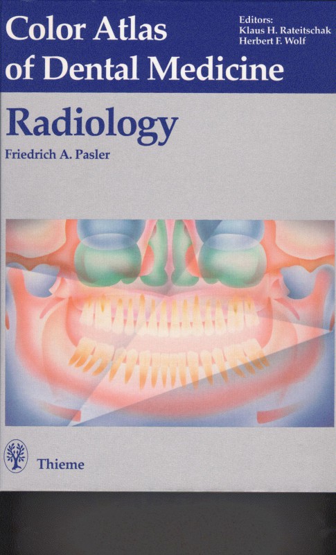
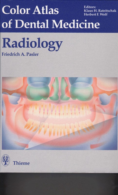
Color Atlas of Dental Medicine Radiology 1st Edition by Friedrich Pasler, Thomas Hassell, Arthur Hefti ISBN 0865774609 9780865774605
$50.00 Original price was: $50.00.$25.00Current price is: $25.00.
Authors:Thieme; (January 1, 1993) , Author sort:Thieme; (January 1, 1993)
Color Atlas of Dental Medicine Radiology 1st Edition by Friedrich Pasler, Thomas Hassell, Arthur Hefti – Ebook PDF Instant Download/Delivery. 0865774609, 978-0865774605
Full download Color Atlas of Dental Medicine Radiology 1st Edition after payment
Product details:
ISBN 10: 0865774609
ISBN 13: 978-0865774605
Author: Friedrich Pasler, Thomas Hassell, Arthur Hefti
Today, for patients of any age, a panoramic radiograph that allows high diagnostic reliability with minimum radiation exposure must be acknowledged as the standard of care. The new, more medically and legally responsible strategy for examination of patients is closely intertwined with a basic principle of patient care. Panoramic radiograph is rapidly becoming a meaningful and normal component of dental practice-oriented prevention. This Atlas presents the proper use of optimum radiographic exposure and contemporary techniques for today’s dental office. Emphasis is placed on the reliable recognition of normal structures in the projected space and clear differentiation of normal features from pathological radiographic signs. These major topics of fundamental importance for correct interpretation are treated more extensively. The section on diagnosis deals in depth with the typical radiographic manifestations of frequently occurring pathological processes. Inherent in that section of the book is the basic understanding that the radiopathologic manifestation of one and the same lesion may vary considerably depending on the patient’s age and sex, on the phase of development of the lesion at the time of documentation, on the localization within different surrounding structures, and according to actual histologic features of the lesion.
Color Atlas of Dental Medicine Radiology 1st Table of contents:
Radiographic Examination of the Patient in the Dental Office
- Examination Strategy and Active Protection Against Radiation Exposure
- Examination Strategies
- Technique for Panoramic Radiography
- Positioning of the Patient in the Apparatus
- Increased Radiographic Quality through Positioning According to Indication
- Typical Incorrect Positioning
- Incorrect Positioning
- Positioning in the Mixed Dentition Stage
- Positioning to Visualize Periodontal Destruction
- Positioning of the Tongue
- Depiction of the Alveolar Ridges
- “Zonarc,” A Special Instrument for Clinics
- Special Radiographs Using the Cephalometric Attachment
- Radiographic Anatomy in the Panoramic Radiograph
- Survey of the Anatomic Structures Visible in a Panoramic Radiograph
- Ventral Portion of the Facial Skeleton
- Ventral Portion of the Facial Skeleton in the Maxilla
- Variations in the Maxillary Sinus
- Retromaxillary Space
- External Ear and Temporomandibular Joint Region
- Palatal Bone in the Shadow of the Coronoid Process
- Tuberosity Region and the Cervical Vertebrae
- Chin Region
- Chin Region and the Body of the Mandible
- Mandibular Canal, Mandibular Rami, and the Cervical Vertebrae
- Mandibular Canal and Retromolar Structures
- Hyoid Bone and Cervical Region
- Hyoid Bone and Subtraction Effect from the Base of the Skull
- Angle of the Mandible and the Styloid Process
- Examination of Children and Adolescents Using the Panoramic Radiograph
- Diagram of Formation and Eruption of the Deciduous Teeth
- Diagram of Formation and Eruption of the Permanent Teeth
Bite-Wing Radiographs
- The Bite-Wing Radiograph
- Examples of Diagnosis Using Bite-Wings
Apical and Periodontal Radiographic Techniques
- Apical and Periodontal Radiographic Technique
- Radiographic Surveys for Patients of Various Ages
- Apical and Periodontal Survey in Adults
- Maxilla
- Mandible
- Third Molars
Radiographic Techniques for Special Regions
- Radiographic Technique with the Occlusal Film
- Intraoral
- Extraoral
Radiographic Anatomy in Periapical and Occlusal Radiographs
- Radiographic Anatomy in Periapical and Occlusal Radiographs
- Radiographic Anatomy During Tooth Development
- Radiographic Anatomy of Special Regions
- Maxillary Anterior Region
- Maxillary Canine Region
- Maxillary Premolar Region
- Maxillary Molar Region
- Mandibular Anterior Region
- Mandibular Canine Region
- Mandibular Premolar Region
- Mandibular Molar Region
Radiographic Anatomy in Occlusal Radiographs
- Radiographic Anatomy in Occlusal Radiographs
- Localization Using Various Methods
- Panoramic Radiography as an Aid in Localization
- Special Localization Problems
- Buccally Impacted Mandibular Canine
- Ectopically Positioned Anterior Teeth
- Apically Displaced Maxillary Third Molars
- Axially Presented Maxillary Third Molars
Errors and Tips for Radiograph Preparation
- Errors in Technique That Reduce Radiograph Quality
- Tips for Preparation of Good Panoramic Radiographs
- Before Positioning the Patient in the Apparatus
- With the Patient in the Apparatus
- Tips for Preparation of Good Radiographs
- Before Exposing the Film
- Immediately Before Exposure
- Summary of Basic Rules for Preparation of High-Quality Radiographs
- Common Errors During Preparation of Periapical Radiographs
- Common Errors During Preparation of Occlusal Radiographs
Processing Techniques and Errors
- Processing Technique and Errors Leading to Poor Quality Radiographs
- Tips for Error-Free Processing
- Tips for Error-Free Development
- Tips for Error-Free Fixation
- Optimum Radiograph
- Reducing Overdeveloped Radiographs
Supplemental Examinations Using Conventional and Modern Imaging Techniques
-
Conventional Skull Films
- First Standard Projection: Posteroanterior Cephalometric Skull Projection
- Second Standard Projection: Lateral Cephalometric Skull Projection
- Special Construction of a High Capacity Cephalometric Instrument
- Third Standard Projection: Full Axial Projection of the Skull
- Lateral Oblique Projection of the Mandible
- Special Positioning for the Lateral Oblique Projection of Mandible
- Zygoma/Cheek Tangential Skull Projections
- Mandibular Posteroanterior Skull Radiograph (Reverse Towne)
- Waters’ Projection Radiograph
-
Supplemental Examination of the Maxillary Sinus with Additional Methods
-
Transcranial Projection: “The Modified Schüller TMJ Film, Open and Closed”
-
Tomography: Supplemental Examination of the Temporomandibular Joint (TMJ)
-
Computed Tomography (CT)
-
Magnetic Resonance Imaging
People also search for Color Atlas of Dental Medicine Radiology 1st:
color atlas of dental medicine orthodontic diagnosis pdf
color atlas of oral and maxillofacial diseases
a color atlas of parasitology pdf
atlas of dental anatomy – pdf
color atlas of anatomy 8th edition

