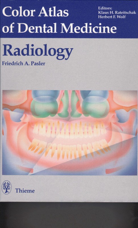
Color Atlas of Dental Medicine Radiology 1st Edition by Friedrich Pasler, Klaus Rateitschak, Herbert Wolf 0865774609 978-0865774605
$50.00 Original price was: $50.00.$25.00Current price is: $25.00.
Authors:Friedrich Pasler , Tags:(Color Atlas of Dental Medicine) , Author sort:Pasler, Friedrich , Publisher:Thieme (1993)
Color Atlas of Dental Medicine Radiology 1st Edition by Friedrich Pasler, Klaus Rateitschak, Herbert Wolf – Ebook PDF Instant Download/Delivery. 0865774609, 978-0865774605
Full download Color Atlas of Dental Medicine Radiology 1st Edition after payment
Product details:
ISBN 10: 0865774609
ISBN 13: 978-0865774605
Author: Friedrich A. Pasler, Klaus H. Rateitschak, Herbert F. Wolf
Today, for patients of any age, a panoramic radiograph that allows high diagnostic reliability with minimum radiation exposure must be acknowledged as the standard of care. The new, more medically and legally responsible strategy for examination of patients is closely intertwined with a basic principle of patient care. Panoramic radiograph is rapidly becoming a meaningful and normal component of dental practice-oriented prevention. This Atlas presents the proper use of optimum radiographic exposure and contemporary techniques for today’s dental office. Emphasis is placed on the reliable recognition of normal structures in the projected space and clear differentiation of normal features from pathological radiographic signs. These major topics of fundamental importance for correct interpretation are treated more extensively. The section on diagnosis deals in depth with the typical radiographic manifestations of frequently occurring pathological processes. Inherent in that section of the book is the basic understanding that the radiopathologic manifestation of one and the same lesion may vary considerably depending on the patient’s age and sex, on the phase of development of the lesion at the time of documentation, on the localization within different surrounding structures, and according to actual histologic features of the lesion.
Color Atlas of Dental Medicine Radiology 1st Table of contents:
Panoramic Radiography for Basic Information and Supplemental Examination Using Special Radiographs
Technique for Panoramio Radiography
Technique for Panoramic Radiography
Positioning of the Patient in the Apparatus
Increased Radiographic Quality through Positioning
According to Indication
Typical Incorrect Positioning
Incorrect Positioning
Positioning in the Mixed Dentition Slage
Positioning to Visualize Periodontal Destruction
Positioning of the Tongue
Depiction of the Alveolar Ridges
“Zonarc,” A Special Instrument for Clinics
Special Radiographs Using the Cephalometric Altachment
Radiographic Anatomy in the Panoramic Radiograph
Survey of the Anatomic Structures Visiblo in a
Panoramic Radiograph
Ventral Portion of the Facial Skeleton
Ventral Portion of the Facial Skeleton in the Maxilla
Variations in the Maxillary Sinus
Retromaxillary Space
External Ear and Temporomandibular Joint Region
Palatal Bone in the Shadow of the Coranoid Process
Tuberosity Region and the Cervical Vertebrae
Chin Region
Chin Region and the Body of the Mandible
Mandibular Canal, Mandibular Rami and the Cervical Vertebrae
Mandibular Canal and Retromolar Structures
Hyoid Bone and Cervical Region
Hyoid Bone and Subtraction Effect from the Base of the Skull
Angle of the Mandible and the Styloid Process
Examination of Children and Adolescents Using the Panoramic Radiograph
Diagram of Formation and Eruption of the Deciduous Teeth
Diagram of Formation and Eruption of the Permanent Teeth
The Bite-Wing Radiograph
Examples of Diagnosis Using Bito-Wings
Apical and Periodontal Radiographic Technique
Radiographic Surveys for Patients of Various Ages
Apical and Periodontal Survey in Adults
Maxilla
Mandible
Third Molars
Radiographic Technique with the Occlusal Film
Intraoral
Extraoral
Radiographic Anatomy in Periapical and Occlusal Radiographs
Radiographic Anatomy During Tooth Development
Radiographic Anatomy of Special Regions
Maxillary Anterior Region
Maxillary Canine Region
Maxillary Premolar Region
Maxillary Molar Region
Mandibular Anterior Region
Mandibular Canine Region
Mandibular Premolar Region
Mandibular Molar Region
Radiographic Anatomy in Ocolusal Radiographs
Localization Using Various Methods
Panoramic Radiography as an Aid in Localization
Special Localization Problems
Buccally Impacted Mandibular Canine
Ectopically Postioned Anterior Teeth
Apically Displaced Maxillary Third Molars
Axially Presented Maxillary Third Molars
Errors in Technique That Reduce Radiograph Quality
Tips for Preparation of Good Panoramic Radiographs Before Positioning the Patient in the Apparatus With the Patient in the Apparatus
Tips for Preparation of Good Radiographs
Before Exposing the Film Immediately Before Exposure
Summary of Basic Rules for Preparation of High Quality
Radiographs
Common Errors During Preparation of Periapical
Radiographs
Common Errors During Preparation of Occlusal
Radiographs
Processing Technique and Errors Leading to Poor Quality Radiographs
Tips for Error-Free Processing
Tips for Error-Free Development
Tips for Error-Free Fixation
Optimum Radiograph
Reducing Overdeveloped Radiographs
Supplemental Examinations Using Conventional and Modern Imaging Techniques
Conventional Skull Films
First Standard Projection: Postercanterior
Cephalometric Skull Projection
Second Standard Projection: Lateral Cephalometric
Skull Projection
Special Construction of a High Capacity
Cephalometric Instrument
Third Standard Projection: Full Axial Projection of the Skull
Lateral Oblique Projection of the Mandible
Special Positioning for the Lateral Oblique Projection of Mandible
Zygoma/Cheek Tangential Skull Projections
Mandibular Postercanterior Skull Radiograph (Reverso Towne
Waters’ Projection Radiograph
Supplemental Examination of the Maxillary Sinus with Additional Methods
Transcranial Projection: “The Modified Schüller
TMJ Film, Open and Closed”
Tomography: Supplemental Examination of the
Temporomandibular Joint (TMJ)
Computed Tomography (CT)
Magnetic Resonance Imaging
Selected Examples of Dental Radiographic Diagnosis
Anomalies of Dental Development and the Teeth
Congenitally Missing Teeth, Retention and Inclusion
Retention, Malocclusion and Resorption
Retention of Supernumerary Teeth, Resorption of
Retained Teeth
Retained Teeth in Special Locations
Retained and Ankylosed Teeth
Mesiodens, Gemination, Taurodontism and Dens in dent
Hypercementosis and Enamel Pearls
Amelogenesis Imperfecta
Dentinogenesis Imperfecta
Additional Dental Dysplasias
Posterior Open Bite with Macroglossia and “Idiopathic
Root Resorption”
Concrements, Calcifications, Ossifications
Regressive Changes in Teeth and Jaws
Inflammation of the Jaws and Osteoradionecrosis
Acute and Chronic Apical Periodontitis
Diffuse Sclerosing Osteomyelitis in Chronic Apical and
Marginal Periodontitis
Sclerosing Osteomyelitis and inflammatory Reactive
Osteosis
Osteomyelitis in Infants and Children
Acute Osteomyelitis
Secondary Chronic Osteomyelitis
Primary Chronic Osteomyelitis, Osteoradionecrosis
Dentogenic Sinus Disorders
Panoramic Radiography of Dentogenic Sinus Pathology
Additional Signs of Dentogenic Infection
Incidental Findings and the Significance of the
Waters’ Projection
Acute Unilateral Dentogenic Sinusitis
Schematic View of Sinus Diagnosis in Waters’ Projection
Radiographs
Acute and Chronic Maxillary Sinusitis
Computed Tomography for Supplemental Diagnosis
Foreign Bodies, Root Fragments and Surgical Defects
Temporomandibular Joint Disturbances
Examination of the Masticatory Organ Using the
Panoramic Radiograph
Temporomandibular Joint Pain with Malocclusion
Film Tomography of the Temporomandibular Joint
Computed Tomography of the Temporomandibular Joint with Direct Lateral Projection
Magnetic Resonance Imaging of the Temporomandibular Joint
Hypoplasia and Exostosis of the Condyles
Hyperplasia and Osteochondral Exostoses
Inflammatory and Degenerative Changes
Cysts and Pseudocysts
Classification
Odontogenic Cysts
Radicular Cyst
Radicular Cyst in the Mandible
Radicular Residual Cyst in the Mandible
Radicular Cyst in the Maxilla
Follicular Cyst
Atypically Localized Follicular Cysts
Nonodontogenic Cysts
Pseudocysts
Odontogenic Tumors and Pseudotumors
Amoloblastoma
Ameloblastic Fibroma
Odontogenic Myxoma
Cementoma
Periapical Cemental Dysplasia
Cementoblastoma
Cementoblastoma, Cementum-forming Fibroma
Odontoma
Complex Odontoma
Transition Forms
Compound Odantoma and Fibro-odontoma
Nonodontogenic Tumors and Pseudotumors
Benign Lesions
Malignant Lesions
Central Reparative Giant Cell Granuloma
Peripheral Reparative Giant Cell Granuloma
Histiocytosis X
Chondroma
Osteochondroma
Desmoplastic Fibroma
Ossilying Fibroma
Fibrous Dysplasia (Jaffe-Lichtenstein)
Fibrous Dysplasia and Cherubism
Osteoid Osteome, Osteoblastoma
Osteoma
Exostoses and Enostoses
Hyperostases and Hypertrophies
Osteoporosis and Alrophy
Bone Marrow Islands and Incorrect Interpretations
Osteogenesis Imperfecta and Osteopetrosis
Osteilis Deformans (Paget’s Disease of Bone)
Hemangioma
Sarcoma
Carcinogenic Infiltration
Mucoepidermoid Tumor
Metastasis
Traumatology
Radiographic Signs of Subluxation
Radiographic Signs of Tooth Fracture
Radiographic Signe of Mandibular Fracture
Mandibular Fracture During Mixed Dentition
Foreign Bodies and Postoperative Conditions
Various Therapeutic Materials Seen in the Radiograph Trauma, Osteosynthesis Material and Implants
Deposition of Filling Materials
Radiography of Root Fragments
Tooth Extraction and Root Fragments
Fractured Bone and Sequestra
Success and Failure with Root Tip Resection
(Apicoectomy)
People also search for Color Atlas of Dental Medicine Radiology 1st:
color atlas of ultrasound anatomy
teeth radiology anatomy
teeth radiology numbering
dental radiology questions and answers

