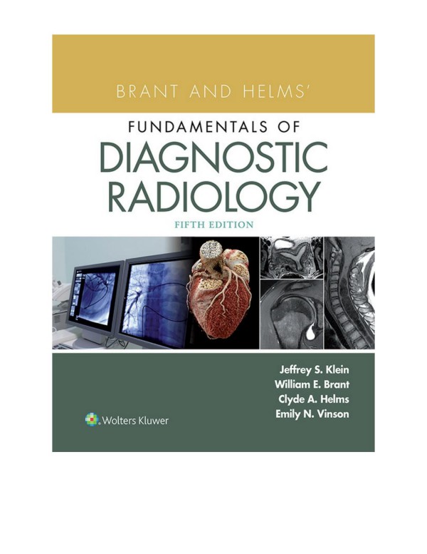
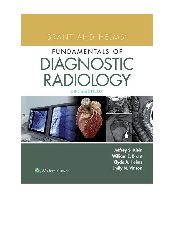
Brant and Helms’ Fundamentals of Diagnostic Radiology 5th edition by Jeffrey Klein,Jennifer Pohl,Emily Vinson,William Brant,Clyde Helms 9781496367426 1496367421
$50.00 Original price was: $50.00.$25.00Current price is: $25.00.
Authors:Jeffrey S. Klein, MD, FACR; William E. Brant, MD, FACR; Clyde A. Helms, MD; Emily N. Vinson, MD , Author sort:Jeffrey S. Klein, MD, FACR & William E. Brant, MD, FACR & Clyde A. Helms, MD & Emily N. Vinson, MD , Published:Published:Jan 2019
Brant and Helms’ Fundamentals of Diagnostic Radiology 5th edition by Jeffrey Klein,Jennifer Pohl,Emily Vinson,William Brant,Clyde Helms – Ebook PDF Instant Download/Delivery.9781496367426,1496367421
Full download Brant and Helms’ Fundamentals of Diagnostic Radiology 5th edition after payment
Product details:
ISBN 10:1496367421
ISBN 13:9781496367426
Author: Jeffrey Klein,Jennifer Pohl,Emily Vinson,William Brant,Clyde Helms
Publisher’s Note: Products purchased from 3rd Party sellers are not guaranteed by the Publisher for quality, authenticity, or access to any online entitlements included with the product.Trusted by radiology residents, interns, and students for more than 20 years, Brant and Helms’ Fundamentals of Diagnostic Radiology, 5th Edition delivers essential information on current imaging modalities and the clinical application of today’s technology. Comprehensive in scope, it covers all subspecialty areas including neuroradiology, chest, breast, abdominal, musculoskeletal imaging, ultrasound, pediatric imaging, interventional techniques, and nuclear radiology. Full-color images, updated content, new self-assessment tools, and dynamic online resources make this four-volume text ideal for reference and review.
Brant and Helms’ Fundamentals of Diagnostic Radiology 5th Table of contents:
SECTION I. BASIC PRINCIPLES
1 DIAGNOSTIC IMAGING METHODS
CONVENTIONAL RADIOGRAPHY
CROSS-SECTIONAL IMAGING TECHNIQUES
Computed Tomography
Magnetic Resonance Imaging
Ultrasonography
RADIOGRAPHIC CONTRAST AGENTS
Iodinated Contrast Agents
Magnetic Resonance Imaging Intravascular Contrast Agents
Gastrointestinal Contrast Agents
Ultrasound Intravascular Contrast Agents
RADIATION RISK AND ENSURING PATIENT SAFETY
RADIOLOGY REPORTING
Suggested Readings
SECTION II. NEURORADIOLOGY
2 INTRODUCTION TO BRAIN IMAGING
LOOKING AT THE BRAIN
CURRENT NEUROIMAGING OPTIONS
IMAGING STRATEGY FOR COMMON CLINICAL SYNDROMES
ANALYSIS OF THE ABNORMALITY
Suggested Readings
3 CRANIOFACIAL TRAUMA
HEAD TRAUMA
Imaging Strategy
Scalp Injury
Skull Fractures
Temporal Bone Fractures
Head Injury Classification
Primary Head Injury: Extra-Axial
Primary Head Injury: Intra-Axial
Secondary Head Injury
Brainstem Injury
Penetrating Trauma
Predicting Outcome After Acute Head Trauma
Child Abuse
FACIAL TRAUMA
Imaging Strategy
Soft Tissue Findings
Nasal Fractures
Maxillary and Paranasal Sinus Fractures
Orbital Trauma
Fractures of the Zygoma
Fractures of the Midface (Le Fort Fractures)
Nasoethmoidal Fractures
Mandibular Fractures
References
Suggested Readings
4 CEREBROVASCULAR DISEASE
ISCHEMIC STROKE
Pathophysiologic Basis for Imaging Changes
Imaging Triage for Emergency Stroke Intervention
Hemorrhagic Transformation of Infarction
Use of Contrast in Ischemic Stroke
Pattern Recognition in Ischemic Stroke
Anterior (Carotid) Circulation
Posterior (Vertebrobasilar) Circulation
Watershed (Borderzone) Infarction
Small Vessel Ischemia
Venous Infarction
HEMORRHAGE
Imaging of Hemorrhage
Subarachnoid Hemorrhage
Parenchymal Hemorrhage
Primary Hemorrhage Versus Hemorrhagic Neoplasm
Suggested Readings
5 CENTRAL NERVOUS SYSTEM NEOPLASMS AND TUMOR-LIKE MASSES
TUMOR CLASSIFICATION
CLINICAL PRESENTATION
NEUROIMAGING PROTOCOL
NEUROIMAGING ANALYSIS
POSTOPERATIVE IMAGING
FOLLOW-UP IMAGING
SPECIFIC NEOPLASMS
Intra-Axial Tumors: Glial
Intra-Axial Tumors: Nonglial
Intra-Axial Tumors: Nonneuroepithelial
Extra-Axial Tumors
Pineal Region Masses
Sellar Region Masses
Masses of Developmental Origin
Suggested Readings
6 CENTRAL NERVOUS SYSTEM INFECTIONS
CONGENITAL INFECTIONS
EXTRA-AXIAL INFECTIONS
Subdural and Epidural Infections
Meningitis
PARENCHYMAL INFECTIONS
Pyogenic Cerebritis and Abscess
Mycobacterial Infections
Fungal Infections
Parasitic Infections
Spirochete Infections
Viral Infections
Creutzfeldt–Jakob Disease
ACQUIRED IMMUNODEFICIENCY SYNDROME–RELATED INFECTIONS
Suggested Readings
7 WHITE MATTER AND NEURODEGENERATIVE DISEASES
DEMYELINATING DISEASES
Primary Demyelination
Ischemic Demyelination
Infection-Related Demyelination
Toxic and Metabolic Demyelination
DYSMYELINATING DISEASES
CEREBROSPINAL FLUID DYNAMICS
NEURODEGENERATIVE DISORDERS
Suggested Readings
8 HEAD AND NECK IMAGING
PARANASAL SINUSES AND NASAL CAVITY
SKULL BASE
Tumors of the Skull Base
Temporal Bone
SUPRAHYOID HEAD AND NECK
Superficial Mucosal Space
Parapharyngeal Space
Carotid Space
Parotid Space
Masticator Space
Retropharyngeal Space
Prevertebral Space
Trans-Spatial Diseases
LYMPH NODES
ORBIT
CONGENITAL LESIONS
Suggested Readings
9 SPINE IMAGING
COMMON CLINICAL SYNDROMES
IMAGING METHODS
INFLAMMATION
INFECTION
Pyogenic Infections
Nonpyogenic Infections
NEOPLASMS
Intramedullary Masses
Intradural-Extramedullary Masses
Extradural Masses
VASCULAR DISEASES
TRAUMA
DEGENERATIVE DISEASES OF THE SPINE
Imaging Methods
Disc Disease
Disc Herniation Location
SPINAL STENOSIS
POSTOPERATIVE CHANGES
Bony Abnormalities
Suggested Readings
SECTION III. CHEST
10 METHODS OF EXAMINATION, NORMAL ANATOMY, AND RADIOGRAPHIC FINDINGS OF CHEST DISEASE
IMAGING MODALITIES
NORMAL CHEST ANATOMY
Frontal Chest Radiograph
Lateral Chest Radiograph
Anatomy of the Normal Mediastinum
Normal Hilar Anatomy
Pleural Anatomy
Chest Wall Anatomy
Diaphragmatic Anatomy
RADIOGRAPHIC FINDINGS IN CHEST DISEASE
Pulmonary Opacity
Pulmonary Lucency
Mediastinal Masses
Mediastinal Widening
Pneumomediastinum and Pneumopericardium
Hilar Disease
Pleural Effusion
Pneumothorax
Localized Pleural Thickening
Diffuse Pleural Thickening
Pleural and Extrapleural Lesions
Chest Wall Lesions
Diaphragmatic Abnormalities
Suggested Readings
11 MEDIASTINUM AND HILA
MEDIASTINAL MASSES
Anterior (Prevascular) Mediastinal Masses (Table 11.2)
Middle (Visceral) Mediastinal Masses (Table 11.3)
Posterior (Paravertebral) Mediastinal Masses (Table 11.5)
DIFFUSE MEDIASTINAL DISEASE (TABLE 11.6)
THE HILA
Unilateral Hilar Enlargement/Increased Density (Table 11.8)
Bilateral Hilar Enlargement
Small Hila
Suggested Readings
12 PULMONARY VASCULAR DISEASE
PULMONARY EDEMA
PULMONARY HEMORRHAGE AND VASCULITIS
PULMONARY EMBOLISM
PULMONARY ARTERIAL HYPERTENSION
Suggested Readings
13 PULMONARY NEOPLASMS AND NEOPLASTIC-LIKE CONDITIONS
THE SOLITARY PULMONARY NODULE
Lesions Presenting as SPNs
LUNG TUMORS
Lung Cancer
NONEPITHELIAL LUNG TUMORS AND TUMOR-LIKE CONDITIONS
TRACHEAL AND BRONCHIAL MASSES
METASTATIC DISEASE TO THE THORAX
Suggested Readings
14 PULMONARY INFECTION
INFECTION IN THE NORMAL HOST
Bacterial Pneumonia
Viral Pneumonia
Fungal Pneumonia
Parasitic Infection
COMPLICATIONS OF PULMONARY INFECTION
INFECTION IN THE IMMUNOCOMPROMISED HOST
Suggested Readings
15 DIFFUSE LUNG DISEASE
THIN-SECTION CT OF THE PULMONARY INTERSTITIUM
Thin-Section CT Signs of Disease
CHRONIC INTERSTITIAL LUNG DISEASE
Chronic Interstitial Pulmonary Edema
Connective Tissue Disease
Idiopathic Chronic Interstitial Pneumonias
Chronic Fibrosing Idiopathic Interstitial Pneumonias
Smoking-Related Idiopathic Interstitial Pneumonias
Acute or Subacute Idiopathic Interstitial Pneumonias
Rare Idiopathic Interstitial Pneumonias
Unclassifiable Idiopathic Interstitial Pneumonia
Other Chronic Interstitial Lung Diseases
INHALATIONAL DISEASE
Pneumoconiosis
Hypersensitivity Pneumonitis
GRANULOMATOUS DISEASES
Sarcoidosis
Berylliosis
Langerhans Cell Histiocytosis of Lung
Granulomatosis With Polyangiitis
EOSINOPHILIC LUNG DISEASE
Idiopathic Eosinophilic Lung Disease
Eosinophilic Lung Disease of Identifiable Etiology
Eosinophilic Lung Disease Associated With Autoimmune Diseases
DRUG-INDUCED LUNG DISEASE
Specific Agents (Table 15.11)
MISCELLANEOUS DISORDERS
References and Suggested Readings
16 AIRWAYS DISEASE AND EMPHYSEMA
INTRODUCTION
TRACHEA AND CENTRAL BRONCHI
Congenital Tracheal and Bronchial Anomalies
Focal Tracheal Disease
Diffuse Tracheal Disease
Tracheal and Bronchial Injury
Broncholithiasis
CHRONIC OBSTRUCTIVE PULMONARY DISEASE
Asthma and Chronic Bronchitis
Bronchiectasis
Emphysema
BULLOUS LUNG DISEASE
SMALL AIRWAYS DISEASE
Suggested Readings
17 PLEURA, CHEST WALL, DIAPHRAGM, AND MISCELLANEOUS CHEST DISORDERS
PLEURA
Anatomy, Physiology, and Pathophysiology
Pleural Effusion
Bronchopleural Fistula
Pneumothorax
Focal Pleural Disease
Diffuse Pleural Disease
Asbestos-Related Pleural Disease
CHEST WALL
Soft Tissues
The Bony Thorax
DIAPHRAGM
CONGENITAL LUNG DISEASE IN ADULTS
TRAUMATIC LUNG DISEASE
ASPIRATION
RADIATION-INDUCED LUNG DISEASE
Suggested Readings
SECTION IV. BREAST RADIOLOGY
18 NORMAL ANATOMY AND HISTOPATHOLOGY
OVERVIEW
SKIN
NIPPLE–AREOLAR COMPLEX
SUBCUTANEOUS TISSUE
MILK DUCTS AND LOBULES
STROMA
CHEST WALL
MALE BREAST
LYMPHATICS
BLOOD SUPPLY
EMBRYOLOGY AND DEVELOPMENT
CHANGES OVER TIME: PREGNANCY, LACTATION, AND MENOPAUSE
PATHOPHYSIOLOGY OF BREAST CANCER
Suggested Readings
19 IMAGING THE SCREENING PATIENT
INTRODUCTION
SCREENING FOR BREAST CANCER
Support for Screening
Screening Guidelines
TECHNICAL FACTORS IN SCREENING
Mammography Physics
Radiation Risk
Positioning of Screening Mammography
Image Quality
BREAST IMPLANTS
Implant Types
Implant Rupture
TOMOSYNTHESIS
INTERPRETING THE MAMMOGRAM
Maximizing Your Interpretation
Breast Density
THE USE OF OTHER IMAGING MODALITIES FOR BREAST CANCER SCREENING
Screening Breast Ultrasound
Functional Imaging
CONCLUSION
Suggested Readings
20 IMAGING THE DIAGNOSTIC PATIENT
INTRODUCTION
DIAGNOSTIC MAMMOGRAPHY
Spot Compression Views
Magnification Views
True Lateral and Rolled CC Views
Tomosynthesis
Ultrasound
EVALUATION OF THE SYMPTOMATIC PATIENT
Palpable Breast Mass
Breast Pain
Nipple Discharge
Inflammation of the Breast
Paget Disease
Axillary Adenopathy
THE MALE BREAST
ASSESSMENT AND RECOMMENDATION
BREAST CANCER STAGING
POSTOPERATIVE SURVEILLANCE
CONCLUSION
Suggested Readings
21 BREAST IMAGING REPORTING AND DATA SYSTEM
INTRODUCTION
MAMMOGRAPHY LEXICON
Breast Composition
Masses
Calcifications
Architectural Distortion
Asymmetries
Associated Features
ULTRASOUND LEXICON
MRI LEXICON
REPORTING
FOLLOW-UP AND OUTCOME MONITORING
Statistical Terms
Medical Audit
Suggested Readings
22 BREAST MAGNETIC RESONANCE IMAGING
INTRODUCTION TO MRI
INDICATIONS
Screening
Extent of Disease
Metastatic Disease With Suspected Breast Primary
Nipple Discharge
Biopsy Planning
Implant Evaluation
TECHNIQUE
Imaging Parameters
Kinetics
Artifacts
INTERPRETATION
Background Parenchymal Enhancement and Fibroglandular Tissue
Findings
WORKUP OF ABNORMAL MRI
RISKS OF MRI
FUTURE OF MR IMAGING
CONCLUSION
Suggested Readings
23 IMAGE-GUIDED BREAST PROCEDURES
INTRODUCTION TO BREAST BIOPSIES
BREAST BIOPSY
Indications
Needle Types
Guidance
Biopsy Markers
Complications
Radiology–Pathology Concordance
Follow-Up After Benign Biopsy
LOCALIZATION
Indications
Localization Devices
Guidance
Specimen Radiograph
GALACTOGRAPHY
CONCLUSION
Suggested Readings
SECTION V. CARDIAC RADIOLOGY
24 INTRODUCTION TO CARDIAC ANATOMY, PHYSIOLOGY, AND IMAGING TECHNIQUES
INTRODUCTION
CARDIAC ANATOMY
CARDIAC PHYSIOLOGY
IMAGING TECHNIQUES
Chest Radiography
Echocardiography
Nuclear Imaging
Cardiac CT
Cardiac MRI
Suggested Readings
25 CORONARY ARTERY ANOMALIES AND DISEASE
CORONARY ARTERY ANATOMY
Left Main Coronary Artery
Left Anterior Descending Coronary Artery
Left Circumflex Coronary Artery
Ramus Intermedius
Right Coronary Artery
Posterior Descending Artery
Posterior Left Ventricular Branch
Conus Branch
Sinoatrial Nodal Branch
Atrioventricular Nodal Branch
CORONARY ARTERY ANOMALIES
ABNORMALITIES IN ORIGIN
Abnormalities in Origin, Benign
Abnormalities in Origin, Possibly Malignant
ABNORMALITIES IN COURSE
Myocardial Bridging
Intracavitary Course
Split (Double) Coronary Artery
ABNORMALITIES IN TERMINATION
Coronary Fistula
CORONARY ARTERY DISEASE
Coronary Artery Calcification
Coronary Plaque and Remodeling
MRI IN CORONARY ARTERY DISEASE
MR Imaging of the Coronary Arteries
MR Assessment of the Myocardium in Coronary Artery Disease
TREATMENT OF CORONARY ARTERY DISEASE
Coronary Stents
Coronary Artery Bypass Grafts
CORONARY ARTERY ANEURYSM AND PSEUDOANEURYSM
CORONARY ARTERY DISSECTION
MECHANICAL COMPLICATIONS OF MYOCARDIAL INFARCTION
ADDITIONAL CONSIDERATIONS
Suggested Readings
26 CARDIAC MASSES
INTRODUCTION
IMAGING TECHNIQUES AND PROTOCOLS
Echocardiography
Cross-Sectional Imaging
BENIGN CARDIAC TUMORS
MALIGNANT CARDIAC TUMORS
Metastatic Disease
Tumorlike Lesions
Suggested Readings
27 VALVULAR DISEASE
VALVE STRUCTURE AND FUNCTION
IMAGING EVALUATION OF VALVE DISEASE
Echocardiography
Radiography and CT
Cardiac MRI
Phase-Contrast MRI
Quantification of Ventricular Stroke Volume
AORTIC VALVE
Congenital Aortic Valve Disease
Acquired Aortic Valve Disease
Aortic Stenosis
Aortic Regurgitation
MITRAL VALVE
Congenital Mitral Valve Disease
Acquired Mitral Valve Disease
TRICUSPID VALVE
Congenital Tricuspid Valve Disease
PULMONARY VALVE
Pulmonary Stenosis
Pulmonary Regurgitation
ACQUIRED DISEASES OF MULTIPLE VALVES
Infective Endocarditis
Nonbacterial Endocarditis
Rheumatic Heart Disease
Carcinoid Valve Disease
POSTOPERATIVE COMPLICATIONS FOLLOWING VALVE SURGERY
Suggested Readings
28 NONISCHEMIC CARDIOMYOPATHIES
HYPERTROPHIC CARDIOMYOPATHY
FABRY DISEASE
CARDIAC AMYLOIDOSIS
LOEFFLER ENDOCARDITIS
DILATED CARDIOMYOPATHIES
ARRHYTHMOGENIC RIGHT VENTRICULAR CARDIOMYOPATHY
MYOCARDITIS
CARDIAC SARCOIDOSIS
LEFT VENTRICULAR NONCOMPACTION
STRESS-INDUCED (TAKOTSUBO) CARDIOMYOPATHY
IRON OVERLOAD CARDIOMYOPATHY
Suggested Readings
29 IMAGING OF THE PERICARDIUM
ANATOMY AND NORMAL APPEARANCE ON IMAGING
Pericardial Sinuses and Recesses
CONGENITAL ANOMALIES OF THE PERICARDIUM
Pericardial Cyst
Pericardial Defect
ACQUIRED PERICARDIAL DISEASES
Pericardial Effusion
Pericardial Tamponade
INFLAMMATION OF THE PERICARDIUM
Acute Pericarditis
Fibrous Pericarditi
People also search for Brant and Helms’ Fundamentals of Diagnostic Radiology 5th:
brant and helms pdf
brant and helms’ fundamentals of diagnostic radiology
brant and helms
brant and helms’ fundamentals of diagnostic radiology 5th edition
fundamentals of diagnostic radiology
You may also like…
eBook PDF
Fundamentals of Oral and Maxillofacial Radiology 1st edition by Sean Hubar 9781119122227 1119122228

