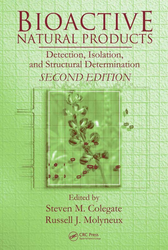
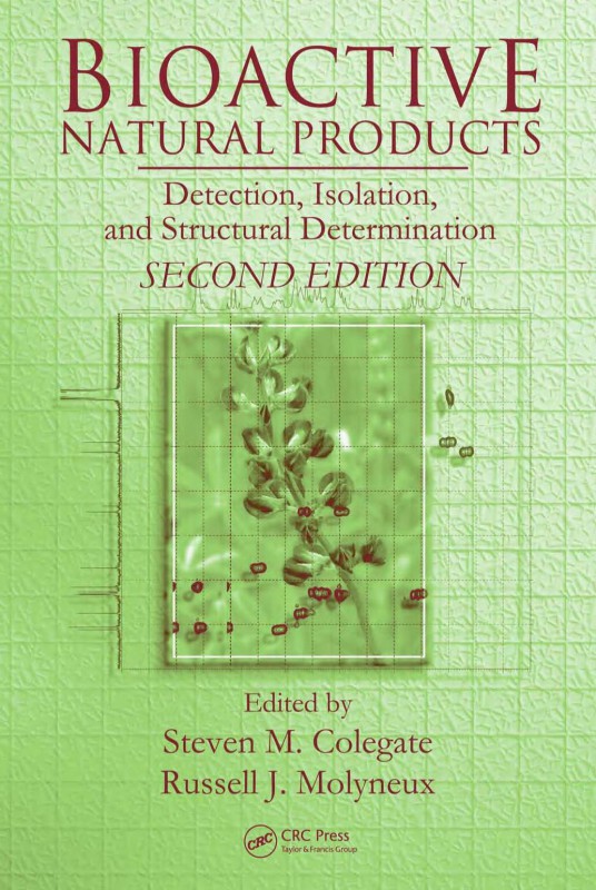
Bioactive Natural Products Detection Isolation and Structural Determination 2nd Edition by Steven Colegate, Russell Molyneux 1498774814 9781498774819
$50.00 Original price was: $50.00.$25.00Current price is: $25.00.
Authors:Steven M. Colegate; Russell J. Molyneux , Series:Science [20] , Tags:Science; Chemistry; General; Medical; Biochemistry; Pharmacology; Life Sciences , Author sort:Colegate, Steven M. & Molyneux, Russell J. , Ids:9780849343728 , Languages:Languages:eng , Published:Published:Sep 1993 , Publisher:CRC Press , Comments:Comments:Bioactive Natural Products covers all the aspects of bioactive natural product research from ethnobotanical investigations to modern, technologically assisted isolation and structural determination of active compounds. An internationally selected group of experts share their knowledge of a wide range of bioactivities and chemical compound classes.Topics in the chapters describing the modern application of detection, isolation, and structural determination techniques are strongly supported by chapters detailing and reviewing research involving various classes of bioactivity. Research areas include the immunomodulatory, antiviral, cytotoxic, anti-inflammatory, and insect behavior classes of bioactivity.Extensive referencing throughout the text is helpful to those readers not familiar with this subject and serves as a critical review for more experienced researchers. The book is also excellent for upper division or post-graduate courses.
Bioactive Natural Products Detection Isolation and Structural Determination 2nd Edition by Steven Colegate, Russell Molyneux – Ebook PDF Instant Download/Delivery. 1498774814, 9781498774819
Full download Bioactive Natural Products Detection Isolation and Structural Determination 2nd Edition after payment
Product details:
ISBN 10: 1498774814
ISBN 13: 9781498774819
Author: Steven M. Colegate; Russell J. Molyneux
Following the successful format of the original, this new edition presents applications of the most recent techniques for the detection, isolation, and structural determination of bioactive natural products. It features new case studies and illustrations that demonstrate applications of techniques covered in the book. Complementing as much as replacing the first edition, most of the contributors are new. The text includes updates on chemical extraction, and NMR-based structure determination, and new contributions on liquid chromatography linked with mass and NMR spectroscopy, dereplication approaches, assessment of source material for natural products and novel bioassay development.
Bioactive Natural Products Detection Isolation and Structural Determination 2nd Table of contents:
Chapter 1 An Introduction and Overview
1.1 Introduction
1.2 The Multidisciplinary Approach
1.3 Why Isolate Biologically Active Natural Products?
1.4 Detection and Isolation
1.5 Structure Determination
1.6 Overview
1.6.1 Methods of Detection, Isolation, and Structural Determination
1.6.1.1 Detection and Isolation
1.6.1.2 Structure Determination
1.6.2 Specific Case Studies
1.7 Conclusions
Chapter 2 Detection and Isolation of Bioactive Natural Products
2.1 Introduction
2.2 Detection of Biologically Active Metabolites
2.3 Screening For Bioactive Metabolites
2.3.1 Primary Screening Assays
2.3.1.1 Brine Shrimp Lethality Test
2.3.1.2 Crown Gall Tumor Bioassay
2.3.1.3 Starfish or Sea Urchin Assay
2.3.1.4 Bioassays for Antibiotic Activity
2.3.1.5 Bioautography
2.3.1.6 Allelopathy
2.3.1.7 Insecticidal Activity
2.3.2 Specialized Screening Assays
2.3.2.1 Antiviral Activity
2.3.2.2 Cytotoxicity, Antitumor, and Antineoplastic Activity
2.3.2.3 Immunosuppressive Activity
2.3.2.4 Antimalarial Activity
2.3.2.5 Amoebicidal Activity
2.3.2.6 Antimycobacterial Activity
2.4 Isolation and Separation
2.4.1 Extraction
2.4.1.1 Dry Biological Material
2.4.1.2 Fresh Material
2.4.1.3 Liquid Culture Broth and Other Biological Fluids
2.4.1.4 Extraction of Water-Soluble Metabolites
2.4.1.5 Removal of Fatty Material
2.4.1.6 Supercritical Fluid and Accelerated Solvent Extraction
2.4.2 Chromatography
2.4.2.1 Liquid-Liquid Chromatography
2.4.2.1.1 Countercurrent Chromatography
2.4.2.1.2 Modern Countercurrent Chromatography
2.4.2.2 Planar Chromatography
2.4.2.2.1 Preparative Thin Layer Chromatography
2.4.2.2.2 Centrifugal Thin Layer Chromatography
2.4.2.2.3 Overpressure Layer Chromatography and Automated Multiple Development
2.4.2.2.4 Multidimensional Planar Chromatography
2.4.2.3 Column Chromatography
2.4.2.3.1 Gel Filtration
2.4.2.3.2 Preparative Column Chromatography
2.4.2.4 Preparative Pressure Liquid Chromatography
2.4.2.4.1 Low-Pressure Lc
2.4.2.4.2 Medium-Pressure Lc
2.4.2.4.3 High-Performance (Pressure) Lc
2.4.3 Separation of Similar Compounds
2.4.4 Separation of Different Classes of Compounds
2.4.5 Other Chromatographic Techniques
2.5 Modern Strategies
2.5.1 High Throughput Screening
2.5.2 Dereplication
2.5.2.1 Biological Screening
2.5.2.2 Chemical Screening
2.5.2.3 New Approach to Natural Products Discovery
2.6 Artifacts
2.6.1 Artifacts from Extraction
2.6.2 Artifacts from Separations
2.7 Concluding Remarks
References
Chapter 3 Nuclear Magnetic Resonance Spectroscopy: Strategies for Structural Determination
3.1 Introduction
3.2 Basic Principles of Nmr Spectroscopy
3.2.1 The Conditions Necessary for Resonance
3.2.2 The Chemical Shift
3.2.3 Spin-Spin Coupling
3.2.4 The Nuclear Overhauser Effect
3.2.5 Pulsed Fourier Transform Nmr Spectroscopy
3.2.6 Pulse Sequences and Two Dimensional Nmr Experiments
3.2.7 The Nmr Spectrometer
3.3 Strategy For Solving Structures
3.4 Obtaining the Basic Information
3.4.1 The 1H Spectrum
3.4.2 The 13C Spectrum
3.4.2.1 The 1H Decoupled 13C Spectrum
3.4.2.2 Determining the Number of Directly Attached Hydrogen Atoms
3.4.2.2.1 Fully Coupled 13C Spectra
3.4.2.2.2 Single-Frequency Off-Resonance Decoupled Spectra
3.4.2.2.3 J-Modulated Spin Echo Procedures
3.4.2.2.4 Methods Involving Polarization Transfer: Dept, Inept
3.5 Establishing the 1H Coupling Networks
3.5.1 Spin-Spin Decoupling Experiments
3.5.2 Two Dimensional Homonuclear Correlation Experiments
3.5.3 One Dimensional Versions of the Cosy Experiment
3.6 Correlating Resonances Due To Directly Bonded 1H and 13C Nuclei
3.6.1 Two Dimensional Heteronuclear Chemical Shift Correlation Experiments
3.6.2 One Dimensional Heteronuclear Chemical Shift Correlation Experiments
3.7 Long-Range Heteronuclear Chemical Shift Correlation
3.7.1 Long-Range Heteronuclear Correlation Techniques
3.7.1.1 Two Dimensional Methods
3.7.1.2 One Dimensional Methods
3.7.2 Relayed Coherence Transfer Experiments
3.8 Establishing Molecular Structure Via 13C-13C Coupling
3.9 Establishing the Proximity of Nuclei Through Space
3.9.1 The Noe Difference Experiment
3.9.2 The Two Dimensional Noesy Experiment
3.9.3 The ge-1D Noesy Experiment
3.9.4 The Roesy Experiment
3.10 Three Dimensional Experiments
3.11 Concluding Remarks
References
Chapter 4 Quantitative Nmr of Bioactive Natural Products
4.1 Introduction
4.1.1 The New Kid on the Block?
4.1.1.1 A New Potential Role for Nmr
4.1.1.2 Definition of Qnmr Terms
4.1.2 The Biological Perspective of Quantitative Nmr
4.1.2.1 Biological Activity and Qnmr Correlation
4.1.2.2 Metabolomics and Biomarkers
4.1.3 The Benefits of Quantitative Nmr
4.1.3.1 The Dual Character of Qnmr
4.1.3.2 The No-Cost Extension Aspect of Qhnmr
4.1.4 General Quantitative Nmr Considerations
4.1.4.1 General Advantages of Qnmr
4.1.4.2 Nuclei Suitable for Qnmr
4.2 The Quantitative Nmr Experiment
4.2.1 General Considerations
4.2.2 Sample Preparation
4.2.3 Signal Proportionality
4.2.3.1 Radio Frequency Excitation and Nmr Observables
4.2.3.2 External Quantitative Analysis
4.2.3.3 Spectrometer Hardware Configuration for Removal of Sidebands, Artifacts, and Satellites
4.2.3.3.1 Sample Spinning
4.2.3.3.2 Removal of 13C Satellites
4.2.3.4 Other Considerations
4.2.4 Common Nmr Workflow
4.2.4.1 Optimization of Qhnmr Acquisition Parameters
4.2.4.1.1 Spectral Window Selection
4.2.4.1.2 Transmitter Offset Selection
4.2.4.1.3 Pulse Width Selection
4.2.4.1.4 Acquisition Time Selection
4.2.4.1.5 Relaxation Delay Selection
4.2.4.1.6 Setting the Number of Scans or Transients
4.2.4.1.7 Receiver Gain Setting
4.2.4.1.8 Steady-State (“Dummy”) Pulses
4.2.4.1.9 Carbon Decoupling
4.2.4.2 Postacquisition Processing
4.3 Quantitative Nmr Applications To Bioactive Natural Products
4.3.1 Basic Considerations
4.3.2 Internal Standards
4.3.2.1 Some Precautions with Internal Standards
4.3.2.2 Use of Solvent Signals as Internal Standards
4.3.3 External Standards
4.3.3.1 Concentric Tubes
4.3.3.2 Separate Analyses of Analytes and Standards
4.3.3.3 Electronic Signals (The Eretic Method)
4.3.4 Quantitative Hnmr Analysis of Ursolic Acid Samples
4.3.5 Sample-Specific Considerations
4.3.5.1 Sample Purity
4.3.5.2 Proton Counts
4.3.5.3 Concentration
4.3.5.4 Proton Exchange
4.3.5.5 Paramagnetism
4.3.5.6 Nuclear Overhauser Enhancements
4.4 Conclusions
4.5 Summary and Outlook
References
Chapter 5 Development and Application of Lc-Nmr Techniques to the Identification of Bioactive Natural Products
5.1 Introduction
5.2 Direct Hyphenation of Lc-Nmr
5.2.1 Principle of Lc-Nmr
5.2.1.1 Nmr Flow Cell Design
5.2.1.2 Experimental Arrangement
5.2.1.3 Sensitivity
5.2.1.4 Dynamic Range and Solvent Suppression
5.2.1.5 Hplc Separation of Crude Plant Extracts
5.2.2 Modes of Operation of Lc-Nmr in Direct Hyphenation
5.2.2.1 On-Flow
5.2.2.2 Stop-Flow
5.2.2.3 Loop Storage
5.2.2.4 Time Slicing
5.2.3 “Hypernation”: Integration of Lc-Nmr in Multiple Hyphenation
5.2.4 Application to the Analysis of Crude Plant Extracts
5.2.4.1 Chemical Screening by On-Flow Lc-Nmr
5.2.4.1.1 Antifungal Isoflavones from E. vogelii
5.2.4.1.2 Tropane Alkaloids from E. vacciniifolium
5.2.4.2 Detailed Structural Investigation by Stop-Flow Lc-Nmr
5.2.4.2.1 Prenylated Flavanones from Monotes englerii
5.2.4.2.2 Oxidation Products of Hyperforin from Hypericum perforatum
5.2.4.3 Study of Unstable Compounds: Iridoids from Jamesbrittenia fodina
5.2.4.4 Study of Epimerization Reactions
5.2.4.5 Online Absolute Configuration Determination
5.2.5 Limitations of the Direct Hyphenation of Lc-Nmr
5.2.5.1 Restricted Observable Nmr
5.2.5.2 Chemical Shift Differences
5.2.5.3 Lack of Sensitivity
5.3 Indirect Hyphenation of Nmr: At-Line Versus Online Approaches
5.3.1 Spe-Nmr
5.3.2 Cap-Nmr
5.3.2.1 Rutin
5.3.2.2 Swertiamarin and Sweroside
5.3.3 De Novo Structural Determination: Antioxidant Flavonoids from E. scheuchzeri
5.4 Review of the Latest Applications of Direct and Indirect Hyphenation of Nmr
5.5 Conclusion
References
Chapter 6 Determination of the Absolute Configuration of Bioactive Natural Products Using Exciton Chirality Circular Dichroism
6.1 Introduction
6.2 Exciton Chirality Method
6.2.1 Basic Principle of the Exciton Chirality Method
6.2.2 Special Features of the Exciton Chirality Method
6.2.2.1 Chromophores Used for Exciton Coupling
6.2.2.2 Additivity of Cd Amplitudes
6.2.3 Applications of the Exciton Chirality Method
6.2.3.1 Exciton Coupling between Different Chromophores
6.2.3.2 Other Applications of the Exciton Chirality Method
6.2.3.2.1 Stereochemical Assignment of Carboxylic Acid Groups
6.2.3.2.2 Stereochemical Assignment of Sulfanyl Groups
6.2.3.3 Absolute Configurational Assignment of Compounds with a Single Stereogenic Center
6.2.3.4 Fluorescence-Detected Exciton-Coupled Circular Dichroism
6.3 Conclusions
References
Chapter 7 Separation of Enantiomeric Mixtures of Alkaloids and Their Biological Evaluation
7.1 Introduction
7.1.1 Enantiomers
7.1.2 Anabasine and Ammodendrine Enantiomers
7.1.3 Optical Rotation Measurements
7.2 Isolation of Anabasine and Ammodendrine Enantiomers
7.2.1 Synthesis and Separation of Anabasine and Ammodendrine-Based Diastereomers
7.2.2 Conversion of Diastereomers to Enantiomers
7.2.3 Enantiomeric Purity
7.2.3.1 Hplc Measurements
7.2.3.2 Optical Rotation Measurements
7.3 Bioactivity
7.3.1 Mouse Toxicity
7.3.2 Human Fetal Nicotinic Acetylcholine Receptor
7.4 Conclusions
7.5 Implications
References
Chapter 8 Dereplication and Discovery of Natural Products by Uv Spectroscopy
8.1 Introduction
8.1.1 The New Chemoinformatic Era of Natural Product Chemistry
8.1.2 The Chemotaxonomic Screening Approach
8.2 Potential and Limitations of Uv Spectroscopy
8.3 A Few Important Fungi and Their Metabolites
8.4 Characteristic Uv Spectra of Fungal Polyketides, Alkaloids, and Terpenoids
8.4.1 Alkaloids
8.4.2 Polyketides
8.4.3 Terpenoids
8.5 Databases and Algorithms
8.5.1 Structure of Hplc-Dad Data
8.5.2 Data Processing/Analysis
8.5.2.1 Noise Reduction
8.5.2.2 Baseline (Background) Correction
8.5.2.3 Chromatographic Alignment
8.5.2.4 Peak Detection
8.5.2.5 Deconvolution of Chromatographic Peaks
8.5.2.6 Peak Purity
8.5.2.7 Standard Library Search Methods
8.5.3 X-Hitting
8.5.3.1 Cross-Hitting
8.5.3.2 New-Hitting
8.6 Conclusions and Perspectives
References
Chapter 9 Liquid Chromatography-Mass Spectrometry in Natural Product Research
9.1 Introduction
9.2 Mass Spectrometry As An Integral Part of An Isolation-Characterization System
9.2.1 Interfaces between Lc and Ms
9.2.1.1 Electrospray Ionization
9.2.1.2 Atmospheric Pressure Chemical Ionization
9.2.1.3 Dual Ionization Mode Capability
9.2.1.4 Limiting Considerations
9.2.2 Common Mass Analyzers for Lc-Ms
9.2.2.1 Quadrupole Analyzer
9.2.2.2 Ion Trap Analyzer
9.2.2.3 Time-of-Flight Analyzer
9.2.2.4 Tandem-in-Space Analyzers
9.3 Applications of Lc-Ms In the Screening and Characterization of Natural Products
9.4 Roles of Lc-Ms In Quality Control of Herbal Products
9.4.1 Fingerprinting for Authentication of Plant Materials
9.4.2 Standardization of Herbal Medicines
9.4.2.1 Uv Detector for Quantification with Ms for Identification
9.4.2.2 Selected Ion Monitoring Mode in Ms
9.4.2.3 Multiple-Reaction Monitoring Mode in Tandem Ms/Ms
9.4.3 Lc-Ms for Detection of Toxic Components and Synthetic Adulterants
9.4.3.1 Detection of Aristolochic Acids I and Ii in Herbal Products
9.4.3.1.1 Solid-Phase Extraction
9.4.3.1.2 Tandem Mass Spectrometric Analysis
9.4.3.2 Sieving out Adulterants from Herbal Products
9.5 Roles of Lc-Ms In Metabolism and Pharmacokinetic Studies of Natural Products
9.6 Summary and Conclusions
References
Chapter 10 Application of High-Speed Countercurrent Chromatography to the Isolation of Bioactive Natural Products
10.1 Introduction
10.2 High-Speed Countercurrent Chromatography
10.2.1 Principle
10.2.2 Apparatus
10.2.3 Methodology
10.3 Application of Hsccc To the Isolation of Bioactive Natural Products
10.3.1 Isocratic Elution
10.3.1.1 One-Step Hsccc Separation Using Isocratic Elution
10.3.1.1.1 Carotenoids and Squalene from Microalgae
10.3.1.1.2 Extraction of Active Components from Herbal Medicines
10.3.1.2 Two-Step Hsccc Separations Using Isocratic Elution
10.3.2 Stepwise or Gradient Elution
10.3.3 Multidimensional Ccc
10.3.4 pH Zone-Refining Ccc
10.3.5 Cross-Axis Ccc
10.3.6 Other Methods
10.3.6.1 Dual-Mode Ccc
10.3.6.2 Elution-Extrusion Ccc
10.3.6.3 Foam Ccc
10.3.6.4 Eluate Detection Systems
10.3.6.5 Small Coil-Volume Ccc
10.3.6.6 Large-Scale Ccc
10.4 Conclusions
References
Chapter 11 Biosensing Approach in Natural Product Research
11.1 Introduction
11.2 Biosensors
11.2.1 Catalytic Biosensors
11.2.2 Affinity Biosensors
11.3 Transduction Principles
11.3.1 Electrochemical Detection
11.3.2 Piezoelectric Detection (Mass-Sensitive Devices)
11.3.3 Optical Detection: Surface Plasmon Resonance-Based Sensing
11.4 Catalytic Biosensors For the Detection of Specific Functional Moieties
11.4.1 Cysteine Sulfoxides
11.4.2 Glucosinolates
11.4.3 Cyanogenic Glycosides
11.4.4 Polyphenols
11.5 Evaluation of Antioxidant Properties
11.5.1 Superoxide Dismutase-Based Biosensor
11.5.2 Laccase-Based Biosensor
11.5.3 Cytochrome c-Based Biosensor
11.5.4 Dna-Based Biosensor
11.6 Affinity Sensors
11.6.1 Immunosensors
11.6.1.1 Optical Sensing for Glycyrrhizin and Paclitaxel
11.6.1.1.1 Surface Plasmon Resonance
11.6.1.1.2 Fluorometric
11.6.1.2 Piezoelectric Sensing for Cocaine
11.6.2 Synthetic Receptors: Molecular Imprinted Polymers for Caffeine Detection
11.6.3 Dna-Based Sensors
11.6.4 Optical Sensing for Antiendotoxins
11.7 Biosensor Based On the Modulation of the Biological Activity of the Receptor
11.7.1 Tissue Biosensors
11.7.2 Microphysiometer to Monitor the Cell Response to Compounds
11.7.3 Enzyme Biosensors
11.8 Future Perspectives
References
Chapter 12 Anticancer Drug Discovery and Development from Natural Products
12.1 Introduction
12.1.1 The Role of Traditional Medicine in Drug Discovery
12.1.2 The Role of Marine Organisms in Drug Discovery
12.1.3 The Role of Microorganisms in Drug Discovery
12.1.4 Microbial Symbionts
12.1.5 The Ongoing Role of Natural Products
12.2 Anticancer Agents From Plant Sources
12.2.1 Plant-Derived Drugs in Clinical Use
12.2.2 Plant-Derived Agents in Clinical Development
12.2.3 Plant-Derived Agents in Preclinical Development
12.3 Anticancer Agents From Marine Sources
12.3.1 Clinically Studied Marine-Derived Anticancer Agents
12.3.1.1 Marine-Derived Agents Now Withdrawn from Clinical Trials
12.3.1.2 Marine-Derived Agents Currently in Clinical Trials
12.3.2 Marine-Derived Agents in Preclinical Development
12.3.2.1 Tubulin Interactive Agents
12.3.2.2 Vacuolar-Atpase Inhibitors
12.3.2.3 Dna Polymerase a Inhibitors
12.3.2.4 Reductive Dna-Cleaving Agents
12.3.2.5 Potential Cyclin-Dependent Kinase (Cdk) Inhibitors
12.3.2.6 Actin-Active Agents
12.3.2.7 Telomerase Inhibitors
12.4 Anticancer Agents From Microbial Sources
12.4.1 Microbial-Derived Drugs in Clinical Use
12.4.2 Microbial-Derived Agents in Clinical Development
12.4.3 Microbial-Derived Agents in Preclinical Development
12.5 Targeted Delivery of Natural Products
12.6 Conclusions
12.7 Personal Insight and Future Directions
References
Chapter 13 Sourcing Natural Products from Endophytic Microbes
13.1 Introduction
13.2 The Need For and Pursuit of Microbial Products
13.2.1 New Medicines
13.2.2 Potential Agricultural and Industrial Needs
13.3 Endophytic Microbes
13.4 rationale for plant selection
13.5 Endophytes and Biodiversity
13.6 Endophytes and Natural Products
13.6.1 Isolation, Preservation, and Storage of Endophytic Cultures for Product Isolation
13.6.2 Some Examples of Bioactive Natural Products from Endophytes
13.6.2.1 Endophytic Fungal Products as Antibiotics
13.6.2.2 Endophytic Bacterial Products as Antibiotics
13.6.2.2.1 Endophytic Pseudomonas Species
13.6.2.2.2 Endophytic Streptomyces Species
13.6.2.3 Volatile Antibiotics from Endophytes
13.6.2.4 Antiviral Compounds from Endophytes
13.6.2.5 Endophytic Fungal Products as Anticancer Agents
13.6.2.6 Endophytic Fungal Products as Antioxidants
13.6.2.7 Endophytic Fungal Products as Immunosuppressive Compounds
13.6.3 Surprising Results from Molecular Biological Studies of P. microspora
13.7 Concluding Statements
Acknowledgments
References
Chapter 14 Isolation of Bioactive Natural Products from Myxobacteria
14.1 Introduction
14.2 Screening Method
14.3 Cystothiazoles
14.3.1 Production and Isolation
14.3.2 Production of Derivatives
14.3.2.1 Large-Scale Fermentation
14.3.2.2 Biotransformation
14.3.2.3 Chemical Synthesis
14.3.3 Biological Activity
14.3.3.1 Anti-Phytophthora Activity
14.3.3.2 Antimicrobial Spectrum
14.3.3.3 Cytotoxicity
14.4 Myxalamide Derivatives
14.4.1 Isolation and Structures
14.4.2 Biological Activity
14.5 Haliangicins
14.5.1 Production and Isolation
14.5.2 Biological Activity
14.6 Summary
References
Chapter 15 Naturally Occurring Glycosidase Inhibitors
15.1 Introduction
15.1.1 The Definition of Naturally Occurring Glycosidase Inhibitors
15.1.2 The Roles of Naturally Occurring Glycosidase Inhibitors
15.1.3 Distribution of Glycosidase Inhibitors
15.1.4 The Therapeutic Value of Alkaloidal Glycosidase Inhibitors
15.1.5 Importance of Alkaloidal Glycosidase Inhibitors in Herbal Medicines and Foods
15.2 Extraction and Processing of Alkaloidal Glycosidase Inhibitors
15.3 Analysis
15.3.1 Gas Chromatography-Mass Spectroscopy
15.3.2 High Performance Liquid Chromatography
15.3.3 Thin Layer Chromatography
15.3.4 High-Voltage Paper Electrophoresis
15.3.5 Glycosidase Inhibition Assay
15.3.6 Nmr
15.4 Personal Insight
References
Chapter 16 Bioassay-Directed Isolation and Identification of Antiaflatoxigenic Constituents of Walnuts
16.1 Introduction
16.2 Bioassay For Antiaflatoxigenic Activity
16.2.1 Fungal Cultures and Inoculation of Media
16.2.1.1 Preparation of Fungal Cultures
16.2.1.2 Preparation of Media
16.2.2 Analysis for Aflatoxins
16.2.2.1 Extraction of Fungal Cultures and Growth Media
16.2.2.2 Hplc Analysis for Aflatoxin B1
16.3 Identification of Aflatoxin Resistance Factors In Tree Nuts
16.3.1 Comparison of Tree Nut Species and Varieties
16.3.2 Tissue Localization of Resistance Factors
16.4 Isolation and Structure Determination
16.4.1 Sequential Extraction and Bioassay of Fractions
16.4.2 Identification of Antiaflatoxigenic Constituents
16.4.3 Analysis of Gallic and Ellagic Acid Content in Walnuts
16.4.3.1 Hplc Analysis of Gallic and Ellagic Acids
16.4.3.2 Contents of Gallic and Ellagic Acid in Walnut Cultivars
16.5 Hydrolyzable Tannin Structure-Activity Considerations
16.6 Mechanism of Antiaflatoxigenic Activity
16.7 Conclusions
References
Chapter 17 Bioactive Peptides in Hen Eggs
17.1 Introduction
17.2 Albumen Peptides
17.2.1 Antimicrobial Activity
17.2.1.1 Lysozyme
17.2.1.2 Ovalbumin
17.2.1.3 Ovotransferrin
17.2.1.4 Avidin
17.2.1.5 Ovomucin
17.2.1.6 Cystatin
17.2.2 Antiadhesive Properties
17.2.3 Immunomodulating Activity
17.2.3.1 Ovalbumin and Ovomucin
17.2.3.2 Lysozyme
17.2.3.3 Ovotransferrin
17.2.3.4 Cystatin
17.2.4 Hypocholesterolemic Activity
17.2.5 Anticancer Activity
17.2.5.1 Lysozyme and Ovomucin
17.2.5.2 Avidin
17.2.5.3 Cystatin
17.2.6 Antihypertensive Activity
17.2.7 Antioxidant Activity
17.2.8 Protease Inhibition
17.2.8.1 Ovomacroglobulin
17.2.8.2 Cystatin
17.2.8.3 Ovomucoid and Ovoinhibitor
17.2.9 Biospecific Ligand Activity
17.3 Egg Yolk Peptides
17.3.1 Antimicrobial Activity
17.3.2 Antiadhesive Properties
17.3.3 Immunomodulating Activity
17.3.4 Antihypertensive Activity
17.3.5 Antioxidant Activity
17.3.6 Nutrient Bioavailability
17.4 Eggshell and Membrane Peptides
References
Chapter 18 Biological Fingerprinting Analysis: Strategy for Screening Bioactive Compounds in Traditional Chinese Medicines
18.1 Introduction
18.2 Biological Fingerprinting Analysis of Traditional Chinese Medicines
18.2.1 Biological Target-Interaction Chromatography
18.2.1.1 Targeting Hsa and Human Serum
18.2.1.2 Targeting Dna
18.2.1.3 Targeting Tublin and Microtubules
18.2.2 Biological Fingerprinting Analysis by Affinity Chromatography with Immobilized Target Molecules
18.2.2.1 Immobilized Liposome and Biomembrane Chromatography
18.2.2.2 Immobilized Plasma Proteins Chromatography
18.2.2.3 Immobilized Dna Chromatography
18.2.3 2D-Hplc: Coupling Affinity Chromatography to Reverse-Phase Hplc
18.2.4 Biological Fingerprinting Analysis Based on In Vitro Metabolism
18.3 Pharmacological Screening With Animal Models
18.3.1 Pharmacological Screening in Biofluids
18.3.2 Pharmacological Screening with Organ and Tissue Models
18.4 Screening Methods With Cellular Models
18.5 Screening Based on the Activity of Receptors and Enzymes
18.6 Application of Mass Spectrometry
18.7 Conclusions and Perspectives
Acknowledgments
References
Chapter 19 Antimalarial Compounds from Traditionally Used Medicinal Plants
19.1 Introduction
19.2 Development of Bioassays For Antimalarial Activity
19.2.1 The Life Cycle of Malaria Infection
19.2.2 Bioassays for the Erythrocytic Stage of Plasmodium
19.2.2.1 Incorporation of Radiolabeled Precursors
19.2.2.2 Colorimetric, Fluorometric, and Flow Cytometry-Based Assays
19.2.2.3 Interlaboratory Variation
19.2.2.4 In Vivo Methods
19.2.3 Bioassays for the Hepatic Stage of Plasmodium
19.2.4 Bioassays for Parasite Targets
19.2.4.1 Inhibition of Heme Polymerization
19.2.4.2 Apicoplast-Based Targets
19.2.4.3 Other Biochemical-Based Targets and Amelioration of Resistance
19.3 Selection of Plants
19.4 Extraction and Isolation
19.4.1 Extraction
19.4.2 Screening for Activity
19.4.3 Isolation of Active Components
19.4.4 Structure Determination
19.5 Examples of Active Compounds
19.5.1 Terpenoids
19.5.1.1 Sesquiterpenoids
19.5.1.2 Diterpenoids
19.5.1.3 Triterpenoids
19.5.1.3.1 Pentacyclic Triterpenes
19.5.1.3.2 Quassinoids
19.5.1.3.3 Limonoids
19.5.2 Phenolics
19.5.2.1 Simple
19.5.2.2 Lignans
19.5.2.3 Aurones
19.5.2.4 Flavonoids
19.5.2.5 Xanthones
19.5.2.6 Quinones
19.5.3 Alkaloids
19.5.3.1 Quinolines
19.5.3.2 Steroidal Alkaloids
19.5.3.3 Bisbenzylisoquinolines
19.5.3.4 Naphthylisoquinolines
19.5.3.5 Resistance-Modulating Alkaloids
19.5.3.6 Indoloquinolines
19.5.3.7 Indolomonoterpenoid Alkaloids
19.5.3.8 Other Alkaloids
19.5.4 Plants and Natural Products Active at the Hepatic Cycle of Infection
19.5.5 Other Sources of Natural Antimalarial Compounds
19.6 Conclusion
References
Chapter 20 Germination Stimulant in Smoke: Isolation and Identification
20.1 Introduction
20.2 Isolation
20.2.1 Germination Bioassay
20.2.2 Types of Smoke That Stimulate Germination
20.2.3 Extraction of the Bioactive Agent
20.3 Separation
20.3.1 Column Chromatography
20.3.2 Preparative Hplc Methods
20.3.3 Hplc-Ms Analysis
20.3.4 Possible Identification: Tropolones
20.3.5 Restart: Increase Initial Concentrations
20.4 Identification
20.4.1 Nmr Spectroscopy
20.4.2 Synthesis
20.4.3 Confirmation of Activity
20.5 Concluding Remarks
References
Chapter 21 Plant-Associated Toxins: Bioactivity-Guided Isolation, Elisa, and Lc-Ms Detection
21.1 Introduction
21.2 The In Vivo Bioactivity-Guided Isolation of Toxic Cucurbitacin Steroidal Glucosides From Stemodia Kingii
21.2.1 Clinical and Pathological Descriptions of the Intoxication of Sheep
21.2.2 Development and Validation of a Mouse Bioassay
21.2.3 Isolation of the Toxins
21.2.3.1 Extraction
21.2.3.2 Chromatography
21.2.3.2.1 Column Chromatography
21.2.3.2.2 Radial Chromatography
21.2.4 Structural Elucidation
21.2.4.1 Physicochemical Analysis
21.2.4.2 High Pressure Liquid Chromatography-Mass Spectrometry
21.2.4.3 Nmr Spectroscopy
21.2.5 Conclusions and Future Research
21.3 The In Vitro Detection of Hepatocytotoxic Fungal Metabolites Potentially Related To Acute Bovine Liver Disease
21.3.1 Description of the Intoxication
21.3.2 Detection of Hepatocytotoxins
21.3.2.1 In Vitro Culture of Drechslera biseptata and Rat Liver Hepatocytes
21.3.2.2 In Vitro Assessment of Extracts
21.3.3 Hplc-Ms of Hepatocytotoxic Extracts
21.3.4 Conclusions and Future Research
21.4 Elisa Detection of Low-Molecular-Weight Bioactive Natural Products
21.4.1 Phomopsins
21.4.1.1 Source and Biological Activity of the Phomopsins
21.4.1.2 Development of a Phomopsin Elisa
21.4.1.2.1 Antibody Production
21.4.1.2.2 Incorporation of Phomopsin Antibodies into an Elisa
21.4.1.3 Application of the Phomopsin Elisa
21.4.2 Corynetoxins
21.4.2.1 Source of Corynetoxins
21.4.2.2 Development of the Corynetoxin Elisa
21.4.2.3 Application of Elisa
21.5 Hplc-Ms Detection and Tentative Identification of Toxic Pyrrolizidine Alkaloids
21.5.1 Toxic Pyrrolizidine Alkaloids
21.5.2 Detection in Plant Extracts
21.5.2.1 Echium plantagineum and Echium vulgare
21.5.2.2 Senecio ovatus and Senecio jacobaea
21.5.3 Detection and Quantitation in Honey
21.6 Personal Insight and Future Directions
People also search for Bioactive Natural Products Detection Isolation and Structural Determination 2nd:
u beauty barrier bioactive treatment
bioactive products
q-naturale 300
j nat prod asap
You may also like…
eBook PDF
Complementary and Alternative Medicine 2nd Edition by Steven Kayne 0853697639 978-0853697633

