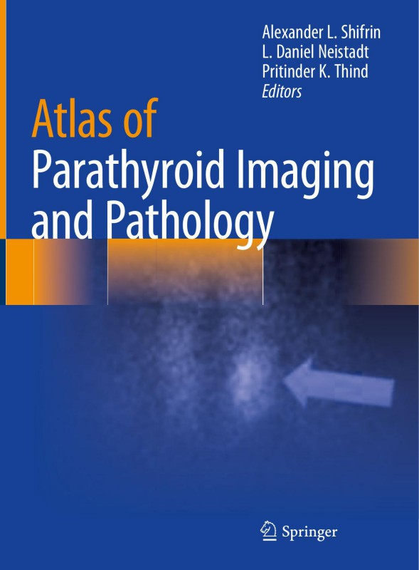
Atlas of Parathyroid Imaging and Pathology 1st edition by Alexander Shifrin, Daniel Neistadt, Pritinder Thind ISBN 3030409619 978-3030409616
$50.00 Original price was: $50.00.$25.00Current price is: $25.00.
Authors:Alexander L. Shifrin, L. Daniel Neistadt, Pritinder K. Thind , Series:Pathology [105] , Author sort:Alexander L. Shifrin, L. Daniel Neistadt, Pritinder K. Thind , Languages:Languages:eng , Published:Published:Sep 2020 , Publisher:Springer
Atlas of Parathyroid Imaging and Pathology 1st edition by Alexander Shifrin, Daniel Neistadt, Pritinder Thind – Ebook PDF Instant Download/Delivery. 3030409619 978-3030409616
Full download Atlas of Parathyroid Imaging and Pathology 1st edition after payment
Product details:
ISBN 10: 3030409619
ISBN 13: 978-3030409616
Author: Alexander Shifrin, Daniel Neistadt, Pritinder Thind
This book provides a visual demonstration of normal and ectopic locations of parathyroid adenomas using different modalities in patients with PHPT and to describe parathyroid gland–related pathology. It includes several modern imaging modalities for localization of parathyroid glands and parathyroid adenomas, such as Sestamibi scan, SPECT/CT Sestamibi scan, neck ultrasound, MRI, thin-cut CT, and 4D CT scans. Written by experts in the field, chapters include pathology images corresponding to radiology imaging for some presented cases (gross and high-power view). Authors have also collected radiological images of difficult-to-localize parathyroid adenomas in ectopic (abnormal) locations. The atlas is organized by location of the adenomas in upper and lower eutopic locations followed by ectopic locations. Each case demonstrates dual or triple modalities such as US, Sestamibi scan, or SPECT/CT Sestamibi scan, thin-cut CT scan, or 4D CT performed on the same patient. A chapter on parathyroid pathology is also included to help the reader understand challenges in pathological interpretation.
Atlas of Parathyroid Imaging and Pathology serves as a valuable reference for radiologists, endocrine surgeons, head and neck surgeons, ENT surgeons, surgical oncologists, endocrinologists, pathologists, nephrologists, students, and all physicians and allayed health practitioners involved in the treatment of patients with primary, secondary, and tertiary hyperparathyroidism.
Atlas of Parathyroid Imaging and Pathology 1st Table of contents:
Section I: Introduction to Parathyroid Pathology
- Anatomy and Physiology of the Parathyroid Glands
- Development and Location of the Parathyroid Glands
- Parathyroid Hormone (PTH) and Its Role in Calcium Homeostasis
- Parathyroid Disease and Its Clinical Manifestations
- Diagnostic Approaches in Parathyroid Pathology
- Imaging Techniques: Ultrasound, CT, MRI, and Scintigraphy
- Biopsy Techniques: Fine Needle Aspiration (FNA) and Core Biopsy
- Histopathology and Immunohistochemistry in Parathyroid Disorders
Section II: Parathyroid Imaging
- Ultrasound Imaging of the Parathyroid Glands
- Technique and Interpretation
- Role of Ultrasound in Primary Hyperparathyroidism
- Identifying Parathyroid Adenomas, Hyperplasia, and Malignancy
- Technetium-99m Sestamibi Scintigraphy
- Principles of Sestamibi Scintigraphy
- Interpretation of Scintigraphy Results in Parathyroid Disease
- Surgical Planning and Use of Sestamibi Imaging
- CT and MRI Imaging in Parathyroid Pathology
- Advantages and Limitations of CT and MRI
- Identifying Parathyroid Adenomas and Ectopic Parathyroid Tissue
- Role in Preoperative Localization
- Role of 4D-CT and PET Imaging
- Advances in Imaging Technology
- 4D-CT in Parathyroid Adenoma Localization
- PET Imaging for Recurrent Hyperparathyroidism
Section III: Parathyroid Pathology
- Parathyroid Adenomas
- Pathologic Features of Parathyroid Adenomas
- Histopathological Diagnosis and Immunohistochemical Markers
- Clinical Presentation and Management
- Primary Hyperparathyroidism
- Etiology and Pathophysiology
- Histopathology of Parathyroid Hyperplasia and Adenomas
- Role of Surgery in Treatment and Outcome
- Parathyroid Carcinoma
- Rare Malignant Parathyroid Tumors
- Diagnostic Challenges and Histopathologic Features
- Prognosis and Treatment
- Parathyroid Hyperplasia
- Primary vs Secondary Hyperplasia
- Histopathologic Criteria for Diagnosis
- Differentiating Hyperplasia from Adenoma
- Ectopic Parathyroid Glands
- Anatomy and Localization of Ectopic Parathyroid Tissue
- Imaging Challenges in Ectopic Parathyroid Disease
- Surgical Management of Ectopic Parathyroid Glands
- Secondary and Tertiary Hyperparathyroidism
- Pathophysiology and Etiology of Secondary Hyperparathyroidism
- Histopathological Changes in Secondary and Tertiary Hyperparathyroidism
- Treatment and Surgical Approaches
- Parathyroid Infections and Inflammatory Lesions
- Parathyroid Infections: Bacterial and Fungal Causes
- Granulomatous Diseases and Their Impact on Parathyroid Function
- Pathologic Features and Clinical Management
Section IV: Clinical Management
- Surgical Treatment of Parathyroid Disorders
- Indications for Parathyroidectomy
- Surgical Techniques and Approaches
- Minimally Invasive Parathyroid Surgery
- Preoperative Imaging and Localization
- Importance of Accurate Preoperative Localization
- Imaging Guidelines for Surgical Planning
- Case Studies and Examples of Imaging Techniques
- Management of Recurrent Hyperparathyroidism
- Causes and Diagnosis of Recurrent Hyperparathyroidism
- Role of Imaging in Recurrent Disease
- Surgical Strategies for Recurrent Parathyroid Disorders
Section V: Advanced Topics
- Molecular Pathology in Parathyroid Disease
- Genetic Mutations and Molecular Markers in Parathyroid Adenomas and Carcinomas
- Advances in Molecular Testing for Parathyroid Disorders
- Implications for Personalized Treatment
- Immunohistochemistry in Parathyroid Pathology
- Use of Immunohistochemical Markers in Diagnosis
- Markers for Parathyroid Adenomas, Hyperplasia, and Carcinoma
- Case Examples of Immunohistochemical Applications
- Endocrine and Parathyroid Disorders in the Pediatric Population
- Pediatric Hyperparathyroidism and Its Etiology
- Imaging and Pathologic Features in Children
- Management Approaches for Pediatric Parathyroid Disease
Appendices
- Glossary of Terms
- Index
People also search for Atlas of Parathyroid Imaging and Pathology 1st:
imaging of parathyroid
atlas of parathyroid surgery
images of thyroid and parathyroid glands
parathyroid images on ultrasound
ct of parathyroid adenoma
You may also like…
eBook PDF
Atlas of Infectious Disease Pathology 1st edition by Bryan Schmitt ISBN B071CKD5KM 978-3319547015
eBook PDF
Atlas of Salivary Gland Pathology 1st edition by JoaquÃn GarcÃa ISBN 3319090208 978-3319090207
eBook PDF
Atlas of Lung Pathology 1st edition by Chen Zhang, Jeffrey Myers ISBN 1493986872 978-1493986873

