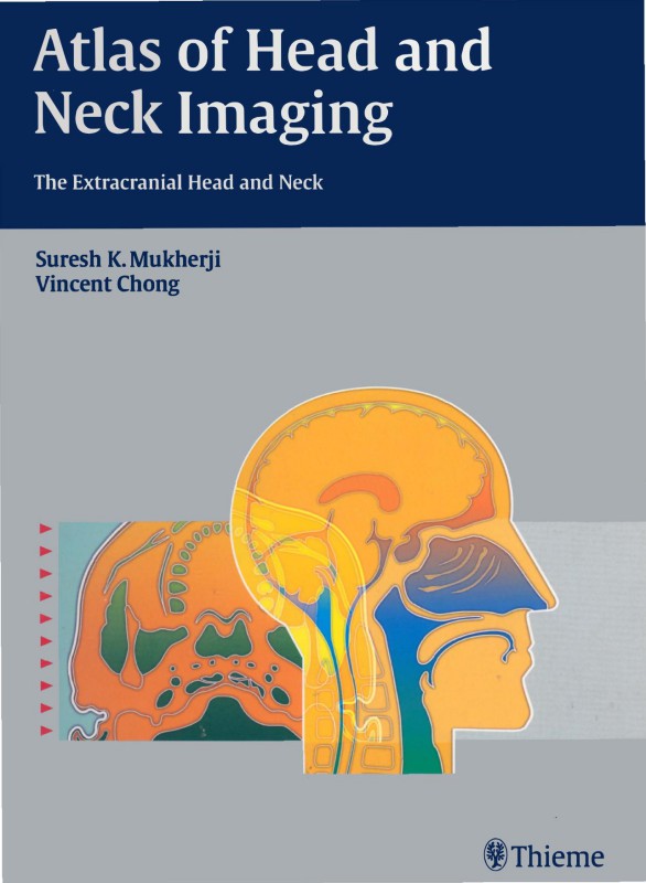
Atlas of Head and Neck Imaging The Extracranial Head and Neck 1st Edition by Suresh Kumar Mukherji, Vincent Chong ISBN B0CDQNPJH9
$50.00 Original price was: $50.00.$25.00Current price is: $25.00.
Authors:Thieme; 1 edition (January 22, 2004) , Author sort:edition, Thieme; 1 , Published:Published:Oct 2011
Atlas of Head and Neck Imaging The Extracranial Head and Neck 1st Edition by Suresh Kumar Mukherji, Vincent Chong – Ebook PDF Instant Download/Delivery. B0CDQNPJH9
Full download Atlas of Head and Neck Imaging The Extracranial Head and Neck 1st Edition after payment
Product details:
ISBN 10: B0CDQNPJH9
ISBN 13:
Author: Suresh Kumar Mukherji, Vincent Chong
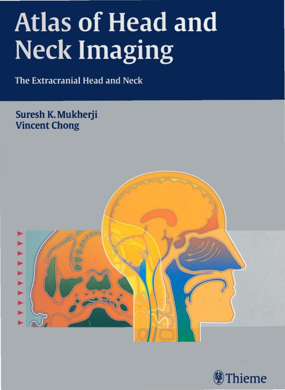
Designed for easy use at the PACS station of viewbox, here is your right-hand tool and pictorial guide for locating, identifying, and accurately diagnosing lesions of the extracranial head and neck. This beautifully produced atlas employs the spaces concept of analysis, which helps radiologists directly visualize complex head and neck anatomy and pathology.
With hundreds of high quality illustrations, this book makes the identification and localization of complex neck masses relatively simple. This book provides CT and MR examples for more than 200 different diseases of the suprahyoid and infrahyoid neck, as well as clear and concise information on the epidemiology, clinical findings, pathology, and treatment guidelines for each disease.
Each space within the head and neck has its own separate section, with examples of the common pathology that arises in this area. A standard format consisting of “Epidemiology, Clinical Presentation, Pathology, Treatment, and Imaging Findings,” allows quick and efficient access to well-structured subjects. This uniform organization streamlines research for radiologists at any level of training.
Although well over 200 pathologies are included within this remarkable text, Atlas of Head and Neck Imaging focuses primarily on the suprahyoid and infrahyoid neck, providing exceptionally detailed information on the most challenging aspects of this field.
Radiologists and radiation oncologists will find this visual text ideal as a quick anatomic reference and diagnostic tool. Radiology residents preparing for board exams and neuroradiology fellows and staff studying for the CAQ exam will also benefit from the wealth of information.
Atlas of Head and Neck Imaging The Extracranial Head and Neck 1st Table of contents:
Chapter 1: Introduction to Head and Neck Imaging
- 1.1 Overview of Head and Neck Anatomy
- 1.2 Imaging Modalities in Head and Neck Imaging
- 1.2.1 Conventional Radiography
- 1.2.2 Computed Tomography (CT)
- 1.2.3 Magnetic Resonance Imaging (MRI)
- 1.2.4 Ultrasound
- 1.2.5 Positron Emission Tomography (PET)
- 1.3 Basic Principles of Imaging and Image Interpretation
- 1.4 Radiologic Anatomy of the Extracranial Head and Neck
Chapter 2: Imaging of the Oral Cavity
- 2.1 Radiographic Anatomy of the Oral Cavity
- 2.2 Common Pathologies of the Oral Cavity
- 2.2.1 Dental Caries
- 2.2.2 Periodontal Disease
- 2.2.3 Oral Tumors and Lesions
- 2.2.4 Cysts and Granulomas
- 2.3 Imaging Techniques for the Oral Cavity
- 2.3.1 Panoramic Radiographs
- 2.3.2 Intraoral Radiographs
- 2.3.3 Cone Beam CT (CBCT)
Chapter 3: Imaging of the Paranasal Sinuses
- 3.1 Normal Anatomy of the Paranasal Sinuses
- 3.2 Sinus Pathologies
- 3.2.1 Sinusitis
- 3.2.2 Tumors and Neoplastic Lesions
- 3.2.3 Trauma and Fractures
- 3.3 Imaging Techniques for the Paranasal Sinuses
- 3.3.1 Plain Film Radiographs
- 3.3.2 CT Imaging of the Sinuses
- 3.3.3 MRI in Sinus Imaging
Chapter 4: Imaging of the Pharynx and Larynx
- 4.1 Radiographic Anatomy of the Pharynx and Larynx
- 4.2 Pathological Conditions of the Pharynx and Larynx
- 4.2.1 Tumors and Malignancies
- 4.2.2 Infections and Inflammatory Conditions
- 4.2.3 Foreign Bodies
- 4.3 Imaging Techniques for the Pharynx and Larynx
- 4.3.1 Lateral Neck X-ray
- 4.3.2 CT and MRI of the Pharynx and Larynx
Chapter 5: Imaging of the Neck Soft Tissues
- 5.1 Radiographic Anatomy of the Neck
- 5.2 Common Neck Masses
- 5.2.1 Lymphadenopathy
- 5.2.2 Inflammatory Lesions
- 5.2.3 Neoplastic Lesions
- 5.2.4 Vascular Abnormalities
- 5.3 Imaging Techniques for Neck Masses
- 5.3.1 Ultrasound of the Neck
- 5.3.2 CT and MRI of the Neck
- 5.3.3 Fine Needle Aspiration (FNA) and Image-Guided Biopsy
Chapter 6: Imaging of the Thyroid Gland
- 6.1 Normal Anatomy of the Thyroid Gland
- 6.2 Pathological Conditions of the Thyroid
- 6.2.1 Benign Tumors
- 6.2.2 Malignant Tumors
- 6.2.3 Thyroiditis and Inflammation
- 6.2.4 Cystic Lesions
- 6.3 Imaging Techniques for the Thyroid
- 6.3.1 Ultrasound of the Thyroid
- 6.3.2 CT and MRI of the Thyroid
- 6.3.3 Nuclear Medicine Imaging of the Thyroid
Chapter 7: Imaging of the Salivary Glands
- 7.1 Normal Anatomy of the Salivary Glands
- 7.2 Salivary Gland Pathologies
- 7.2.1 Sialolithiasis
- 7.2.2 Tumors (Benign and Malignant)
- 7.2.3 Inflammatory Conditions
- 7.2.4 Cysts and Abscesses
- 7.3 Imaging Techniques for the Salivary Glands
- 7.3.1 Sialography
- 7.3.2 Ultrasound and MRI of the Salivary Glands
- 7.3.3 CT in Salivary Gland Imaging
Chapter 8: Imaging of the Temporomandibular Joint (TMJ)
- 8.1 Radiographic Anatomy of the TMJ
- 8.2 Common TMJ Disorders
- 8.2.1 Degenerative Diseases
- 8.2.2 Internal Derangement
- 8.2.3 Inflammatory Conditions
- 8.3 Imaging Techniques for TMJ Disorders
- 8.3.1 Plain Film Radiography
- 8.3.2 MRI and CT for TMJ Imaging
- 8.3.3 3D Imaging Techniques for the TMJ
Chapter 9: Imaging of the Head and Neck in Trauma
- 9.1 Overview of Trauma in the Head and Neck
- 9.2 Imaging Techniques for Trauma Diagnosis
- 9.2.1 CT Imaging for Trauma
- 9.2.2 MRI in Soft Tissue Trauma
- 9.2.3 Plain Film Radiographs
- 9.3 Management of Head and Neck Trauma with Imaging
- 9.4 Post-Traumatic Imaging and Complications
Chapter 10: Imaging of Head and Neck Cancer
- 10.1 Overview of Head and Neck Cancer
- 10.2 Radiographic Evaluation of Head and Neck Tumors
- 10.2.1 Squamous Cell Carcinoma
- 10.2.2 Lymphomas
- 10.2.3 Sarcomas
- 10.2.4 Metastatic Tumors
- 10.3 Imaging Techniques in Cancer Staging and Follow-up
- 10.3.1 CT and MRI for Tumor Staging
- 10.3.2 PET Imaging in Cancer Detection
- 10.3.3 Lymph Node Evaluation
Chapter 11: Advanced Imaging Techniques in Head and Neck Imaging
- 11.1 3D Imaging and Its Applications
- 11.2 Role of Cone Beam CT in Head and Neck Imaging
- 11.3 Functional Imaging Techniques
- 11.3.1 Diffusion Tensor Imaging (DTI)
- 11.3.2 PET and PET/CT Fusion Imaging
- 11.4 Future Directions in Head and Neck Imaging
People also search for Atlas of Head and Neck Imaging The Extracranial Head and Neck 1st:
head neck imaging extracranial head nck
head neck radiology
imaging of head
head neck imaging
head and neck imaging anatomy

