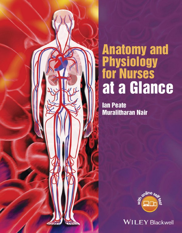
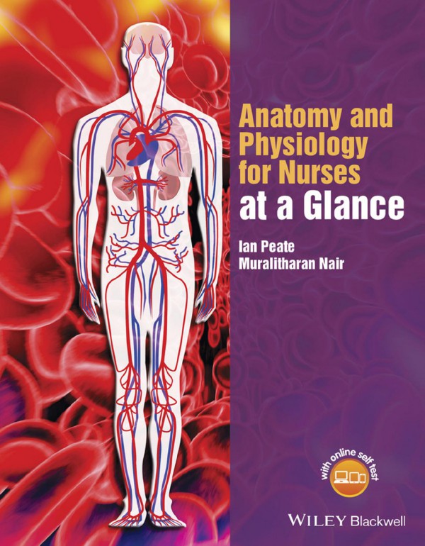
Anatomy and Physiology for Nurses at a Glance 1st Edition by Ian Peate, Muralitharan Nair 1118746317 9781118746318
$50.00 Original price was: $50.00.$25.00Current price is: $25.00.
Authors:Ian Peate; Muralitharan Nair , Series:Anatomy [227] , Tags:Medical; Nursing; General; Critical Care , Author sort:Peate, Ian & Nair, Muralitharan , Ids:9781118746318 , Languages:Languages:eng , Published:Published:Apr 2015 , Publisher:John Wiley & Sons , Comments:Comments:Anatomy and Physiology for Nurses at a Glance is the perfect companion for study and revision for pre-registration nursing and healthcare students, from the publishers of the market-leading at a Glance series. Combining superb illustrations with accessible and informative text, this book covers all the body systems and key concepts encountered from the start of the pre-registration nursing or healthcare programme, and is ideal for anyone looking for an overview of the human body. Providing a concise, visual overview of anatomy and physiology and the related biological sciences, this book will help students develop practical skills, enabling them to become caring, kind and compassionate nurses. Superbly illustrated, with full colour illustrations throughout Breaks down complex concepts in an accessible way Written specifically for nursing and healthcare students with all the information they need Includes access to a companion website with self-assessment questions for each chapter Available in a range of digital formats- perfect for ‘on the go’ study and revision
Anatomy and Physiology for Nurses at a Glance 1st Edition by Ian Peate, Muralitharan Nair – Ebook PDF Instant Download/Delivery. 1118746317, 9781118746318
Full download Anatomy and Physiology for Nurses at a Glance 1st Edition after payment
Product details:
ISBN 10: 1118746317
ISBN 13: 9781118746318
Author: Ian Peate, Muralitharan Nair
From the authors of the successful and acclaimed ‘Fundamentals of Anatomy and Physiology for Student Nurses’, comes this easy-to-read, comprehensive and highly visual overview of all the key anatomy and physiology a healthcare student needs to know.
Anatomy and Physiology for Nurses at a Glance 1st Table of contents:
Part 1 Foundations
1 The genome
Figure 1.1 DNA double helix with nucleotides in situ
Figure 1.2 RNA molecule
Figure 1.3 A nucleotide with its three parts
Figure 1.4 A chromosome
Figure 1.5 Splitting of DNA and transcription by RNA
Genetics
The double helix of DNA
RNA
Nucleotides
Bases
Chromosomes
Protein synthesis
Transcription
Translation
Gene transference
Mitosis
Meiosis
2 Homeostatic mechanisms
Figure 2.1 Components of a negative feedback system
Figure 2.2 Negative feedback of raised blood pressure
Figure 2.3 Negative feedback of raised temperature
Figure 2.4 Positive feedback of childbirth
Homeostasis
Feedback mechanisms
Receptor
Control centre
Effector
Negative feedback
What happens when the body is too hot?
What happens when the body is too cold?
Positive feedback
3 Fluid compartments
Figure 3.1 Distribution of body water
Table 3.1 Fluid intake and losses
Total body water
Intracellular fluid
Extracellular fluid
The plasma membrane
Fluid regulation
4 Cells and organelles
Figure 4.1 A typical cell
Figure 4.2 Mitochondrion
Figure 4.3 Cell membrane
Cells
Cell membrane
Mitochondria
Endoplasmic reticulum
Nucleus
Cytoplasm
Lipid bilayer
Membrane proteins
Functions of the plasma membrane
5 Transport systems
Figure 5.1 Osmosis
Figure 5.2 Simple diffusion
Figure 5.3 Active transport
Figure 5.4 Facilitated diffusion
Figure 5.5 Endocytosis
Figure 5.6 Exocytosis
Osmosis
Diffusion
Facilitated diffusion
Active transport
Secondary active transport
Endocytosis and exocytosis
Purpose of exocytosis
6 Blood
Figure 6.1 Red blood cell shape
Figure 6.2 Haemoglobin molecule
Figure 6.3 Recycling of red blood cells (RBC)
Figure 6.4 White blood cells
Figure 6.5 Platelets
Blood
Formation of blood cells
RBC
Haemoglobin
Recycling of RBC
WBC
Neutrophils
Eosinophils
Basophil
Monocytes
Lymphocytes
Platelets
Blood plasma
7 Inflammation and immunity
Figure 7.1 The cells of the immune system
Figure 7.2 Types of acquired immunity
Table 7.1 Types of antibodies
Immune system
Types of immunity
Innate immunity
Inflammation
Acquired immunity
Natural and artificial acquired immunity
8 Tissues
Figure 8.1 Levels of organisation
Figure 8.2 Types of cells
Figure 8.3 Human body tissues
Tissues
Epithelial tissue
Nervous tissue
Connective tissue
Muscle tissue
Part 2 The nervous system
9 The brain and nerves
Figure 9.1 The brain
Figure 9.2 A Neuron
Figure 9.3 Cranial nerves
Figure 9.4 The meninges
Brain
Meninges
Cerebrospinal fluid
Neuron
Axon
Dendrite
Cell body
Myelin sheath
Cranial nerves
10 Structures of the brain
Figure 10.1 The cerebrum
Figure 10.2 The cerebellum
Figure 10.3 Limbic system
Structures of the brain
Cerebrum
Diencephalon
Thalamus
Hypothalamus
Epithalamus
Brain stem
Midbrain
Pons
Medulla oblongata
Cerebellum
Limbic system
Ventricles of the brain
Cerebrospinal fluid
11 The spinal cord
Figure 11.1 The spinal cord and spinal nerves
Figure 11.2 Spinal cord layers
Spinal cord
Pia matter
Arachnoid matter
Dura matter
Spinal cord sections
Functions of the spinal cord
Reflex actions
Spinal nerves
12 The blood supply
Figure 12.1 Circle of Willis
Figure 12.2 Blood brain barrier
Circle of Willis
Function of the circle of Willis
Blood–brain barrier (BBB)
Function of BBB
13 The autonomic nervous system
Figure 13.1 The sympathetic and parasympathetic nervous system
ANS
Sympathetic nervous system
Functions
Parasympathetic nervous system
Functions
Autonomic control by the CNS
Autonomic reflexes
14 Peripheral nervous system
Figure 14.1 Somatic nervous system
Figure 14.2 Motor pathway
PNS
Peripheral nervous system connections
Sensory division
Somatic
Visceral
Motor division
Part 3 The heart and vascular system
15 The heart
Figure 15.1 Location of the heart
Figure 15.2 Walls of the heart
Figure 15.3 Myocardium cells
Figure 15.4 Chambers of the heart
Heart
Walls of the heart
Pericardium
Fibrous pericardium
Parietal pericardium
Myocardium
Endocardium
Chambers of the heart
Valves of the heart
Blood vessels of the heart
16 Blood flow through the heart
Figure 16.1 Blood flow through the heart
Figure 16.2 Blood vessels of the heart
Figure 16.3 Posterior view of the coronary veins
Blood flow
Pulmonary circulation
Systemic circulation
Coronary circulation
Coronary arteries
Coronary veins
17 The conducting system
Figure 17.1 Conducting system of the heart
Figure 17.2 Cardiac cycle
Cardiac conduction
SA node
AV node
Bundle of His
Left and right bundle branches
Purkinje fibres
Cardiac cycle
First diastole phase
First systole phase
Second diastole phase
Second systole phase
18 Nerve supply to the heart
Figure 18.1 Cardioregulatory centre
Autonomic nervous system
Chemical regulation of the heart
Hormones
Ions
Baroreceptors
Cardioinhibitory centre
Vasomotor centre
Other factors in heart regulation
Body temperature
Body fluid level
19 Structure of the blood vessels
Figure 19.1 Blood vessels
Figure 19.2 Layers of the blood vessel
Figure 19.3 Comparison of an artery, vein and capillary
Figure 19.4 Capillary network
Blood vessels
Structure
Arteries
Veins
Capillaries
20 Blood pressure
Figure 20.1 Baroreceptors
Figure 20.2 Blood pressure measurement
What is blood pressure?
Physiological factors
Control of blood pressure
Baroreceptors
Chemoreceptors
Circulating hormones
Renin-angiotensin system
Hypothalamus
Taking blood pressure
21 Lymphatic circulation
Figure 21.1 The lymphatic system
Figure 21.2 Lymphatic capillaries
Figure 21.3 Lymph node
Lymphatic system
What is lymph?
Lymph nodes
Lymphatic organs
Spleen
Thymus gland
Functions of the lymphatic system
Part 4 The respiratory system
22 The respiratory tract
Figure 22.1 Respiratory organs
Figure 22.2 Detail structures of the respiratory tract
Figure 22.3 Lower respiratory tract
Figure 22.4 Bronchial tree
Respiratory system
Upper respiratory tract
Lower respiratory tract
Larynx
Trachea
Bronchi and bronchioles
Lungs
Blood supply
23 Pulmonary ventilation
Figure 23.1 Boyle’s Law
Figure 23.2 Inspiration and expiration
Breathing
Inspiration
Exhalation
Factors affecting pulmonary ventilation
Surface tension of alveolar fluid
Airway resistance
Compliance of the lungs
Lung volumes
24 Control of breathing
Figure 24.1 Respiratory centre
Figure 24.2 Peripheral chemoreceptors
Pneumotaxic area
Apneustic area
Medullary rhythmicity area
Central chemoreceptors
Peripheral chemoreceptors
Inflation reflex
Other influences on respiration
Limbic system
Temperature
Pain
Irritation of airways
25 Gas exchange
Figure 25.1 External respiration
Figure 25.2 Gas exchange in the lungs
Figure 25.3 Internal respiration
External respiration
Exchange of gases in the lungs
Fick’s Law
Internal respiration
Factors affecting pulmonary and systemic gas exchange
Partial pressure difference of gases
Surface area available for gas exchange
Diffusion distance
Solubility of gases
Transport of gases
Part 5 The gastrointestinal tract
26 The upper gastrointestinal tract
Figure 26.1 Upper and lower GI tract
Figure 26.2 The tongue
Figure 26.3 Swallowing action
Figure 26.4 Stomach
The mouth (oral cavity)
Lips and cheeks
Tongue
Palate
Salivary glands
Oesophagus
Swallowing (deglutition)
Stomach
27 The lower gastrointestinal tract
Figure 27.1 Small intestine
Figure 27.2 Large intestine
Small intestine
Duodenum
Jejunum
Ileum
Large intestine (colon)
28 The liver, gallbladder and biliary tree
Figure 28.1 The liver, gallbladder and pancreas
Figure 28.2 Liver lobules
Figure 28.3 Bile production and secretion
Liver
Segments of the liver
Ligaments of the liver
Blood supply
Liver functions
Gallbladder
Function of bile
Biliary tree (tract)
29 Pancreas and spleen
Figure 29.1 The pancreas
Pancreas
Composition of pancreatic juice
Functions of pancreatic juice
Spleen
Function of the spleen
30 Digestion
Figure 30.1 Digestion and absorption
Mechanical
Mastication
Peristalsis
Chemical
Digestion and absorption
Part 6 The urinary system
31 The kidney: microscopic
Figure 31.1 A nephron
Figure 31.2 Bowman’s capsule
Nephrons
Glomerular capsule
Glomerulus
Proximal convoluted tubule
Loop of Henle
Distal convoluted tubule
Collecting ducts
32 The kidney: macroscopic
Figure 32.1 External layers of the kidney
Figure 32.2 Blood flow through the kidney
Kidney
Renal cortex
Renal medulla
Renal pelvis
Blood supply
Nerve supply
33 The ureter, bladder and urethra
Figure 33.1 Blood supply of the ureter
Figure 33.2 Bladder
Figure 33.3 Male urethra
Figure 33.4 Female urethra
Ureters
Abdominal ureter
Pelvic ureter
Blood supply
Urinary bladder
Layers of the bladder
Vessels and nerves
Urethra
Male urethra
Female urethra
34 Formation of urine
Figure 34.1 Renal filtration
Figure 34.2 Renin-angiotension pathway
Filtration
Selective reabsorption
Secretion
Hormonal control
ADH
Angiotension
Aldosterone
Atrial natriuretic peptide (ANP)
Part 7 The male reproductive system
35 External male genitalia
Figure 35.1 Left testis and epididymis
Figure 35.2 Sagittal view of penis and male pelvis
Figure 35.3 The anatomy of the penis
The testes
Spermatogenesis
The penis
The epididymis
The vas deferens, ejaculatory ducts and spermatic cord
36 The prostate gland
Figure 36.1 The zones of the prostate gland
The gland
The zones of the prostate gland
The peripheral zone
The transition zone
Central zone
The surfaces of the prostate gland
The base
The apex
The anterior, inferolateral and posterior surfaces
The function of the prostate gland
Prostatic fluid
Some structures around the prostate
Prostate-specific antigen (PSA)
37 Spermatogenesis
Figure 37.1 Spermatogenesis
Figure 37.2 Primary and secondary sex characteristics
Spermatogenesis
Male sex hormones
Part 8 The female reproductive system
38 Female internal reproductive organs
Figure 38.1 The female internal reproductive organs
Table 38.1 The layers of the uterus
The internal female reproductive organs
The ovaries
The ovarian cortex
Graafian follicles
Corpus luteum
The ovarian medulla
Oogenesis
Female sex hormones
The uterus
The fallopian tubes
The vagina
The cervix
39 External female genitalia
Figure 39.1 The female external genitalia
The mons veneris
Labia majora
Labia minora
Clitoris
The urethra
Hymen
Blood supply
Arterial supply of the female external genitalia
Venous drainage of the female external genitalia
Lymph drainage
Nerve supply
40 The breast
Figure 40.1 The breast
Figure 40.2 The breast and surrounding structures
Figure 40.3 Lobules and ducts
Figure 40.4 Breast lymph
Figure 40.5 Axillary lymph nodes
The breast
Function
Structure
Lobules and ducts
Fat, ligaments and connective tissue
Nerve supply
Arteries and capillaries
Lymph nodes and lymph ducts
Breast development
Hormones and the breast
41 The menstrual cycle
Figure 41.1 Hormonal regulation of the changes in the ovary and uterus
Figure 41.2 Changes in the concentration of anterior pituitary and ovarian hormones
The reproductive cycle
The pituitary gland
The follicular phase
The ovulatory phase
The luteal phase
The menstrual cycle
Part 9 The endocrine system
42 The endocrine system
Figure 42.1 The major endocrine glands
Figure 42.2 The pituitary gland and surrounding structures
Table 42.1 Hormones released by the hypothalamus and anterior pituitary gland
The endocrine glands
The pituitary gland and the hypothalamus
The pineal gland
The anterior pituitary lobe
The posterior pituitary lobe
43 The thyroid and adrenal glands
Figure 43.1 The thyroid and parathyroid glands
Table 43.1 Some effects associated with an abnormal secretion of thyroid hormones
Figure 43.2 Control of thyroid hormone production — negative feedback
The thyroid gland
Parathyroid glands
The adrenal glands
Hormones of the adrenal cortex
Hormones of the adrenal medulla
44 The pancreas and gonads
Figure 44.1 The pancreas
Figure 44.2 Insulin and glucagon effects on blood glucose
Table 44.1 Other endocrine glands
The pancreas
The exocrine pancreas
The endocrine pancreas
Insulin
Glucagon
Somatostatin
The gonads
The ovaries
The testes
Other endocrine glands
Part 10 The musculoskeletal system
45 Bone structure
Figure 45.1 Bone remodelling
Figure 45.2 Bone growth
Figure 45.3 Osteon, haversian canal
Bone remodelling and modelling
Ossification
Osteoclasts
Osteoblasts
The skeleton
46 Bone types
Figure 46.1 The skeleton: axial and appendicular
Figure 46.2 Compact bone (long bone): found in the toes, legs, fingers and arms
Figure 46.3 Short bone
Figure 46.4 Flat bone
Figure 46.5 Irregular bone
Figure 46.6 Sesamoid bone
Skeleton divisions
The axial skeleton
The appendicular skeleton
Bone types
Long bones
Short bones
Flat bones
Irregular bones
Sesamoid bones
47 Joints
Figure 47.1 Joint types
Movements
Fibrous joints
Cartilaginous joints
Synovial joints
Freely movable (synovial) joints
Hinge
Pivot
Ball and socket
Saddle
Condyloid
Gliding
48 Muscles
Figure 48.1 Muscle tissues
Muscles
Muscle tissue
Skeletal muscle tissue
Cardiac muscle tissue
Smooth muscle tissue
Properties of muscle tissue
Part 11 The skin
49 The skin layers
Figure 49.1 The skin and associated structures
Figure 49.2 Skin types
Figure 49.3 The layers of the epidermis
The skin
The layers of the skin
The epidermis
The dermis
The subcutaneous tissues
50 The skin appendages
Figure 50.1 The hair and associated glands
Figure 50.2 The nails
The dermal appendages
The hair
The nail
The glands
The sebaceous glands
The sudoriferous glands
The ceruminous glands
51 Epithelialisation
Figure 51.1 Epidermal and deep wound healing
Epithelialisation
Wounds
How wounds heal
Types of wound healing
Primary closure
Secondary closure
Delayed primary closure
Wound closure
52 Granulation
Figure 52.1 Summary of wound healing events
Granulation tissue
Tissue repair
Over-granulation
Part 12 The senses
53 Sight
Figure 53.1 The eye
Table 53.1 The eye
Figure 53.2 The lacrimal apparatus
Sight
The orbit
The eye lids, eye lashes and eye brows
The lacrimal apparatus
The sclera
The cornea
The aqueous humour
The iris
The lens and ciliary muscle
The retina
The visual system pathways to the brain
The visual cortex
54 Hearing
Figure 54.1 The ear
Figure 54.2 The outer ear
Figure 54.3 The middle ear
Figure 54.4 The inner ear
Hearing
The ear
The outer (external ear)
The middle ear
Tympanic membrane
Ossicles
Auditory tube
The inner ear
The vestibule
The semicircular canals
The cochlea
Blood supply to the inner ear
The organ of Corti
55 Olfaction
Figure 55.1 Pathway of smell
Figure 55.2 Olfactory epithelium and olfactory receptor cells
Figure 55.3 The olfactory bulb (nerve)
Physiology of olfaction
Olfactory nerve and the cribriform plate
Olfactory bulb
Olfactory tract
56 Gustation
Figure 56.1 The tongue
Figure 56.2 Taste
Gustation
The tongue
Anatomy and physiology
Taste bud anatomy
Back Matter
Appendices
Appendix 1: Cross-references to chapters in Pathophysiology for Nurses at a Glance
Appendix 2: Normal physiological values
Appendix 3: Prefixes and suffixes
Appendix 4: Glossary
Further reading
Index
WILEY END USER LICENSE AGREEMENT
People also search for Anatomy and Physiology for Nurses at a Glance 1st:
physiology for nurses
anatomy and physiology for nursing students chapter 3
pns 112 galen
quizlet nursing anatomy and physiology
You may also like…
eBook PDF
MENS HEALTH The Practice Nurses Handbook 1st Edition by Ian Peate 0470035552 9780470035559
eBook PDF
Physiology at a Glance 4th Edition by Jeremy Ward, Roger Linden ISBN 1119247314 9781119247319
eBook PDF
Caring for Children and Families 1st Edition by Ian Peate, Lisa Whiting 0470019700 9780470019702

