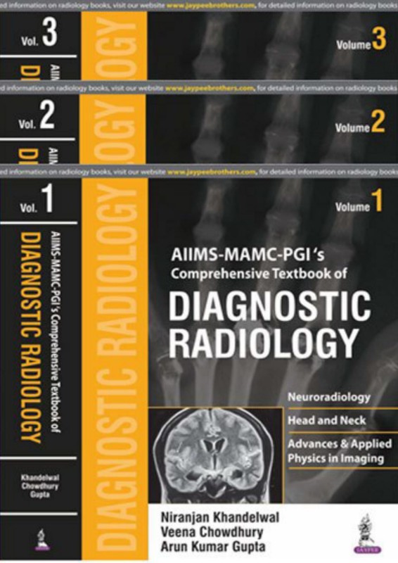
AIIMS MAMC PGI’s Comprehensive Textbook of Diagnostic Radiology 1st Edition by Niranjan Khandelwal, Arun Kumar Gupta, Veena Chowdhury ISBN 935250187X 978-9352501878


Original price was: $50.00.$25.00Current price is: $25.00.
Authors:Niranjan Khandelwal, Arun Kumar Gupta, Veena Chowdhury , Tags:Vol 1; Vol 2; Vol 3 , Author sort:Niranjan Khandelwal, Arun Kumar Gupta, Veena Chowdhury , Ids:9789352501878 , Published:Published:Apr 2017 , Comments:Comments:AIIMS MAMC – PGI’s Comprehensive Textbook of Diagnostic Radiology is an extensive three volume review of conventional and novel imaging techniques for the diagnosis of a wide range of diseases.The first volume of this book covers neuroradiology, including head and neck, recent advances and applied physics in imaging. The second volume covers chest and cardiovascular, gastrointestinal and hepatobiliary, and genitourinary imaging. The final volume covers paediatric imaging, musculoskeletal imaging, including information on imaging for bone, soft tissue and joint diseases, and breast imaging including the role of mammography.AIIMS MAMC – PGI’s Comprehensive Textbook of Diagnostic Radiology includes discussion on various imaging modalities and the potential for their future use. The book is enhanced by over 6250 images and illustrations, making this an ideal visual resource for radiology residents and practising radiologists.Key PointsThree volume review of diagnostic imaging techniquesVolumes cover neuroradiology, chest, cardiovascular, gastrointestinal, hepatobiliary, genitourinary, paediatric musculoskeletal, and breast imaging6254 images and illustrations
AIIMS-MAMC PGI’s Comprehensive Textbook of Diagnostic Radiology 1st Edition by Niranjan Khandelwal, Arun Kumar Gupta, Veena Chowdhury – Ebook PDF Instant Download/Delivery. 935250187X 978-9352501878
Full download AIIMS-MAMC PGI’s Comprehensive Textbook of Diagnostic Radiology 1st Edition after payment
Product details:
ISBN 10: 935250187X
ISBN 13: 978-9352501878
Author: Niranjan Khandelwal, Arun Kumar Gupta, Veena Chowdhury
AIIMS MAMC – PGI’s Comprehensive Textbook of Diagnostic Radiology is an extensive three volume review of conventional and novel imaging techniques for the diagnosis of a wide range of diseases. The first volume of this book covers neuroradiology, including head and neck, recent advances and applied physics in imaging. The second volume covers chest and cardiovascular, gastrointestinal and hepatobiliary, and genitourinary imaging. The final volume covers paediatric imaging, musculoskeletal imaging, including information on imaging for bone, soft tissue and joint diseases, and breast imaging including the role of mammography. AIIMS MAMC – PGI’s Comprehensive Textbook of Diagnostic Radiology includes discussion on various imaging modalities and the potential for their future use. The book is enhanced by over 6250 images and illustrations, making this an ideal visual resource for radiology residents and practising radiologists. Key Points: Three volume review of diagnostic imaging techniques: Volumes cover neuroradiology, chest, cardiovascular, gastrointestinal, hepatobiliary, genitourinary, paediatric musculoskeletal, and breast imaging. 6254 images and illustrations.
AIIMS-MAMC PGI’s Comprehensive Textbook of Diagnostic Radiology 1st Table of contents:
1. Introduction to Chest X-Ray Interpretation
-
Overview of Chest X-ray: A brief introduction to the chest X-ray as a diagnostic tool, explaining the importance of chest radiography in clinical practice, its uses, and limitations.
-
Basic Principles of X-ray Imaging: Describes the fundamentals of X-ray imaging, including how X-rays are produced, how images are formed, and the basic interpretation techniques for beginners.
-
Understanding X-ray Anatomy: A guide to the normal anatomical structures visible on a chest X-ray, including the lungs, heart, diaphragm, ribs, and mediastinum, to establish a baseline for identifying abnormal findings.
2. Normal Chest X-Ray
-
Interpretation of a Normal Chest X-ray: This section likely focuses on understanding the normal appearances in a chest X-ray. It would include typical findings in healthy individuals and the key things to look for when reviewing normal radiographs.
3. Common Chest Pathologies: 100 Practice Cases
-
Case 1-10: Pulmonary Infections: These cases would feature conditions such as pneumonia, tuberculosis, and other respiratory infections, focusing on identifying characteristic signs such as consolidation, cavitation, or infiltrates.
-
Case 11-20: Cardiovascular Conditions: Chest X-rays in conditions like heart failure, cardiomegaly, pulmonary edema, and pericardial effusion. It might focus on recognizing enlarged cardiac silhouette or signs of fluid overload in the lungs.
-
Case 21-30: Pulmonary Embolism and Vascular Abnormalities: Interpretation of chest X-rays in patients with pulmonary embolism, deep vein thrombosis, and vascular abnormalities such as aortic dissection or aneurysms.
-
Case 31-40: Trauma and Fractures: Radiographs showing injuries like rib fractures, pneumothorax, hemothorax, and other chest trauma, with emphasis on how to differentiate between fractures, soft tissue injuries, and other causes of trauma-related radiographic changes.
-
Case 41-50: Neoplastic Conditions: These cases would feature tumors such as lung cancer, metastasis, and benign lung lesions, with a focus on identifying masses, nodules, and structural changes related to malignancy.
-
Case 51-60: Chronic Obstructive Pulmonary Disease (COPD) and Emphysema: Radiographs in COPD, emphysema, and other chronic lung diseases, helping learners identify hyperinflation, flattened diaphragms, and changes in lung tissue.
-
Case 61-70: Interstitial Lung Diseases: This section would cover conditions like pulmonary fibrosis, sarcoidosis, and interstitial lung diseases, emphasizing patterns such as reticular changes, honeycombing, and ground-glass opacities.
-
Case 71-80: Pleural Effusion and Pneumothorax: Radiographs showing pleural effusion (fluid accumulation between the pleura), pneumothorax (air between the pleura), and other pleural conditions, focusing on the characteristic signs of each.
-
Case 81-90: Post-surgical and Post-procedural Chest X-rays: Postoperative and post-procedural radiographs showing changes after surgery, including complications such as infection, atelectasis, or improper placement of medical devices (e.g., pacemakers, central lines).
-
Case 91-100: Pediatric Chest X-rays: Cases involving pediatric patients, with a focus on congenital anomalies, foreign body aspiration, and other conditions unique to children.
4. How to Read Chest X-rays: Step-by-Step Approach
-
Systematic Approach to Chest X-ray Interpretation: A guide to the structured method of reading chest X-rays, breaking down the process into steps such as assessing the quality of the image, analyzing the anatomy, and evaluating for abnormalities.
-
Red Flags to Look For: Common signs of severe or urgent conditions, such as large masses, signs of lung collapse, or pneumothorax, that require immediate clinical attention.
-
Common Pitfalls and Mistakes: This section likely discusses common errors in interpreting chest X-rays, such as missing subtle signs of pathology or mistaking normal variants for abnormalities.
5. Full X-Ray Reports and Annotations
-
Case Report 1-100: For each of the 100 cases, the book provides full X-ray reports, including a detailed interpretation of the image. These reports would likely include a step-by-step description of findings, diagnosis, differential diagnosis, and clinical correlation.
-
Clinical Context and Recommendations: Each case would also include clinical context and recommendations for further diagnostic work-up or management, providing practical insight into how chest X-rays influence clinical decision-making.
6. Further Reading and Resources
-
Recommended Textbooks and Journals: A list of further resources for in-depth study of radiology and chest imaging.
-
Radiology Societies and Educational Websites: A guide to professional societies, online courses, and other platforms for ongoing learning in radiology and chest X-ray interpretation.
7. Conclusion and Final Thoughts
-
Mastering Chest X-ray Interpretation: A summary of key takeaways, reinforcing the importance of systematic reading and the value of practice in mastering chest X-ray interpretation.
People also search for AIIMS-MAMC PGI’s Comprehensive Textbook of Diagnostic Radiology 1st:
aiims mamc pgi’s comprehensive textbook of diagnostic radiology
is mamc better than aiims
what is the fees of aiims medical college
which is better aiims delhi or mamc
largest aiims campus
You may also like…

eBook PDF
Manufacturing Processes 2nd edition by Gupta, Gupta, Arun Mittal ISBN 8122427359 97881224273596