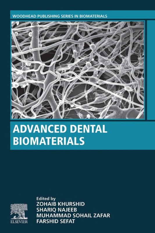
Advanced Dental Biomaterials 1st Edition by Zohaib Khurshid, Shariq Najeeb ISBN 0081024770 9780081024775
$50.00 Original price was: $50.00.$25.00Current price is: $25.00.
Authors:Zohaib Khurshid; Shariq Najeeb , Series:Dentistry [130] , Author sort:Khurshid, Zohaib & Najeeb, Shariq , Ids:Goodreads , Languages:Languages:eng , Published:Published:May 2019 , Publisher:Woodhead Publishing , Comments:Comments:Advanced Dental Biomaterialsis an invaluable reference for researchers and clinicians within the biomedical industry and academia. The book can be used by both an experienced researcher/clinician learning about other biomaterials or applications that may be applicable to their current research or as a guide for a new entrant into the field who needs to gain an understanding of the primary challenges, opportunities, most relevant biomaterials, and key applications in dentistry.
Advanced Dental Biomaterials 1st Edition by Zohaib Khurshid, Shariq Najeeb – Ebook PDF Instant Download/Delivery. 0081024770, 9780081024775
Full download Advanced Dental Biomaterials 1st Edition after payment
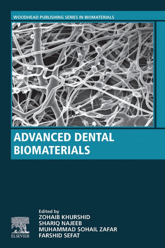
Product details:
ISBN 10: 0081024770
ISBN 13: 9780081024775
Author: Zohaib Khurshid, Shariq Najeeb
Advanced Dental Biomaterials is an invaluable reference for researchers and clinicians within the biomedical industry and academia. The book can be used by both an experienced researcher/clinician learning about other biomaterials or applications that may be applicable to their current research or as a guide for a new entrant into the field who needs to gain an understanding of the primary challenges, opportunities, most relevant biomaterials, and key applications in dentistry.
- Provides a comprehensive review of the materials science, engineering principles and recent advances in dental biomaterials
- Reviews the fundamentals of dental biomaterials and examines advanced materials’ applications for tissues regeneration and clinical dentistry
- Written by an international collaborative team of materials scientists, biomedical engineers, oral biologists and dental clinicians in order to provide a balanced perspective on the field
Advanced Dental Biomaterials 1st Table of contents:
1 Introduction to dental biomaterials and their advances
References
Further reading
2 Properties of dental biomaterials
2.1 Introduction
2.2 Optical properties (color)
2.3 Thermal properties
2.3.1 Temperature
2.3.2 Transition temperatures
2.3.3 Heat of fusion (L)
2.3.4 Thermal conductivity (K)
2.3.5 Specific heat (Cp)
2.3.6 Thermal diffusivity (Δ)
2.3.7 Coefficient of thermal expansion (α)
2.4 Viscosity
2.5 Electrical conductivity and resistivity
2.6 Mechanical properties and characterization methods
2.7 Limitation of mechanical testing methods
2.8 Biological properties
2.8.1 Biocompatibility
2.8.2 In vitro testing
2.8.3 In vivo testing
2.8.4 Usage tests
2.9 Toxicity and cytotoxicity
2.10 Cytotoxicity tests
2.11 Fluoride and caries
2.11.1 Fluoride toxicity
2.12 Carcinogenicity
2.13 Biodegradation
2.14 Bioactivity
2.15 Osseointegration
2.16 Osteoinduction
2.17 Foreign body reaction
2.18 Conclusive remarks
References
3 Dental gypsum and investments
3.1 Introduction
3.2 Desirable properties of gypsum products
3.3 Production of calcium sulfate hemihydrate
3.4 Types of gypsum products
3.5 The setting and manipulation characteristics of gypsum products
3.5.1 Mixing technique
3.5.1.1 Measuring the water
3.5.1.2 Measuring the powder
3.5.1.3 Adding powder and water
3.5.2 Pouring the impression
3.5.3 The setting processes
3.5.3.1 Stages of setting
3.5.3.2 The rate of setting reaction
3.5.3.3 Water/powder ratio
3.5.3.4 Spatulation
3.5.3.5 Temperature
3.5.3.6 Modifying agents
3.5.3.7 Fineness
3.5.3.8 Effect of pH
3.5.4 Setting time
3.5.4.1 Initial setting time
3.5.4.2 Final setting time
3.5.4.3 Gilmore test for final setting time
3.6 Setting expansion–hygroscopic setting expansion
3.6.1 Reproduction of detail
3.6.2 Compressive strength
3.6.3 Tensile strength
3.6.4 Surface hardness and abrasion resistance
3.6.5 Dimensional stability
3.7 Dies and models produced from digital data
3.8 Conclusion
References
4 Ceramic materials in dentistry
4.1 Introduction
4.1.1 Glass ceramics
4.1.1.1 Feldspathic porcelain
4.1.1.2 Leucite-reinforced porcelain
4.1.1.3 Fluorine-containing glass ceramics
4.1.1.4 Lithium silicate
4.1.2 Oxide ceramics
4.1.2.1 Glass-infiltrated aluminium-oxide ceramic
4.1.2.2 Densely sintered aluminium-oxide ceramic
4.1.2.3 Zirconia
4.1.3 Polymer-containing ceramics
4.2 Ceramic bonding
4.2.1 Mechanism
4.2.1.1 Chemical surface conditioning
Hydrofluoric acid etching
Primer
4.2.1.2 Mechanical surface conditioning
Grit-blasting
Laser
Polishing/grinding
4.2.2 Bond strength evaluation
4.2.3 Fatigue
4.3 Ceramic biological interaction
4.3.1 Surface chemistry
4.3.2 Physical parameters
4.3.3 Sterilization methods
4.4 Conclusion
References
5 Acrylic denture base materials
5.1 Introduction
5.2 Ideal properties of a denture base material
5.3 Acrylic denture base materials
5.3.1 Development of denture base materials
5.3.2 Chemical structure and mechanism of polymerization
5.3.2.1 Curing mechanisms of acrylic denture base materials
Heat-cured acrylic resins
Cold-cured acrylic resins
Visible light–cured resins
Microwave-cured acrylic resins
Pour-type denture resins
5.3.3 Commercial forms and composition
5.3.3.1 Powder–liquid form
Powder
Liquid
5.3.3.2 Gel form
5.4 Modified and novel denture base materials and manufacturing technologies
5.4.1 Rubber-reinforced resins
5.4.2 Fiber-reinforced resins
5.4.2.1 Position and placement of fibers
5.4.3 Particulate-reinforced resins
5.4.3.1 Alumina
5.4.3.2 Zirconia
5.4.3.3 Titanium
Silver
Hydroxyapatite fillers
Silicon dioxide
Silica-based filler
5.4.4 Hybrid reinforcement
5.4.5 Hypoallergenic resins
5.4.6 Thermoplastic resins
5.4.6.1 Thermoplastic nylon
5.4.6.2 Thermoplastic acetal
5.4.6.3 Thermoplastic acrylic
5.4.6.4 Thermoplastic polycarbonate
5.4.7 Novel technologies in manufacturing removable denture base
5.5 Denture lining materials
5.5.1 Clinical indication
5.5.2 Hard relining
5.5.3 Soft relining
5.5.3.1 Methacrylate resin liners (heat-activated)
5.5.3.2 Methacrylate resin liners (chemically activated)
5.5.3.3 Silicone-based liners (heat-activated)
5.5.3.4 Silicone-based liners (chemically activated)—room temperature vulcanized silicones
5.5.4 Tissue conditioners
5.6 Acrylic artificial teeth
5.7 Conclusion
References
Further reading
6 Dental amalgam
6.1 Introduction
6.2 Dental filling biomaterials
6.2.1 Gold fillings
6.2.2 Dental composites
6.2.3 Amalgam
6.3 History of amalgam
6.4 Composition of amalgam
6.4.1 Low-copper dental amalgam
6.4.2 High-copper dental amalgam
6.5 Amalgam bonding
6.5.1 Nonbonded amalgam restorations
6.5.2 Bonded amalgam restorations
6.5.3 Nonbonded versus adhesively bonded amalgam restorations
6.6 Material properties of amalgam
6.6.1 Compressive and tensile strength
6.6.2 Creep
6.6.3 Tarnish and corrosion
6.6.3.1 Marginal sealing
6.7 Dimensional change
6.8 Hardness
6.9 Young’s modulus
6.10 Failure mode
6.11 Biocompatibility
6.11.1 Toxicology of mercury
6.12 Conclusion
References
Further reading
7 Resin-based dental composites for tooth filling
7.1 Introduction
7.2 General composition
7.2.1 The resin matrix
7.2.2 Fillers
7.2.2.1 Filler size and filler loading
7.2.2.2 Prepolymerized filler particles
7.2.2.3 Nanofilled resin composite
7.2.2.4 Fiber-reinforced composites
7.2.3 Silane coupling agent
7.2.4 Initiator–accelerator system
7.2.4.1 Self-cured composites
7.2.4.2 Light-cured composites
Photoinitiators
Lucirin—TPO (2,4,6-trimethylbenzoyldiphenylphosphine oxide) and phenyl-propanedione photoinitiator
7.2.4.3 Dual-cured composite
7.2.5 Pigments and other components
7.3 Classification of resin composites
7.3.1 According to the fillers size and distribution
7.3.2 According to the composite consistency
7.3.3 According to the packing (placement) technique
7.3.4 According to the curing techniques
7.4 Clinical indications of resin composites
7.5 Properties and limitations
7.5.1 Degree of conversion
7.5.1.1 Effect of resin shade
7.5.1.2 Effect of resin increment thickness
7.5.1.3 Effect of light-curing system
7.5.1.4 Effect of light-curing tip distance from RBC surface
7.5.1.5 Effect of cavity location
7.5.1.6 Effect of light-curing duration
7.5.2 Polymerization shrinkage and polymerization shrinkage stresses
7.5.2.1 Polymerization shrinkage stress
7.5.2.2 Factors affecting polymerization shrinkage stresses in dental composites
Volumetric shrinkage
Viscoelastic behavior and polymerization kinetics
C-factor and substrate compliance
Water sorption
7.5.2.3 Management of polymerization shrinkage stress
Light-curing modes
Incremental layering technique
Low elastic modulus liners
Lower shrinkage stress monomer chemistry
Preheating
7.5.3 Physical properties
7.5.3.1 Thermal properties
7.5.3.2 Solubility
7.5.4 Esthetic properties
7.5.5 Mechanical properties
7.5.6 Biocompatibility
7.5.7 Degradation
7.5.8 Clinical durability
7.6 Attempts for resin composite improvement
7.6.1 Regarding material formulation
7.6.1.1 Low-shrinkage composites
Ring-opening epoxy siloxane (Silorane)
High molecular-weight resin (DX-511, Kalore, GC)
Dimer acid–based dimethacrylate monomer (N’Durance, Septodont)
Low-shrinkage TCD urethane–based monomer (Venus Diamond, Heraeus Kulzer)
7.6.1.2 Smart composites
Bioactive remineralizing composites
Antibacterial composites
Polyacid modified composite (compomer)
Self-healing composites
7.6.2 Regarding manipulation
7.6.2.1 Layering technique
7.6.2.2 Composite preheating
7.6.2.3 Use of lining material
7.6.3 Regarding both material formulation and manipulation
7.6.3.1 Self-adhesive composites
7.6.3.2 Bulk-fill composites
Indirect composite
7.7 Guidelines and recommendations for future laboratory and clinical researches
7.7.1 Guidelines for laboratory evaluation of resin composite (mechanical behavior and technique sen
7.7.2 Recommendations for future clinical studies
References
8 Glass-ionomer cement: chemistry and its applications in dentistry
8.1 Introduction
8.2 Development of glass-ionomer cements
8.3 Components of glass-ionomer cements
8.3.1 Composition and nature of the glass component
8.3.2 Composition and nature of the acid component
8.3.3 Water: the reaction medium
8.4 Chemistry of the setting reaction
8.4.1 Decomposition of the glass powder
8.4.2 Gelation phase
8.4.3 Maturation phase
8.5 Fluoride release from glass-ionomer cements
8.5.1 Source of fluoride
8.5.2 Mechanism of fluoride release
8.5.3 Factors effecting fluoride release
8.6 Mechanical properties
8.6.1 Compressive strength
8.6.2 Flexural strength
8.7 Esthetics
8.8 Chemical adhesion with tooth
8.9 Moisture sensitivity of glass-ionomer cements
8.10 Use of glass-ionomer cements in alternative restorative technique
8.11 Nanoapatite-filled glass ionomers
8.12 Thermo-cured glass ionomers
8.13 Resin-modified glass-ionomer cements
8.14 Glass ionomer as a “nondental” cement
References
Further reading
9 Impression materials for dental prosthesis
9.1 Introduction
9.2 Elastic impression materials
9.2.1 Polyethers
9.2.2 Polysulfide
9.2.3 Alginate
9.2.4 Agar
9.2.5 Silicones
9.2.5.1 Polysiloxanes
9.2.5.2 Condensation silicone
9.2.5.3 Vinyl polyether siloxane
9.3 Inelastic impression materials
9.3.1 Impression wax
9.3.2 Impression compound
9.3.3 Impression plaster
9.3.4 Metallic oxide pastes (zinc oxide–eugenol impression paste)
9.4 Characteristics of impression materials
9.4.1 Dimensional accuracy/dimensional stability
9.4.2 Wettability
9.4.3 Elastic recovery/flexibility
9.4.4 Mechanical properties
9.4.5 Miscellaneous
9.5 Conclusion and future perspective
References
10 Nano glass ionomer cement: modification for biodental applications
10.1 Introduction
10.2 Applications of glass ionomer cements
10.3 Nanomodifications of glass ionomer cement powders
10.3.1 Powder-based nanomodification of glass ionomer cements
10.3.2 Nanohydroxyapatite and ionomers
10.3.3 Glass ionomer cements modified with other nanoparticles
10.3.4 Nanomodified resin-modified glass ionomer cements
10.4 Conclusion
References
11 Enamel etching and dental adhesives
11.1 Introduction
11.2 Indications of adhesives
11.3 Composition of adhesives
11.3.1 Etchant
11.3.2 Primer
11.3.3 Bonding
11.4 Types of enamel etching
11.4.1 Acid etching
11.4.1.1 Phosphoric acid
11.4.1.2 Citric acid
11.4.1.3 Ferric chloride solution
11.4.1.4 Sodium hypochlorite
11.4.1.5 Ethylenediamine tetra-acetic acid
11.4.2 Laser etching
11.4.3 Self-etching
11.5 Classifications of adhesives
11.5.1 Classification based on generations
11.5.1.1 First-generation adhesives
11.5.1.2 Second-generation adhesives
11.5.1.3 Third-generation adhesives
11.5.1.4 Fourth-generation adhesives
11.5.1.5 Fifth-generation adhesives
11.5.1.6 Sixth-generation adhesives
11.5.1.7 Seventh-generation adhesives
11.5.1.8 Eighth-generation adhesives
11.5.2 Classification based on clinical steps
11.5.2.1 Three-step etch-and-rinse systems
11.5.2.2 Two-step systems
11.5.2.3 One-step self-etch system
11.5.3 Classification based on interaction with smear layer
11.5.3.1 Smear layer removing systems
11.5.3.2 Smear layer dissolving systems
11.6 Dentin bonding
11.7 Advancement in adhesives
11.7.1 Antibacterial properties
11.7.2 Bioactive properties
11.8 Conclusion
References
12 Endodontic materials: from old materials to recent advances
12.1 Introduction
12.2 Materials used in vital pulp therapy
12.2.1 Mineral trioxide aggregates
12.2.2 Biodentine (Septodont, Saint-Maur-des-Fosses, France)
12.2.3 Bioaggregate (Innovative Bioceramix, Vancouver, BC, Canada)
12.2.4 Mineral Trioxide Aggregate Angelus (Londrina, PR, Brazil)
12.2.5 Endosequence (Brasseler USA, Savanah, Georgia, United States)
12.3 Materials used as root canal irrigants
12.3.1 Sodium hypochlorite
12.3.2 Ethylenediamine tetra-acetic acid
12.3.3 Chlorhexidine
12.3.4 Citric acid
12.3.5 MTAD
12.3.6 Tetraclean
12.3.7 Hydrogen peroxide
12.3.8 Iodine potassium iodide
12.3.9 1-Hydroxyethylidene-1,1-bisphosphonate
12.3.10 QMiX
12.4 Intracanal medicaments
12.4.1 Calcium hydroxide
12.4.2 Chlorhexidine
12.4.3 Ledermix
12.4.4 Triple antibiotics pastes
12.4.5 Bioactive glass
12.5 Root canal obturation materials
12.5.1 Core obturation materials
12.5.1.1 Silver points (or cones)
12.5.1.2 Gutta-percha
12.5.1.3 Resilon
12.5.2 Root canal sealers (cementing medium)
12.5.2.1 Zinc oxide eugenol sealers
12.5.2.2 Calcium hydroxide sealers
12.5.2.3 Glass ionomer sealers
12.5.2.4 Resin sealers
12.5.2.5 Bioceramic-based sealers
12.5.2.6 Other sealers
12.6 Root-end filling materials
12.6.1 Amalgam
12.6.2 Zinc oxide eugenol cements
12.6.3 Composite resins (Retroplast)
12.6.4 Glass ionomer cements
12.6.5 Diaket (3M/ESPE, Seefeld, Germany)
12.6.6 Resin–ionomer suspension and compomer
12.6.7 Other types of cement
12.7 Perforation repair materials
12.8 Summary
References
Further reading
13 Fiber-reinforced composites
13.1 Introduction
13.2 Anatomy and physiology of teeth
13.2.1 Enamel
13.2.2 Dentin
13.2.3 Dental pulp
13.2.4 Cementum
13.2.5 Tooth development
13.3 Mechanical properties of teeth
13.4 Biomaterials used in dentistry
13.4.1 Metals
13.4.2 Ceramics
13.4.3 Composites
13.5 Fiber-reinforced composites
13.5.1 Fiber-reinforced composite composition
13.5.1.1 Fiber types
13.5.1.2 Glass
13.5.1.3 Carbon
13.5.1.4 Polyethlene
13.5.2 Influencing factors on mechanical properties
13.5.2.1 Fiber quantity
13.5.2.2 Fiber distribution
13.5.2.3 Fiber orientation
13.5.2.4 Fiber length
13.5.2.5 Adhesion of fibers to polymer matrix
13.5.2.6 Impregnation of fibers with polymer matrix
13.5.2.7 Water sorption
13.5.2.8 Polymerization shrinkage
13.6 Clinical applications of fiber-reinforced composites
13.6.1 Tooth restoration
13.6.2 Implants
13.6.3 Endodontics
13.6.4 Prosthodontics
13.6.5 Orthodontic
13.6.6 Periodontal
13.7 Conclusion
References
Further reading
14 Zirconium in dentistry
14.1 Introduction
14.2 Classification
14.2.1 Feldspathic ceramics
14.2.1.1 Commercial examples
14.2.2 Leucite-based ceramics
14.2.2.1 Commercial examples
14.2.3 Lithium disilicate–based ceramics
14.2.3.1 Commercial examples
14.2.4 Alumina-based ceramics
14.2.4.1 Commercial examples
14.2.5 Zirconia-based ceramics
14.2.5.1 Commercial examples
14.3 Zirconia in dentistry
14.4 Yttrium-stabilized tetragonal zirconia
14.5 Zirconia-toughened alumina
14.6 Surface topography, clinical treatments of zirconia surface, and adhesion to zirconia in dental
14.7 Failure and fractographic analysis of zirconia restorations
14.8 Mechanical testing of zirconia ceramics
14.9 Limitations and challenges
Further reading
15 Natural and synthetic bone replacement graft materials for dental and maxillofacial applications
15.1 Introduction
15.2 Rationale behind use of bone replacement graft materials
15.3 Natural tissues and synthetic biomaterials used for bone grafting
15.3.1 Autografts
15.3.2 Allografts
15.3.2.1 Fresh or frozen allografts
15.3.2.2 Mineralized freeze-dried bone allografts
15.3.2.3 Demineralized freeze-dried bone allogeneic grafts
15.3.3 Xenografts
15.3.4 Alloplasts
15.3.4.1 Polymers
15.3.4.2 Calcium phosphates
Hydroxyapatite
Tricalcium phosphate
Dicalcium phosphates
15.3.4.3 Bioactive glasses
15.3.4.4 Calcium polyphosphate
15.3.4.5 Glass ionomers
15.3.4.6 Calcium sulfate
15.3.4.7 Magnesium-based biodegradable materials
15.4 Biocompatibility of bone replacement graft materials and their degradation products
15.5 Biodegradation of implanted graft materials and bone formation
15.6 Future of bone tissue graft materials
References
16 Calcium orthophosphates as a dental regenerative material
16.1 Introduction
16.2 General definitions and knowledge
16.3 Brief information on current biomedical applications of CaPO4
16.4 CaPO4 for dental caries prevention and in dentifrices
16.4.1 Toothpastes
16.4.2 Chewing gums
16.4.3 Teeth remineralization
16.4.4 Dentin hypersensitivity treatments
16.5 Clinical applications of CaPO4 in dentistry
16.5.1 Classification according to the existing CaPO4
16.5.1.1 Monocalcium phosphate monohydrate and monocalcium phosphate anhydrous
16.5.1.2 Dicalcium phosphate dehydrate and dicalcium phosphate anhydrous
16.5.1.3 Octacalcium phosphate
16.5.1.4 Amorphous calcium phosphates
16.5.1.5 α-Tricalcium phosphate and β-tricalcium phosphate
16.5.1.6 Apatites (hydroxyapatite, calcium-deficient hydroxyapatite, and fluorapatite)
16.5.1.7 Tetracalcium phosphate
16.5.1.8 Biphasic and multiphasic CaPO4 formulations
16.5.2 Classification according to the dental specialties
16.5.2.1 Endodontics
16.5.2.2 Oral and maxillofacial surgery
16.5.2.3 Orthodontics
16.5.2.4 Prosthodontics
16.5.2.5 Periodontics (periodontology)
16.5.2.6 Other types of oral applications
16.6 Tissue engineering approaches
16.7 Conclusion
References
Further reading
17 Bioactive glasses—structure and applications
17.1 Introduction
17.2 Bioactivity of glasses
17.2.1 Mechanism of action
17.2.2 Solubility
17.3 Factors affecting apatite formation
17.4 Composition of different bioactive glasses
17.4.1 Silicate-based bioactive glasses
17.4.2 Borate-based bioactive glasses
17.5 Methods of synthesis
17.6 Clinical applications of bioactive glasses
17.6.1 Bone graft substitute
17.6.2 Bone regeneration
17.6.3 Drug delivery system
17.6.4 Coating of implants
17.6.5 Use in toothpastes
17.6.6 Antibacterial activity
17.6.7 Role in minimal invasive dentistry
17.6.8 Bioactive glass scaffolds
17.6.9 Particle size of bioactive glasses and its effect on various clinical applications
17.7 Future of bioactive glasses
17.8 Conclusion
References
Further reading
18 Nanotechnology and nanomaterials in dentistry
18.1 Introduction
18.2 Natural biomaterials and nanoscience
18.3 General properties of nanomaterials
18.4 Dental applications of nanobiomaterials
18.4.1 Nanobiomaterials for preventive dentistry
18.4.1.1 Preventive nanocomposites surface coatings
18.4.1.2 Casein phosphopeptide and amorphous calcium phosphate nanocomplexes
18.4.1.3 Nanohydroxyl apatite toothpaste
18.4.2 Nanomaterials for periodontics
18.4.3 Nanomaterials for dental implants
18.4.3.1 Nanozirconia and alumina materials
18.4.3.2 Titanium and silica nanocomposites
18.4.3.3 Calcium phosphorous nanoparticles
18.4.3.4 Hydroxyapatite nanocrystals
18.4.3.5 Nanodiamond coatings
18.4.4 Restorative nanobiomaterials
18.4.4.1 Nanocomposites
18.4.4.2 Nanoglass ionomers (nanoionomers)
18.4.5 Endodontic nanobiomaterials
18.4.5.1 Nanoparticles-based endodontic sealer
18.4.5.2 GuttaFlow sealer
18.4.5.3 Antibacterial nanoparticle–modified endodontic sealer
18.4.5.4 Nanohydroxyapatite gutta percha
18.4.5.5 Bioactive nanoscale glass for root canal disinfection
18.4.6 Nanomaterials and endodontic regeneration
18.4.7 Nanomaterials and tissue engineering
18.4.8 Electrospun nanomaterials
18.5 Potential of nanomaterials
18.6 Conclusive remarks
References
19 Digital dentistry
19.1 Introduction
19.2 Digital radiography and magnetic resonance imaging
19.2.1 Intraoral, extraoral, including cone beam computed tomography
19.2.2 Clinical applications
19.2.3 Limitations
19.3 Caries detection
19.4 Photography and shade selection
19.5 Computer-aided design–computer-aided manufacturing systems in dentistry
19.5.1 Chairside milling
19.5.2 Laboratory and industrial milling
19.5.3 Machining of the restorations
19.5.4 Three-dimensional printing
19.6 Computer-supported implant dentistry
19.6.1 Three-dimensional printing in implant dentistry
19.6.2 Recent advances in implant technologies
19.6.3 Computer-guided implant surgery
19.6.4 Computer-navigated implant surgery
19.6.5 Computer-aided design–computer-aided manufacturing systems in implant restorative dentistry
19.6.6 Prosthetic abutments
19.6.7 Computer-aided design–computer-aided manufacturing abutments in implant dentistry
19.6.8 Materials used
19.6.9 Computer-aided design–computer-aided manufacturing custom implant abutments
19.7 Lasers and dental applications
19.7.1 History of lasers in dentistry
19.7.2 Types of lasers
19.7.2.1 Carbon dioxide laser
19.7.2.2 Neodymium:yttrium–aluminum–garnet laser
19.7.2.3 Erbium laser
19.7.2.4 Diode laser
19.7.3 Mechanism of laser action
19.8 Technology and dental education
References
Further reading
20 Biomaterials used in orthodontics: brackets, archwires, and clear aligners
20.1 Introduction
20.2 Orthodontic brackets
20.2.1 Metal brackets
20.2.1.1 Stainless steel brackets
Classifications of stainless steel alloys
Austenitic stainless steel (300 series)
Precipitation-hardenable steels
Ferrite steel
Martensitic steels (400 series)
Duplex stainless steel (SAF 2205)
20.2.1.2 Cobalt–chromium brackets
20.2.1.3 Titanium brackets
20.2.1.4 Precious metal brackets
20.2.2 Plastic brackets
20.2.3 Ceramic brackets
20.2.3.1 Polycrystalline zirconia
20.2.3.2 Alumina ceramic brackets
20.3 Orthodontic archwires
20.3.1 Properties of orthodontic archwires
20.3.2 Classification of orthodontic archwires
20.3.2.1 Gold
20.3.2.2 Stainless steel
Australian stainless steel wires
Braided or twisted wires (or multistranded stainless steel wires)
20.3.2.3 Cobalt–chromium-based archwires (also known as Elgiloy)
20.3.2.4 Nickel–titanium wires
Thermoelasticity of NiTi alloys
Different generations of NiTi alloys
20.3.2.5 Pure titanium wires and other titanium-based wires
β-Titanium or titanium–molybdenum alloys
Titanium–niobium wires
Timolium wires or titanium–vanadium wires
20.4 Clear aligners
20.4.1 Material composition
20.4.2 The thermoforming process
20.4.3 Forces of thermoplastic aligners
20.4.3.1 Differences in force generation between clear aligner therapy and fixed appliances
20.4.3.2 Material factors affecting force delivery
20.4.4 Mechanical properties
20.4.4.1 Elastic modulus
20.4.4.2 Creep resistance
20.4.4.3 Stress relaxation
20.4.4.4 Water absorption
20.4.4.5 Wear, abrasion, and intraoral aging
20.4.5 Attachments
20.4.6 Cytotoxicity
20.5 Final remarks
References
21 Dental implants materials and surface treatments
21.1 Introduction
21.2 Osseointegration: cellular and biomaterial aspects
21.3 Biomaterial properties and implant surface characteristics
21.4 Biomechanical properties of dental implants
21.5 Surface properties
21.6 Type of dental implant material
21.6.1 Alveolar bone properties
21.6.2 Influence of oral health and systemic disease on implant survival
21.7 Modification of the dental implants
21.7.1 Modification of titanium implants
21.7.1.1 Titanium plasma spraying
21.7.1.2 Grit-blasting
21.7.1.3 Nanostructured titanium implant surfaces
21.7.1.4 Acid-etching of titanium surfaces
21.7.1.5 Calcium phosphate–coated titanium surfaces
21.8 Functionally graded/hierarchical dental implant surfaces
21.9 Modification of the polyetheretherketone dental implants
21.10 Modification of zirconia implants
21.11 Conclusion
References
22 Graphene to improve the physicomechanical properties and bioactivity of the cements
22.1 Introduction
22.1.1 Graphene and its derivatives
22.2 Graphene to improve cementitious materials
22.3 Conclusion
References
23 Biomaterials for maxillofacial prosthetic rehabilitation
23.1 Highlights
23.2 Historical background
23.3 Ideal properties of maxillofacial material
23.4 Search for ideal materials for maxillofacial rehabilitation
23.4.1 Acrylic resins (1940–60)
23.4.2 Polyvinylchloride and copolymer
23.4.3 Chlorinated polyethylene
23.4.4 Polyurethane elastomers (1970–90)
23.4.5 Thermoset urethane elastomers
23.4.6 Silicones (1960–70)
23.5 Silicones
23.5.1 Polymer structures (Deanin, 1972)
23.6 Classification of maxillofacial silicones
23.6.1 Classification of silicones according to application (Beumer et al., 2011)
23.6.1.1 Implant grade
23.6.1.2 Medical grade
23.6.1.3 Food grade
23.6.1.4 Industrial grade
23.6.1.5 Maxillofacial silicones
23.6.1.6 Room temperature vulcanizing silicone elastomer
Cross-linking by condensation reaction (Mitra et al., 2014)
Cross-linking by addition reaction (Mitra et al., 2014)
23.6.1.7 High-temperature vulcanizing silicone elastomer
23.7 Types of maxillofacial silicones
23.7.1 Most common room temperature vulcanizing silicones
23.7.2 Medical grade liquid silicone elastomers
23.7.3 Recommendations
23.7.4 Medical grade VerSiTal silicone elastomers
23.8 M-511 platinum silicone rubber
23.8.1 Silicone fluids
23.8.2 Properties
23.8.3 Types of silicone fluids
23.9 Primers
23.10 Soft liners and tissue conditioners
23.10.1 Soft liner
23.10.2 Coe-Comfort and Coe-Soft
23.10.3 Sculpturing clays and waxes
23.11 Coloring agents
23.11.1 Colored flocking
23.11.2 Intrinsic stains
23.11.3 Extrinsic colors
23.11.4 Acetoxy silastic adhesives
23.12 Skin adhesives
References
24 Biomaterials for craniofacial tissue engineering and regenerative dentistry
24.1 Introduction
24.1.1 Scaffolds for bone tissue engineering
24.1.2 Functions and features of scaffolds
24.1.3 Classification of biomaterials
24.2 Natural biomaterials
24.2.1 Collagen
24.2.2 Fibrin
24.2.3 Alginate
24.2.4 Silk
24.2.5 Hyaluronate
24.2.6 Chitosan
24.2.7 Agarose
24.2.8 Elastin
24.3 Synthetic biomaterials
24.3.1 Polyethyleneglycol
24.3.2 Poly-e-caprolactone
24.3.3 Polyglycolic acid
24.4 Bioceramics
24.4.1 Tricalcium phosphate
24.4.2 Hydroxyapatite
24.4.3 Tricalcium phosphate/hydroxyapatite biphasic ceramics (biphasic calcium phosphate)
24.4.4 Bioactive glasses
24.5 Metals
24.5.1 Biodegradable metal scaffolds
24.5.2 Titanium
24.5.3 Zirconia
24.6 Bioactive restorative materials
24.6.1 Mineral trioxide aggregate
24.6.2 Biodentine
24.7 Three-dimensional printed scaffolds
24.8 Conclusion
References
25 Applications of silver diamine fluoride in management of dental caries
25.1 Introduction
25.2 Brief history
25.3 Clinical effects of silver diamine fluoride applications on caries management
25.3.1 Management of coronal caries in children
25.3.2 Management of coronal caries in adults
25.3.3 Management of root caries in the elderly
25.4 Cariostatic mechanism of silver diamine fluoride
25.4.1 Cariostatic effects of silver diamine fluoride on dental mineral
25.4.2 Cariostatic effects of silver diamine fluoride on cariogenic bacteria
25.4.3 Cariostatic effects of silver diamine fluoride on organic content of dentine
25.5 Safety of silver diamine fluoride treatment
25.6 Conclusion
People also search for Advanced Dental Biomaterials 1st:
advanced dental biomaterials
advanced dental biomaterials pdf
advanced dental materials lake mary
advanced dental materials llc
advanced dental materials
You may also like…
eBook PDF
Biocompatibility of Dental Biomaterials 1st edition by Richard Shelton ISBN 0081009437 9780081009437

