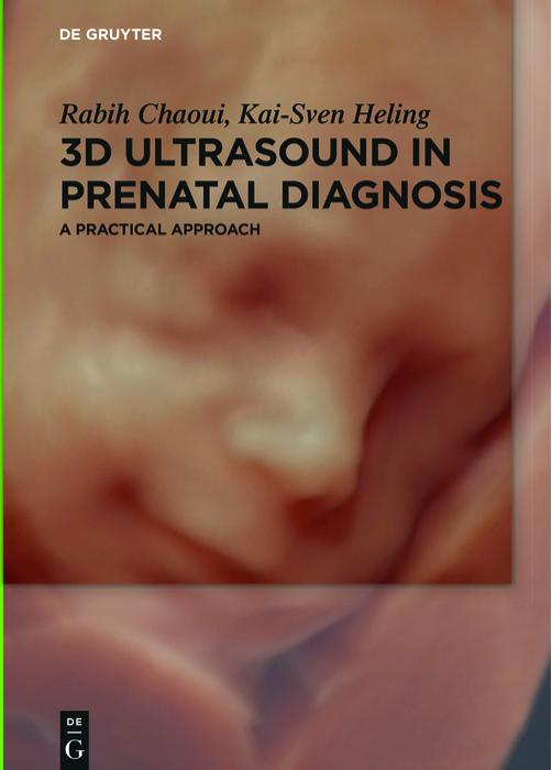
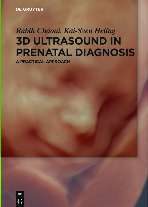
3D Ultrasound in Prenatal Diagnosis: A Practical Approach 1st Edition by Rabih Chaoui, Kai-Sven Heling – Ebook PDF Instant Download/Delivery. 3110496518 978-3110496512
Full download 3D Ultrasound in Prenatal Diagnosis: A Practical Approach 1st Edition after payment

Product details:
ISBN 10: 3110496518
ISBN 13: 978-3110496512
Author: Rabih Chaoui, Kai-Sven Heling
In the last decade there was a widespread use of 3D ultrasound in obstetrical imaging. It is estimated that more than half of the obstetrical clinics are currently using ultrasound equipment with 3D capabilities. Initially known for its beautiful images of the faces of babies, 3D ultrasound has, however, become an important tool in prenatal diagnosis for its ability to image fetal organs in normal and abnormal conditions. This book is a state-of-the-art work conceived as a practical guide to the application of 3D ultrasound in obstetrics. The book is illustrated with images reflecting the clinical utility of 3D ultrasound in prenatal diagnosis. The book has three sections: one section on the technical principles of 3D ultrasound, a second section on various 3D rendering tools with a step-by-step explanation of its use. The third section is dedicated to the clinical use of 3D in the examination of the fetal organs. The authors of this book have extensive expertise in 3D ultrasound that spans for more than 15 years.
3D Ultrasound in Prenatal Diagnosis: A Practical Approach 1st Table of contents:
Part I: Basics of 3D Ultrasound
1 Basics of 3D and 4D Volume Acquisition
1.1 Introduction
1.2 Preparing the volume acquisition
1.3 Types of volume acquisition
1.4 Conclusions
2 Orientation and Navigation within a Volume
2.1 Introduction
2.2 Storing and exporting volume data sets
2.3 Orientation in the three orthogonal planes
2.4 Navigation within the orthogonal planes
2.5 Artifacts in the multiplanar mode
2.6 Conclusions
Part II: Methods of 3D Rendering
3 3D Rendering of a Volume
3.1 Introduction
3.2 The render box and the orientation within a 3D volume
3.3 Artifacts in 3D rendering
3.4 Different rendering modes and the mixing of modes
3.5 Special effects in 3D: dynamic depth 3D rendering and light source
3.6 Threshold, transparency, brightness and color scales
3.7 Magicut, the electronic scalpel
3.8 Multiple light sources and „HD-live studio“
3.9 Conclusions
4 Volume Contrast Imaging (VCI)
4.1 Introduction
4.2 Principle of VCI
4.3 Static VCI
4.4 4D with VCI-Omniview
4.5 4D with VCI-A
4.6 Conclusions
5 Multiplanar Display I – Orthogonal Mode and Omniview Planes
5.1 Principle
5.2 Multiplanar reconstruction and different ways of displaying cross-sectional images
5.3 Practical approach in orthogonal mode
5.4 Practical approach in getting an „anyplane“ using Omniview tool
5.5 Typical applications of Omniview planes
5.6 Conclusions
6 Multiplanar Display II: Tomographic Mode
6.1 Principle
6.2 Practical approach
6.3 Typical applications in tomographic mode
6.4 Conclusions
7 Surface Mode Rendering and HD-Live
7.1 Principle
7.2 Practical approach
7.3 Typical applications of surface mode
7.4 Conclusions
8 Maximum Mode Rendering
8.1 Principle
8.2 Practical approach
8.3 Typical applications of maximum mode
8.4 Conclusions
9 The Minimum Mode
9.1 Principle
9.2 Practical approach
9.3 Typical applications of minimum mode
9.4 Conclusions
10 The Inversion Mode
10.1 Introduction
10.2 Practical approach
10.3 Typical applications of inversion mode
10.4 Conclusions
11 The Silhouette Tool
11.1 Principle
11.2 Practical application
11.3 Typical applications of silhouette tool
11.4 Conclusions
12 The Glass-Body Mode and HD-Live Flow
12.1 Principle
12.2 Practical approach
12.3 Glass-body mode with HD-live flow function
12.4 Typical applications in the glass-body mode
12.5 HD-live flow using the color silhouette tool
12.6 Conclusions
13 The B-Flow Mode
13.1 Principle
12.2 Practical approach
13.3 Typical applications of the B-flow mode
13.4 Conclusions
14 Biplane Display using the Electronic Matrix Transducer
14.1 Principle
14.2 Practical approach
14.3 Typical applications of biplane mode
14.4 Conclusions
15 Calculation of 3D Volumes
15.1 Principle
15.2 Practical approach
15.3 Clinical application of volume calculation
15.4 Conclusions
Part III: Clinical Applications of Prenatal Diagnosis
16 3D Fetal Neurosonography
16.1 Introduction
16.2 Fetal neurosonography with 3D ultrasound
16.3 3D visualization of specific brain structures
16.4 Reconstruction of fetal brain structures in 3D rendering
16.5 The intracranial vascular system in color Doppler
16.6 Fetal neurosonography before 14 weeks of gestation
16.7 Conclusions
17 3D of the Fetal Skeleton
17.1 Limitations in the assessment of the fetal skeleton using 2D ultrasound
17.2 3D of fetal spine and ribs
17.3 3D of the fetal limbs
17.4 3D of the facial and cranial bones
17.5 Conclusions
18 3D of the Fetal Face
18.1 The sonographic examination of the face in 2D and 3D ultrasound
18.2 The face in multiplanar display
18.3 The normal face in 3D/4D surface mode
18.4 The abnormal face in 3D/4D
18.5 The facial bones in 3D/4D
18.6 Conclusions
19 3D Intrathoracic and Intraabdominal Organs
19.1 Introduction
19.2 Intrathoracic organs
19.3 Intraabdominal organs
19.4 Conclusions
20 STIC and 3D/4D Fetal Echocardiography
20.1 The sonographic assessment of the heart in two-dimensional ultrasound
20.2 Acquiring cardiac volumes
20.3 Fetal echocardiography in 3D/4D multiplanar reconstruction
20.4 Fetal heart in 3D/4D volume rendering
20.5 Conclusions
21 3D in Early Pregnancy
21.1 Background
21.2 3D volume rendering in early gestation
21.3 Multiplanar display in early gestation
21.4 Conclusions
People also search for 3D Ultrasound in Prenatal Diagnosis: A Practical Approach 1st:
3d ultrasound in prenatal diagnosis a practical approach
3d ultrasound in prenatal diagnosis a practical approach 2nd edition
3d prenatal ultrasound near me
do you get 3d ultrasound during pregnancy
what is 3d ultrasound for pregnancy