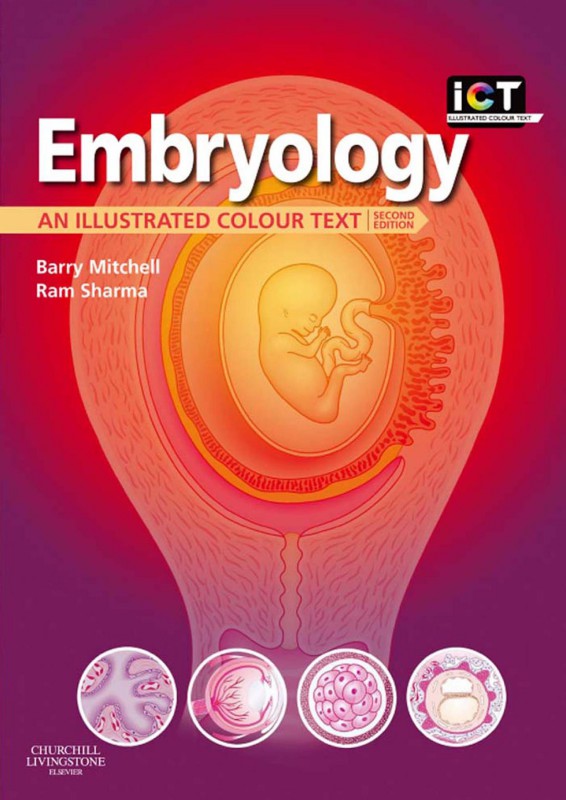
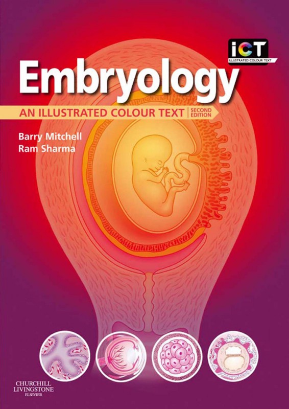
Embryology An Illustrated Colour Text 2nd Edition by Barry Mitchell, Ram Sharma ISBN 070206890X 9780702068904
$50.00 Original price was: $50.00.$25.00Current price is: $25.00.
Authors:Barry Mitchell; Ram Sharma , Series:Anatomy [53] , Tags:Medical; Embryology , Author sort:Mitchell, Barry & Sharma, Ram , Ids:9780702050817 , Languages:Languages:eng , Published:Published:Jan 2012 , Publisher:Elsevier Health Sciences , Comments:Comments:EMBRYOLOGY provides a concise and highly illustrated text, which confines its descriptions to those that are relevant for modern undergraduate and postgraduate medical courses, and similar courses in other related disciplines. An appreciation of embryology is essential to understand topological relationships in gross anatomy and to explain many congenital anomalies. Each chapter is supplemented by clinical point ‘boxes’ and by key revision points.Text in concise Illustrated Colour Text style, so core information on embryology can be quickly recognised and digested.Clear full colour diagrams and pictures make the embryological concepts clear and easily assimilated.Clinical boxes highlight essential points of importance to medical students.
Embryology An Illustrated Colour Text 2nd Edition by Barry Mitchell, Ram Sharma – Ebook PDF Instant Download/Delivery. 070206890X, 9780702068904
Full download Embryology An Illustrated Colour Text 2nd Edition after payment

Product details:
ISBN 10: 070206890X
ISBN 13: 9780702068904
Author: Barry Mitchell, Ram Sharma
EMBRYOLOGY provides a concise and highly illustrated text, which confines its descriptions to those that are relevant for modern undergraduate and postgraduate medical courses, and similar courses in other related disciplines. An appreciation of embryology is essential to understand topological relationships in gross anatomy and to explain many congenital anomalies. Each chapter is supplemented by clinical point ‘boxes’ and by key revision points.
- Text in concise Illustrated Colour Text style, so core information on embryology can be quickly recognised and digested.
- Clear full colour diagrams and pictures make the embryological concepts clear and easily assimilated.
- Clinical boxes highlight essential points of importance to medical students.
Embryology An Illustrated Colour Text 2nd Table of contents:
Chapter 1: How does an embryo form?
The 1st week—Fertilization and formation of the blastocyst
The 2nd week—Implantation and formation of bilaminar embryonic disc
The 3rd week—Further development of the embryo and formation of trilaminar embryonic disc
The 4th week—Folding of the embryo
Chapter 2: How do the placenta and fetal membranes form?
What are the fetal membranes?
Development of the uteroplacental circulation
Further development of chorionic villi
Formation of the placenta and placental circulation
Placental membrane and placental functions
Structure of the full-term placenta
The umbilical cord
Twins and their fetal membranes
Chapter 3: The body cavities and the diaphragm
Septum transversum and intra-embryonic coelom
Division of the intra-embryonic coelom into four cavities
The diaphragm
Chapter 4: The integumentary, skeletal and muscular systems
The integumentary system
The musculoskeletal system
The skeletal system
The muscular system
Chapter 5: The respiratory system
Development of the respiratory system
Chapter 6: The cardiovascular system
Heart tube formation
Septation of the heart tube
Septum formation in the ventricle
Septum formation in the outflow tracts of the heart
Development of the venous drainage into the heart
Valve formation in the atrioventricular canal and truncus arteriosus
Development of the arterial system
Development of the venous system
Changes in the vascular system at birth
Chapter 7: The digestive system
Primitive gut tube
Foregut
Other foregut derivatives
Midgut
Hindgut
Chapter 8: The urinary system
The urinary system
Kidneys and ureters
Urinary bladder and urethra
Chapter 9: The reproductive system
Development of the indifferent gonad
Development of the testis (Fig. 9.3)
Development of the ovary (Fig. 9.4)
Genital ducts
Descent of the testis and development of the inguinal canal
External genitalia
Chapter 10: The nervous system
Development of the spinal cord (Fig. 10.2A, B)
Development of the brain
Development of the meninges
Formation of the pituitary gland
Chapter 11: Development of the head and neck, the eye and ear
Pharyngeal arches
Pharyngeal clefts
Pharyngeal pouches
Development of the tongue
Development of the thyroid gland
Development of the face
Development of the nasal cavity and paranasal sinuses
Formation of the palate
Development of the eye and ear
People also search for Embryology An Illustrated Colour Text 2nd:
embryology an illustrated colour text
embryology an illustrated colour text pdf
embryology coloring book
illustrated dental embryology histology and anatomy workbook answers
illustrated dental embryology histology and anatomy 5th edition


