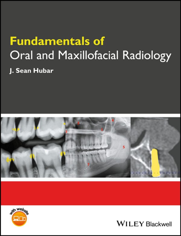
Fundamentals of Oral and Maxillofacial Radiology 1st edition by Sean Hubar 9781119122227 1119122228
$50.00 Original price was: $50.00.$25.00Current price is: $25.00.
Authors:J. Sean Hubar , Series:Dentistry [297] , Tags:Medical; Dentistry; General , Author sort:Hubar, J. Sean , Ids:Goodreads; Google; 9781119122210 , Languages:Languages:eng , Published:Published:May 2017 , Publisher:Wiley-Blackwell , Comments:Comments:Fundamentals of Oral and Maxillofacial Radiologyprovides a concise overview of the principles of dental radiology, emphasizing their application to clinical practice.Distills foundational knowledge on oral radiology in an accessible guide Uses a succinct, easy-to-follow approach Focuses on practical applications for radiology information and techniques Presents summaries of the most common osseous pathologic lesions and dental anomalies Includes companion website with figures from the book in PowerPoint and x-ray puzzles
Fundamentals of Oral and Maxillofacial Radiology 1st edition by Sean Hubar – Ebook PDF Instant Download/Delivery.9781119122227,1119122228
Full download Fundamentals of Oral and Maxillofacial Radiology 1st edition after payment
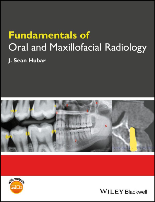
Product details:
ISBN 10:1119122228
ISBN 13:9781119122227
Author:Sean Hubar
Fundamentals of Oral and Maxillofacial Radiology provides a concise overview of the principles of dental radiology, emphasizing their application to clinical practice.
- Distills foundational knowledge on oral radiology in an accessible guide
- Uses a succinct, easy-to-follow approach
- Focuses on practical applications for radiology information and techniques
- Presents summaries of the most common osseous pathologic lesions and dental anomalies
- Includes companion website with figures from the book in PowerPoint and x-ray puzzles
Fundamentals of Oral and Maxillofacial Radiology 1st Table of contents:
Part One: Fundamentals
A Introduction
What is dental radiology?
What are x rays?
What’s the big deal about x‐ray images?
B History
Discovery of x rays
Who took the world’s first “dental” radiograph?
Dr. C. E. Kells, Jr., a New Orleans dentist and the early days of dental radiography
C Generation of X Rays
D Exposure Controls
Voltage (V)
Amperage (A)
Exposure timer
E Radiation Dosimetry
Exposure
Absorbed dose
Equivalent dose
Effective dose
F Radiation Biology
What happens to the dental x‐ray photons that are directed at a patient?
Determinants of biologic damage from x‐radiation exposure
G Radiation Protection
1. RADIATION PROTECTION: PATIENT
Protective apron
Collimation
Filtration
Digital versus analog
Exposure settings
Operator technique
2. RADIATION PROTECTION: OFFICE PERSONNEL
How much occupational radiation exposure is permitted?
H Patient Selection Criteria
I Film versus Digital Imaging
Film
Digital imaging
Imaging software
J What do Dental X‐ray Images Reveal?
Alterations to the dentition
Periodontal disease
Growth and development
Alterations to periapical tissues
Osseous pathology
Temporomandibular joint disorder
Implant assessment (pre‐ and post‐placement)
Identification of a foreign body
K Intraoral Imaging Techniques
1. PARALLELING TECHNIQUE
Maxillary incisors paralleling projection (Fig. K4)
Maxillary cuspid paralleling projection (Fig. K5)
Maxillary bicuspid paralleling projection (Fig. K6)
Maxillary molar paralleling projection (Fig. K7)
Mandibular incisor paralleling projection (Fig. K8)
Mandibular cuspid paralleling projection (Fig. K9)
Mandibular bicuspid paralleling projection (Fig. K10)
Mandibular molar paralleling projection (Fig. K11)
2. BISECTING ANGLE TECHNIQUE
Maxillary incisor bisecting angle projection
Maxillary cuspid bisecting angle projection
Maxillary bicuspid bisecting angle projection
Maxillary molar bisecting angle projection
Mandibular incisor bisecting angle projection
Mandibular cuspid bisecting angle projection
Mandibular bicuspid bisecting angle projection
Mandibular molar bisecting angle projection
3. BITEWING TECHNIQUE
Bicuspid bitewing (Fig. K14)
Molar bitewing (Fig. K15)
Anterior bitewing projection (Fig. K16)
4. DISTAL OBLIQUE TECHNIQUE
5. OCCLUSAL IMAGING TECHNIQUE
Maxillary occlusal projection
Mandibular occlusal projection
L Intraoral Technique Errors
Cone‐cut
Apex missing
Elongation
Foreshortening
Overlapped contacts
Missing contacts
Overexposure and underexposure
Motion artifact
Foreign object
M Extraoral Imaging Techniques
1. PANORAMIC IMAGING
Positioning the patient
Exposure settings
Advantages and disadvantages
Technique errors
Anatomic landmarks
2. LATERAL CEPHALOGRAPH IMAGING
3. CONE BEAM COMPUTED TOMOGRAPHY
Introduction
Anatomic landmarks
N Quality Assurance
O Infection Control
Excerpt from “CDC Guidelines for Infection Control in Dental Health‐Care Settings”
General instructions for cleaning and disinfecting a solid‐state receptor (courtesy of Sirona™)
P Occupational Radiation Exposure Monitoring
Q Hand‐held X‐ray Systems
Dental radiographic examinations: recommendations for patient selection and limiting radiation exposure
Commentary
Part Two: Interpretation
R Localization of Objects (SLOB Rule)
S Recommendations for Interpreting Images
T X‐ray Puzzles
U Radiographic Anatomy
1. DENTAL ANATOMY
2. ANATOMIC LANDMARKS OF THE MAXILLARY REGION
Radiopaque landmarks
Radiolucent landmarks
3. ANATOMIC LANDMARKS OF THE MANDIBULAR REGION
Radiopaque landmarks
Radiolucent landmarks
V Dental Caries
Limitations to visualizing caries on x‐ray images
Classification of caries
W Dental Anomalies
Number
Size
Shape
Developmental factors
Environmental factors
X Osseous Pathology (Alphabetic)
Y Lagniappe (Miscellaneous Oddities)
People also search for Fundamentals of Oral and Maxillofacial Radiology 1st:
what does an oral and maxillofacial radiologist do
oral and maxillofacial radiology programs
fundamentals of oral and maxillofacial radiology pdf
oral and maxillofacial radiology job description
oral and maxillofacial radiology procedures
You may also like…
eBook PDF
Atlas of Oral and Maxillofacial Radiology 1st Edition by Bernard Koong ISBN 1118939646 9781118939642

