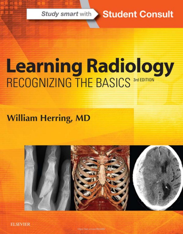
Learning Radiology Recognizing the Basics 1st edition by William Herring 9780323567299 0323567290
Original price was: $50.00.$25.00Current price is: $25.00.
Authors:William Herring , Tags:Medical; Allied Health Services; Imaging Technologies; General; Learning Radiology; Third Edition (2016) 451pp. 978-0-323-38851-1 , Author sort:Herring, William , Ids:Google; 9780323388511 , Languages:Languages:eng , Published:Published:Apr 2015 , Publisher:Elsevier Health Sciences , Comments:Comments:A must-have for anyone who will be required to read and interpret common radiologic images, Learning Radiology: Recognizing the Basics is an image-filled, practical, and easy-to-read introduction to key imaging modalities. Skilled radiology teacher William Herring, MD, masterfully covers exactly what you need to know to effectively interpret medical images of all modalities. Learn the latest on ultrasound, MRI, CT, patient safety, dose reduction, radiation protection, and more, in a time-friendly format with brief, bulleted text and abundant high-quality images. Identify a wide range of common and uncommon conditions based upon their imaging findings. Arrive at diagnoses by following a pattern recognition approach, and logically overcome difficult diagnostic challenges with the aid of decision trees. Quickly grasp the fundamentals you need to know through more than 700 images and an easy-to-use format and pedagogy, including: bolding of key points and icons designating special content; Diagnostic Pitfalls; Really, Really Important Points; Weblinks; and Take-Home Points. Gauge your mastery of the material and build confidence with extra images, bonus content, interactive self-assessment exercises, and USMLE-style Q&A that provide effective chapter review and quick practice for your exams. Apply the latest recommendations on patient safety, dose reduction and radiation protection Benefit from the extensive knowledge and experience of esteemed author Dr. William Herring—a skilled radiology teacher and the host of his own specialty website, www.learningradiology.com. Stay current in the latest advancements and developments with meticulous updates throughout including a new chapter on Pediatric Radiology as well as more than 60 new and updated photos, many highlighting newer imaging modalities.
Learning Radiology Recognizing the Basics 1st edition by William Herring – Ebook PDF Instant Download/Delivery.9780323567299,0323567290
Full download Learning Radiology Recognizing the Basics 1st edition after payment

Product details:
ISBN 10:0323567290
ISBN 13:9780323567299
Author:William Herring
A must-have for anyone who will be required to read and interpret common radiologic images, Learning Radiology: Recognizing the Basics is an image-filled, practical, and easy-to-read introduction to key imaging modalities. Skilled radiology teacher William Herring, MD, masterfully covers exactly what you need to know to effectively interpret medical images of all modalities. Learn the latest on ultrasound, MRI, CT, patient safety, dose reduction, radiation protection, and more, in a time-friendly format with brief, bulleted text and abundant high-quality images. Then ensure your mastery of the material with additional online content, bonus images, and self-assessment exercises at Student Consult.
- Identify a wide range of common and uncommon conditions based upon their imaging findings.
- Arrive at diagnoses by following a pattern recognition approach, and logically overcome difficult diagnostic challenges with the aid of decision trees.
- Quickly grasp the fundamentals you need to know through more than 700 images and an easy-to-use format and pedagogy, including: bolding of key points and icons designating special content; Diagnostic Pitfalls; Really, Really Important Points; Weblinks; and Take-Home Points.
- Gauge your mastery of the material and build confidence with extra images, bonus content, interactive self-assessment exercises, and USMLE-style Q&A that provide effective chapter review and quick practice for your exams
Learning Radiology Recognizing the Basics 1st Table of contents:
Chapter 1 Recognizing Anything
Many Shades of Gray
Conventional Radiography (Plain Films)
The Five Basic Densities
Computed Tomography
Ultrasound
Magnetic Resonance Imaging
Fluoroscopy
Nuclear Medicine
Conventions Used in This Book
Chapter 2 Recognizing a Technically Adequate Chest Radiograph
Evaluating the Chest Radiograph for Technical Adequacy
Penetration
Inspiration
Rotation
Magnification
Angulation
Chapter 3 Recognizing Normal Pulmonary Anatomy
The Normal Frontal Chest Radiograph
The Normal Lateral Chest Radiograph
Normal CT Anatomy of the Chest
Chapter 4 Recognizing Normal Cardiac Anatomy
Evaluating the Heart on Chest Radiographs
General Principles
Evaluating the Heart on Cardiac CT
Uses of Cardiac CT
Cardiac MRI
Chapter 5 Recognizing Airspace Versus Interstitial Lung Disease
Classifying Parenchymal Lung Disease
Characteristics of Airspace Disease
Some Causes of Airspace Disease
Characteristics of Interstitial Lung Disease
Some Causes of Interstitial Lung Disease
Chapter 6 Recognizing the Causes of an Opacified Hemithorax
Atelectasis of the Entire Lung
Massive Pleural Effusion
Pneumonia of an Entire Lung
Postpneumonectomy
Chapter 7 Recognizing Atelectasis
What Is Atelectasis?
Signs of Atelectasis
Types of Atelectasis
How Atelectasis Resolves
Chapter 8 Recognizing a Pleural Effusion
Normal Anatomy and Physiology of the Pleural Space
Modalities for Detecting Pleural Effusions
Causes of Pleural Effusions (Table 8.1)
Types of Pleural Effusions
Side-Specificity of Pleural Effusions
Recognizing the Different Apperances of Pleural Effusions
Chapter 9 Recognizing Pneumonia
General Considerations
General Characteristics of Pneumonia
Patterns of Pneumonia
Lobar Pneumonia
Segmental Pneumonia (Bronchopneumonia)
Interstitial Pneumonia
Round Pneumonia
Cavitary Pneumonia
Aspiration
Localizing Pneumonia
How Pneumonia Resolves
Chapter 10 Recognizing the Correct Placement of Lines and Tubes And Their Potential Complications
Endotracheal and Tracheostomy Tubes
Intravascular Catheters
Peripherally Inserted Central Catheters: PICC
Multiple Lumen Catheters: “Quinton Catheters,” Hemodialysis Catheters
Pleural Drainage Tubes (Chest Tubes, Thoracotomy Tubes)
Cardiac Devices: Pacemaker, Automatic Implantable Cardiac Defibrillator (AICD), Intra-aortic Balloon Pump (IABP)
Automatic Implantable Cardiac Defibrillators (AICD)
Intra-aortic counterpulsation balloon pump (IABP or IACB)
Gi Tubes and Lines: Nasogastric Tubes, Feeding Tubes
Chapter 11 Recognizing Other Diseases of the Chest
Mediastinal Masses
Anterior Mediastinum
Middle Mediastinal Masses
Posterior Mediastinal Masses
Solitary Nodule/Mass in the Lung
Bronchogenic Carcinoma
Metastatic Neoplasms in the Lung
Pulmonary Thromboembolic Disease (PE)
Chronic Obstructive Pulmonary Disease
Bullae, Cysts, and Cavities
Bronchiectasis
Chapter 12 Recognizing Adult Heart Disease
Recognizing an Enlarged Cardiac Silhouette
Recognizing Common Cardiac Diseases
Chapter 13 Recognizing the Normal Abdomen and Pelvis
Recognizing the Normal Abdomen: What to Look For
Acute Abdominal Series: The Views and What They Show
Recognizing the Normal Abdomen: Extraluminal Air
Recognizing the Normal Abdomen: Calcifications
Recognizing the Normal Abdomen: Organomegaly
Chapter 14 Recognizing the Normal Abdomen and Pelvis on Computed Tomography
Introduction to Abdominal and Pelvic CT
Intravenous Contrast in CT Scanning
Oral Contrast in CT Scanning
Abdominal CT: General Considerations
Abdominal CT: By organ
Chapter 15 Recognizing Bowel Obstruction and Ileus
Abnormal Gas Patterns
Laws of the Gut
Functional Ileus: Localized—Sentinel Loops
Functional Ileus: Generalized Adynamic Ileus
Mechanical Obstruction: Small Bowel Obstruction
Mechanical Obstruction: Large Bowel Obstruction (LBO)
Volvulus of the Colon
Intestinal Pseudoobstruction (Ogilvie Syndrome)
Chapter 16 Recognizing Extraluminal Gas in the Abdomen
Signs of Free Intraperitoneal Air
Signs of Extraperitoneal Air (Retroperitoneal Air)
Signs of Air in the Bowel Wall
Signs of Air in the Biliary System
Chapter 17 Recognizing Abnormal Calcifications and Their Causes
Patterns of Calcification
Location of Calcification
Chapter 18 Recognizing Gastrointestinal, Hepatobiliary, and Urinary Tract Abnormalities
Barium Studies of the Gastrointestinal Tract
Esophagus
Stomach and Duodenum
Small and Large Bowel
Large Bowel
Pancreas
Hepatobiliary Abnormalities
Biliary System
Urinary Tract
Pelvis
Urinary Bladder
Adenopathy
Chapter 19 Ultrasonography
How It Works
Types of Ultrasound
Adverse Effects or Safety Issues
Medical Uses of Ultrasonography
Female Pelvic Organs
Pregnancy
Abdominal Hernias
Appendicitis
Ascites
Musculoskeletal System
Contrast-Enhanced Ultrasound
Chapter 20 Vascular, Pediatric, and Point-of-Care Ultrasound
Vascular Ultrasound
Arterial Stenosis
Pseudoaneurysm
Deep Vein Thrombosis (DVT)
Pediatrics
Point-of-Care Ultrasound
Chapter 21 Magnetic Resonance Imaging
How MRI Works
Hardware That Makes Up an MRI Scanner
What Happens Once Scanning Begins
How Can You Identify a T1-Weighted or T2-Weighted Image?
MRI Contrast: General Considerations
MRI Safety Issues
Diagnostic Applications of MRI
Chapter 22 Recognizing Nontraumatic Abnormalities of the Appendicular Skeleton Including Arthritis
Conventional Radiography, CT, and MRI in Bone Imaging
Normal Bone and Joint Anatomy
Diseases That Affect Bone Density
Diseases That Increase Bone Density
Diseases That Decrease Bone Density
Diseases of the Joints: An Approach to Arthritis
Chapter 23 Recognizing Nontraumatic Abnormalities of the Spine
The Normal Spine (Fig. 23.1)
Normal MRI Appearance of the Spine
Back Pain
Chapter 24 Recognizing Trauma to the Bony Skeleton
Recognizing an Acute Fracture
Recognizing Dislocations and Subluxations
Describing Fractures
Avulsion Fractures
Salter-Harris Fractures: Epiphyseal Plate Fractures in Children
Child Abuse
Stress Fractures
Common Fracture Eponyms
Some Subtle Fractures or Dislocations
Fracture Healing
Spinal Trauma
Pathologic Fractures
Chapter 25 Recognizing the Imaging Findings of Trauma to the Chest
Chest Trauma
Chest Wall Trauma
Pleural Abnormalities: Pneumothorax
Imaging Modalities Used to Diagnose a Pneumothorax
Trauma-Related Parenchymal Lung Abnormalities
Aortic Trauma
Chapter 26 Recognizing the Imaging Findings of Trauma to the Abdomen and Pelvis
Abdominal Trauma
Pelvic Trauma
Less Common Abdominal Injuries
Chapter 27 Recognizing Some Common Causes of Intracranial Pathology
Normal Anatomy (Fig. 27.1)
Mri and the Brain
Head Trauma
Intracranial Hemorrhage
Diffuse Axonal Injury
Increased Intracranial Pressure
Stroke
Ruptured Aneurysms
Hydrocephalus
Cerebral Atrophy
Brain Tumors
Other Diseases
Terminology
Chapter 28 Recognizing Pediatric Diseases
Diseases Discussed in This Chapter
Newborn Respiratory Distress
Childhood Lung Disease
Soft Tissues of the Neck
Ingested Foreign Bodies
Other Diseases
Salter-Harris Fractures: Epiphyseal Plate Fractures in Children
Child Abuse
Necrotizing Enterocolitis (NEC)
Esophageal Atresia With/Without Tracheoesophageal Fistula (TEF)
Chapter 29 Using Image-Guided Interventions in Diagnosis and Treatment
Arterial Access and Arteriography
Central Venous Access: Image-Guided Venous Access
Pulmonary Embolism: Thrombolysis
Pulmonary Embolism: Inferior Vena Caval (IVC) Filter Placement
Pulmonary Nodule: Image-Guided Biopsy
Hepatic/Renal/Pulmonary Tumor: Thermal Ablation
Portal Hypertension: Transjugular Intrahepatic Portosystemic Shunt (TIPS)
Abscess: Percutaneous Abscess Aspiration and Drain Placement
Gastrointestinal (GI) Bleeding: Arteriography and Embolization
Obstructive Uropathy: Percutaneous Nephrostomy (PCN)/ Nephroureterostomy (PCNU)
Aortic Aneurysm: Endovascular Aneurysm Repair (EVAR)
Uterine Fibroids: Uterine Fibroid Embolization (UFE)
Acute Ischemic Stroke: Mechanical Thrombectomy
Chapter 30 Recognizing the Findings in Breast Imaging
Breast Imaging Modalities: Overview
Mammography: Screening Versus Diagnostic
Fundamental Mammography Findings
Ultrasound
Magnetic Resonance Imaging
Management of Breast Abnormalities
Special Considerations
People also search for Learning Radiology Recognizing the Basics 1st:
learning radiology e book
learning radiology recognizing the basics 4th edition
learning radiology recognizing the basics 5th edition
learning radiology book
learning radiology recognizing the basics ebook
You may also like…


eBook EPUB
Emergent Medicine and the Law 1st edition by Chau, Jonathan Herring 9783030602086 3030602087