The Unofficial Guide to Radiology 100 Practice Chest X Rays with Full Colour Annotations and Full X Ray Reports 1st Edition by Mohammed Rashid Akhtar, Rebecca Best, Lydia Shackshaft, Mark A Rodrigues, Zeshan Qureshi ISBN 1910399019 978-1910399019

Original price was: $50.00.$25.00Current price is: $25.00.
Authors:Mark Rodrigues; Zeshan Qureshi , Author sort:Rodrigues, Mark & Qureshi, Zeshan , Published:Published:Jan 2017
The Unofficial Guide to Radiology: 100 Practice Chest X-Rays, with Full Colour Annotations and Full X-Ray Reports 1st Edition by Mohammed Rashid Akhtar, Rebecca Best, Lydia Shackshaft, Mark A Rodrigues, Zeshan Qureshi – Ebook PDF Instant Download/Delivery. 1910399019 978-1910399019
Full download The Unofficial Guide to Radiology: 100 Practice Chest X-Rays, with Full Colour Annotations and Full X-Ray Reports 1st Edition after payment
Product details:
ISBN 10: 1910399019
ISBN 13: 978-1910399019
Author: Mohammed Rashid Akhtar, Rebecca Best, Lydia Shackshaft, Mark A Rodrigues, Zeshan Qureshi
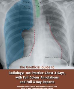
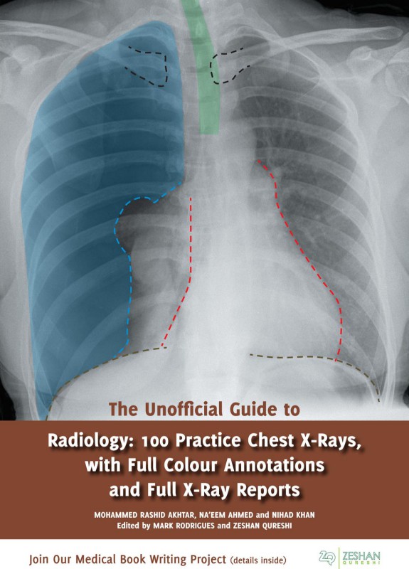
The Unofficial Guide to Radiology: 100 Practice Chest X Rays is a companion to the Unofficial Guide to Radiology. This book teaches systematic analysis of Chest X Rays. The layout is designed to make the book as relevant to clinical practice as possible; the X-rays are presented in the context of a real life scenario. The reader is asked to interpret the X-ray before turning over the page to reveal a model report accompanied by a fully colour annotated version of the X-ray. Uniquely, all cases provide realistic high quality X Ray images, are annotated in full colour, and are fully reported, following international radiology reporting guidelines. This means the X Rays are explained comprehensively, but with clear annotation so that a complete beginner can follow the thinking of the expert. This book has relevance beyond examinations, for post graduate further education and as a day-to-day reference for professionals.
The Unofficial Guide to Radiology: 100 Practice Chest X-Rays, with Full Colour Annotations and Full X-Ray Reports 1st Table of contents:
1. Introduction to Chest X-Ray Interpretation
- Overview of Chest X-ray: A brief introduction to the chest X-ray as a diagnostic tool, explaining the importance of chest radiography in clinical practice, its uses, and limitations.
- Basic Principles of X-ray Imaging: Describes the fundamentals of X-ray imaging, including how X-rays are produced, how images are formed, and the basic interpretation techniques for beginners.
- Understanding X-ray Anatomy: A guide to the normal anatomical structures visible on a chest X-ray, including the lungs, heart, diaphragm, ribs, and mediastinum, to establish a baseline for identifying abnormal findings.
2. Normal Chest X-Ray
- Interpretation of a Normal Chest X-ray: This section likely focuses on understanding the normal appearances in a chest X-ray. It would include typical findings in healthy individuals and the key things to look for when reviewing normal radiographs.
3. Common Chest Pathologies: 100 Practice Cases
- Case 1-10: Pulmonary Infections: These cases would feature conditions such as pneumonia, tuberculosis, and other respiratory infections, focusing on identifying characteristic signs such as consolidation, cavitation, or infiltrates.
- Case 11-20: Cardiovascular Conditions: Chest X-rays in conditions like heart failure, cardiomegaly, pulmonary edema, and pericardial effusion. It might focus on recognizing enlarged cardiac silhouette or signs of fluid overload in the lungs.
- Case 21-30: Pulmonary Embolism and Vascular Abnormalities: Interpretation of chest X-rays in patients with pulmonary embolism, deep vein thrombosis, and vascular abnormalities such as aortic dissection or aneurysms.
- Case 31-40: Trauma and Fractures: Radiographs showing injuries like rib fractures, pneumothorax, hemothorax, and other chest trauma, with emphasis on how to differentiate between fractures, soft tissue injuries, and other causes of trauma-related radiographic changes.
- Case 41-50: Neoplastic Conditions: These cases would feature tumors such as lung cancer, metastasis, and benign lung lesions, with a focus on identifying masses, nodules, and structural changes related to malignancy.
- Case 51-60: Chronic Obstructive Pulmonary Disease (COPD) and Emphysema: Radiographs in COPD, emphysema, and other chronic lung diseases, helping learners identify hyperinflation, flattened diaphragms, and changes in lung tissue.
- Case 61-70: Interstitial Lung Diseases: This section would cover conditions like pulmonary fibrosis, sarcoidosis, and interstitial lung diseases, emphasizing patterns such as reticular changes, honeycombing, and ground-glass opacities.
- Case 71-80: Pleural Effusion and Pneumothorax: Radiographs showing pleural effusion (fluid accumulation between the pleura), pneumothorax (air between the pleura), and other pleural conditions, focusing on the characteristic signs of each.
- Case 81-90: Post-surgical and Post-procedural Chest X-rays: Postoperative and post-procedural radiographs showing changes after surgery, including complications such as infection, atelectasis, or improper placement of medical devices (e.g., pacemakers, central lines).
- Case 91-100: Pediatric Chest X-rays: Cases involving pediatric patients, with a focus on congenital anomalies, foreign body aspiration, and other conditions unique to children.
4. How to Read Chest X-rays: Step-by-Step Approach
- Systematic Approach to Chest X-ray Interpretation: A guide to the structured method of reading chest X-rays, breaking down the process into steps such as assessing the quality of the image, analyzing the anatomy, and evaluating for abnormalities.
- Red Flags to Look For: Common signs of severe or urgent conditions, such as large masses, signs of lung collapse, or pneumothorax, that require immediate clinical attention.
- Common Pitfalls and Mistakes: This section likely discusses common errors in interpreting chest X-rays, such as missing subtle signs of pathology or mistaking normal variants for abnormalities.
5. Full X-Ray Reports and Annotations
- Case Report 1-100: For each of the 100 cases, the book provides full X-ray reports, including a detailed interpretation of the image. These reports would likely include a step-by-step description of findings, diagnosis, differential diagnosis, and clinical correlation.
- Clinical Context and Recommendations: Each case would also include clinical context and recommendations for further diagnostic work-up or management, providing practical insight into how chest X-rays influence clinical decision-making.
6. Further Reading and Resources
- Recommended Textbooks and Journals: A list of further resources for in-depth study of radiology and chest imaging.
- Radiology Societies and Educational Websites: A guide to professional societies, online courses, and other platforms for ongoing learning in radiology and chest X-ray interpretation.
7. Conclusion and Final Thoughts
- Mastering Chest X-ray Interpretation: A summary of key takeaways, reinforcing the importance of systematic reading and the value of practice in mastering chest X-ray interpretation.
People also search for The Unofficial Guide to Radiology: 100 Practice Chest X-Rays, with Full Colour Annotations and Full X-Ray Reports 1st:
the unofficial guide to radiology pdf free download
the unofficial guide to radiology pdf download
the unofficial guide to radiology 2nd edition
the unofficial guide to radiology 2nd edition pdf
the unofficial guide to radiology book
You may also like…