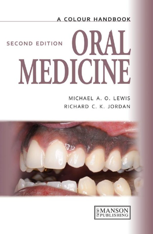
Oral Medicine 2nd Edition by Michael Lewis, Richard Jordan ISBN 1482228742 9781482228748
$50.00 Original price was: $50.00.$25.00Current price is: $25.00.
Authors:Michael A.O.Lewis, Richard C.K. Jordan , Author sort:Michael A.O.Lewis, Richard C.K. Jordan
Oral Medicine 2nd Edition by Michael Lewis, Richard Jordan – Ebook PDF Instant Download/Delivery. 1482228742, 9781482228748
Full download Oral Medicine 2nd Edition after payment
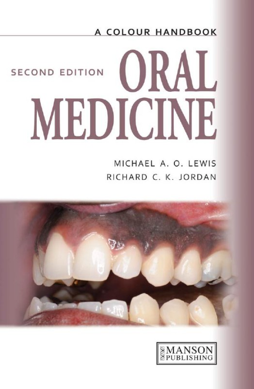
Product details:
ISBN 10: 1482228742
ISBN 13: 9781482228748
Author: Michael Lewis, Richard Jordan
This is a revised edition of a bestselling handbook. The authors have fully updated the text to include the most up to date treatment options, have added a section on head and neck imaging (CT/MRI), a series of self-test clinical cases, and 100 new photographs. The book uses a symptom-based approach to assist the clinician in the diagnosis and mana.
Oral Medicine 2nd Table of contents:
CHAPTER 1 Introduction
A symptom-based approach to diagnosis
History
Clinical examination
EXTRAORAL EXAMINATION
INTRAORAL EXAMINATION
1 Bi-manual palpation of the left submandibular gland.
2 Dorsum of the tongue.
3 Left lateral margin of the tongue.
4 Right lateral margin of the tongue.
5 Left buccal mucosa.
6 Right buccal mucosa.
7 Soft and hard palate.
8 Floor of the mouth.
Normal structures
9 Circumvallate papillae at the junction of the anterior two-thirds and the posterior third of the tongue.
10 Foliate papillae on the posterior lateral margin of the tongue.
11 Fissured tongue.
12 Lingual varicosities.
13 Lingual varicosities.
14 Ectopic sebaceous glands.
Special investigation of orofacial disease
HEMATOLOGICAL ASSESSMENT
MICROBIOLOGICAL INVESTIGATION
Smear
Plain swab
15 Smear from an angle of the mouth stained by Gram’s method showing epithelial cells, candidal hyphae, and spores (blue).
16 Swab of an angle of the mouth.
17 Plain swab and viral transport medium.
Imprint culture
Concentrated oral rinse
TISSUE BIOPSY
18 Imprint being taken from the dorsum of the tongue.
19 Method for concentrated oral rinse sampling of the oral flora.
20 Placement of local anesthesia adjacent to the lesion.
27 Tissue supported on filter paper prior to placement in formal saline.
Exfoliate cytology and brush biopsy
Vital staining
28 Cytobrush.
29 Blue-stained mucosa in the palate following topical application of tolonium chloride.
Salivary gland investigations
SALIVARY FLOW RATES
SIALOGRAPHY
30 Carlson–Crittenden cup.
31 Carlson–Crittenden cup placed over each parotid papilla.
32 Lateral oblique sialogram of the right parotid gland showing the normal gland and duct architecture.
SCINTISCANNING
LABIAL GLAND BIOPSY
SCHIRMER TEAR TEST
33 Scintiscan showing normal uptake of technetium in the thyroid glands and salivary tissues.
34 Labial gland biopsy.
35 Schirmer tear (lacrimal flow) test.
Imaging techniques
ULTRASOUND
36 Ultrasound image showing a tumor within the parotid salivary gland.
37 Ultrasound being used to guide a fine needle aspiration of a tumor in the parotid salivary gland.
COMPUTED TOMOGRAPHY
POSITRON EMISSION TOMOGRAPHY
MAGNETIC RESONANCE IMAGING
38 CT scan showing a malignant soft tissue mass in right angle of the mandible.
39 Cone beam CT scan showing position of the right mental foramen in the mandible.
40 Cone beam CT scan showing the inferior dental canal close to the lower border of the mandible.
41 PET scan showing a tumor in the right body of the mandible.
42 MR image showing a malignant mass in the left ethmoid sinus penetrating the left orbit.
CHAPTER 2 Ulceration
General approach
Table 1 Patterns of ulceration and differential diagnosis
Traumatic ulceration
ETIOLOGY AND PATHOGENESIS
CLINICAL FEATURES
DIAGNOSIS
MANAGEMENT
43 Ulcer on the lateral margin of the tongue induced by trauma from the edge of a fractured restoration in the first lower molar.
44 Traumatic ulcer on the right lateral margin of the tongue.
45 Irregular ulcer that was self-induced by the patient.
46 Diffuse ulceration in the palate due to the placement of salicylic acid gel by the patient onto the fitting surface of her upper denture.
Recurrent aphthous stomatitis
ETIOLOGY AND PATHOGENESIS
CLINICAL FEATURES
DIAGNOSIS
MANAGEMENT
47 Small round ulcer (MiRAS) affecting the labial mucosa.
48 Small round and oval ulcers (MiRAS) affecting the soft palate.
49 Large round ulcer (MaRAS) in the buccal mucosa.
50 Multiple small round and oval ulcers (HU) in the soft palate.
Behçet’s disease
ETIOLOGY AND PATHOGENESIS
CLINICAL FEATURES
DIAGNOSIS
MANAGEMENT
51 An ulcer of major recurrent aphthous stomatitis in the palate of a patient with Behçet’s disease.
52 Scarring following the resolution of major recurrent aphthous stomatitis.
Cyclic neutropenia
ETIOLOGY AND PATHOGENESIS
CLINICAL FEATURES
DIAGNOSIS
MANAGEMENT
53, 54 Minor aphthous stomatitis in cyclic neutropenia.
Squamous cell carcinoma
ETIOLOGY AND PATHOGENESIS
CLINICAL FEATURES
DIAGNOSIS
MANAGEMENT
55–60 Squamous cell carcinoma presenting as an ulcer with rolled indurated margins on the lip and at a variety of intraoral sites.
Necrotizing sialometaplasia
ETIOLOGY AND PATHOGENESIS
CLINICAL FEATURES
DIAGNOSIS
MANAGEMENT
61 Necrotizing sialometaplasia presents initially as a swelling.
62 Necrotizing sialometaplasia presenting as swelling with central ulceration.
63 Ulceration in necrotizing sialometaplasia.
Tuberculosis
ETIOLOGY AND PATHOGENESIS
CLINICAL FEATURES
DIAGNOSIS
MANAGEMENT
64 Tuberculous ulcer on the tongue.
65 Radiograph showing a radiopaque mass due to tuberculosis within the submandibular lymph nodes.
Syphilis
ETIOLOGY AND PATHOGENESIS
CLINICAL FEATURES
DIAGNOSIS
MANAGEMENT
66 Ulcerated nodular lesion of primary syphilis.
67 Ulceration of secondary syphilis.
68 Notch deformity of the incisors due to congenital syphilis (Hutchinson’s incisors).
69 Hypoplastic deformity of the first molar due to congenital syphilis (Moon’s molar, mulberry molar).
Epstein–Barr virus-associated ulceration
ETIOLOGY AND PATHOGENESIS
CLINICAL FEATURES
DIAGNOSIS
MANAGEMENT
70 Epstein–Barr virus ulceration on the lateral border of the tongue in an immunosuppressed patient.
Bisphosphonate-related osteonecrosis of the jaw
ETIOLOGY AND PATHOGENESIS
CLINICAL FEATURES
DIAGNOSIS
MANAGEMENT
71, 72 Osteonecrosis 2 weeks following extraction of lower anterior teeth.
Acute necrotizing ulcerative gingivitis
ETIOLOGY AND PATHOGENESIS
CLINICAL FEATURES
DIAGNOSIS
MANAGEMENT
73, 74 Gingival ulceration and loss of the interdental papillae in the lower incisor region.
75 Smear from acute necrotizing ulcerative gingivitis stained by Gram’s method showing large numbers of fusiform bacteria and spirochetes.
Erosive lichen planus
ETIOLOGY AND PATHOGENESIS
CLINICAL FEATURES
DIAGNOSIS
MANAGEMENT
76–78 Erosive lichen planus presenting as ulceration in the buccal mucosa and floor of the mouth.
Lichenoid reaction
ETIOLOGY AND PATHOGENESIS
CLINICAL FEATURES
DIAGNOSIS
MANAGEMENT
79 Ulceration caused by a lichenoid reaction to an antihypertensive drug.
80 Ulceration induced by nicorandil drug therapy.
81, 82 Lichenoid reaction on the lateral margin of the tongue following systemic chemotherapy.
Graft versus host disease
ETIOLOGY AND PATHOGENESIS
CLINICAL FEATURES
DIAGNOSIS
83, 84 Graft versus host disease presenting as swelling and diffuse erosion of the buccal mucosa and tongue.
MANAGEMENT
Radiotherapy-induced mucositis
ETIOLOGY AND PATHOGENESIS
CLINICAL FEATURES
DIAGNOSIS
MANAGEMENT
85 Mucositis following adjunctive radiotherapy.
Osteoradionecrosis
ETIOLOGY AND PATHOGENESIS
CLINICAL FEATURES
DIAGNOSIS
MANAGEMENT
86, 87 Osteoradionecrosis.
CHAPTER 3 Blisters
General approach
Table 2 Patterns of blistering and differential diagnosis
Primary herpetic gingivostomatitis
ETIOLOGY AND PATHOGENESIS
CLINICAL FEATURES
DIAGNOSIS
MANAGEMENT
88–90 Vesicles, blood crusting of the lips, and multiple intraoral ulcers characteristic of primary herpes simplex virus infection.
91 Blistering of the upper lip caused by herpes simplex virus.
92 Swelling of the interdental papillae and gingival margins caused by herpes simplex virus.
Recurrent herpes simplex infection
ETIOLOGY AND PATHOGENESIS
CLINICAL FEATURES
93 Vesicles of herpes labialis.
94 Vesicle stage of recurrent herpes labialis.
95 Crusting of a healing herpes labialis.
96 Crusting stage of multiple recurrent herpes labialis.
DIAGNOSIS
MANAGEMENT
97, 98 Multiple ulcers in the hard palate, a frequent site of recurrent herpes simplex virus infection.
99 Ulceration of the gingival margins due to recurrent herpetic infection.
Chickenpox and shingles
ETIOLOGY AND PATHOGENESIS
CLINICAL FEATURES
DIAGNOSIS
MANAGEMENT
100, 101 Unilateral lesions of recurrent varicella zoster virus infection.
102 Vesicular eruption of herpes zoster on the skin.
Hand, foot, and mouth disease
ETIOLOGY AND PATHOGENESIS
CLINICAL FEATURES
DIAGNOSIS
MANAGEMENT
103–105 Vesicular eruptions of hand, foot, and mouth disease.
Herpangina
ETIOLOGY AND PATHOGENESIS
CLINICAL FEATURES
DIAGNOSIS
MANAGEMENT
Epidermolysis bullosa
ETIOLOGY AND PATHOGENESIS
CLINICAL FEATURES
DIAGNOSIS
MANAGEMENT
106 Blisters of herpangina in the palate.
107 Blister of epidermolysis bullosa on upper labial mucosa.
Mucocele
ETIOLOGY AND PATHOGENESIS
CLINICAL FEATURES
DIAGNOSIS
MANAGEMENT
108–110 Mucocele presenting as a localized fluctuant swelling inside the lower lip and retromolar region.
Angina bullosa hemorrhagica
ETIOLOGY AND PATHOGENESIS
CLINICAL FEATURES
DIAGNOSIS
MANAGEMENT
111 Angina bullosa hemorrhagica in the palate.
112 Angina bullosa hemorrhagica on the tongue.
113, 114 Bullae of angina bullosa hemorrhagica rupture early to leave blood at the periphery of the lesion.
Erythema multiforme
ETIOLOGY AND PATHOGENESIS
CLINICAL FEATURES
DIAGNOSIS
MANAGEMENT
115, 116 Blood-crusted lips.
117–120 Widespread mucosal erosions of erythema multiforme.
121–123 Cutaneous ‘target lesions’.
Mucous membrane pemphigoid
ETIOLOGY AND PATHOGENESIS
CLINICAL FEATURES
DIAGNOSIS
MANAGEMENT
124, 125 Gingival involvement in mucous membrane pemphigoid.
126, 127 Mucous membrane pemphigoid-producing lesions of the full width of the attached gingivae.
128 Blood-filled vesicle characteristic of mucous membrane pemphigoid.
129 H&E stain of a biopsy of oral mucosa showing a subepithelial split.
130 Direct immunofluorescence showing linear deposition of IgG along the basement membrane.
131 Symblepheron due to scar formation in the conjunctivae of a patient with mucous membrane pemphigoid. Both eyes were similarly affected.
132 Symblepheron causing eye closure and loss of sight.
Pemphigus
ETIOLOGY AND PATHOGENESIS
CLINICAL FEATURES
DIAGNOSIS
MANAGEMENT
137 Bulla of pemphigus.
138 H&E stain of a biopsy of mucosa showing an intraepithelial split.
139 Direct immunofluorescence showing intercellular deposition of IgG.
Linear IgA disease
ETIOLOGY AND PATHOGENESIS
CLINICAL FEATURES
DIAGNOSIS
MANAGEMENT
140 Erosion of the attached gingivae due to linear Ig A disease.
Dermatitis herpetiformis
ETIOLOGY AND PATHOGENESIS
CLINICAL FEATURES
DIAGNOSIS
MANAGEMENT
141, 142 Extensive erosions on the tip of the tongue and lower lip of a patient with dermatitis herpetiformis.
CHAPTER 4 White patches
General approach
Table 3 Patterns of white patches and differential diagnosis
Lichen planus
ETIOLOGY AND PATHOGENESIS
143, 144 Symmetrical reticular lichen planus in both buccal mucosae.
CLINICAL FEATURES
145, 146 Bilateral and symmetrical reticular lichen planus.
147, 148 Reticular lichen planus in the right (147) and left (148) buccal mucosa of the same patient.
149 Plaque-like lichen planus on the dorsum of the tongue.
150 Atrophic lichen planus on the dorsum of the tongue.
151 Atrophic lichen planus on the gingivae.
DIAGNOSIS
MANAGEMENT
152 Erosive lichen planus on the buccal mucosa.
153 Erosive lichen planus on the gingivae.
154–157 Chronological change in the appearance of the tongue over a 4-year period in a patient with lichen planus.
158 Cutaneous lesions of lichen planus on the flexor surface of the wrists.
Lichenoid reaction
ETIOLOGY AND PATHOGENESIS
CLINICAL FEATURES
DIAGNOSIS
MANAGEMENT
159 Lichenoid drug reaction with asymmetric distribution on the tongue.
160 Lichenoid drug reaction with asymmetric distribution on the buccal mucosa.
161 Lichenoid drug reaction on the buccal mucosa related to hypersensitivity to amalgam.
162 Lichenoid reaction in the right buccal mucosa related to amalgam restoration in the lower molar tooth.
Lupus erythematosus
ETIOLOGY AND PATHOGENESIS
CLINICAL FEATURES
DIAGNOSIS
MANAGEMENT
163 Radiating white patches with erythema in the buccal sulcus, which is characteristic of the oral lesions of lupus erythematosus.
Chemical burn
ETIOLOGY AND PATHOGENESIS
CLINICAL FEATURES
DIAGNOSIS
MANAGEMENT
164 Aspirin burn seen as white slough adjacent to a grossly carious molar tooth. The patient had placed an aspirin tablet, at regular intervals, adjacent to the carious molar tooth in an attempt to relieve extreme pain.
Pseudomembranous candidosis (thrush, candidiasis)
ETIOLOGY AND PATHOGENESIS
CLINICAL FEATURES
DIAGNOSIS
MANAGEMENT
165, 166 Widespread pseudomembranous candidosis (candidiasis).
167 Pseudomembranous candidosis (candidiasis) in the soft palate related to steroid inhaler use.
Chronic hyperplastic candidosis (candidal leukoplakia)
ETIOLOGY AND PATHOGENESIS
CLINICAL FEATURES
DIAGNOSIS
MANAGEMENT
168 Chronic hyperplastic candidosis (candidiasis) presenting as an indurated ulcer with slough. The appearance raised some concern clinically that the lesion was a carcinoma.
169 Chronic hyperplastic candidosis (candidiasis) in the commisure region.
170 Chronic hyperplastic candidosis (candidiasis) presenting as a persistent white patch in the midline of the tongue.
171 Persistent white patches on the lips of a patient with chronic mucocutaneous syndrome.
White sponge nevus
ETIOLOGY AND PATHOGENESIS
CLINICAL FEATURES
DIAGNOSIS
MANAGEMENT
172–174 Widespread involvement of the oral mucosa in a patient with white sponge nevus.
Dyskeratosis congenita
ETIOLOGY AND PATHOGENESIS
CLINICAL FEATURES
DIAGNOSIS
MANAGEMENT
175 Dysplastic leukoplakia on the tongue of a patient with dyskeratosis congenita.
Frictional keratosis
ETIOLOGY AND PATHOGENESIS
CLINICAL FEATURES
DIAGNOSIS
MANAGEMENT
176, 177 Frictional keratosis presenting as bilateral linear white patches in the buccal mucosa at the occlusal line.
178 Frictional keratosis on an edentulous ridge.
179, 180 Keratosis of the buccal mucosa due to a cheek biting habit.
Leukoplakia
ETIOLOGY AND PATHOGENESIS
CLINICAL FEATURES
181–185 Various appearances of intraoral leukoplakia.
186 Leukoplakia on the dorsum of the tongue. There is also an element of scalloping along the lateral margins.
187 Leukoplakia on the left lateral margin of the tongue.
188 Leukoplakia on the attached gingivae.
189 Leukoplakia in the soft palate.
190 Speckled leukoplakia in the floor of the mouth.
DIAGNOSIS
MANAGEMENT
191, 192 Leukoplakia in the right buccal and palatal mucosa that underwent malignant change.
Nicotinic stomatitis (smoker’s keratosis)
ETIOLOGY AND PATHOGENESIS
CLINICAL FEATURES
DIAGNOSIS
MANAGEMENT
193 Nicotinic stomatitis in the palate.
194 Nicotinic stomatitis in the posterior region of the palate with a lack of involvement in the anterior tissues, which are normally covered by an upper denture.
Squamous cell carcinoma
ETIOLOGY AND PATHOGENESIS
CLINICAL FEATURES
DIAGNOSIS
MANAGEMENT
195, 196 Contrasting appearance of squamous cell carcinoma as a smooth white patch on the tongue (195) compared with an exophytic lesion in the palate (196).
Skin graft
ETIOLOGY AND PATHOGENESIS
CLINICAL FEATURES
DIAGNOSIS
MANAGEMENT
197 Split skin graft in the buccal mucosa.
198 Skin graft in the floor of the mouth with hair.
199 Skin graft in the floor of the mouth following ‘haircut’.
Hairy leukoplakia
ETIOLOGY AND PATHOGENESIS
CLINICAL FEATURES
DIAGNOSIS
MANAGEMENT
200 Classic presentation of hairy leukoplakia as a corrugated white patch on the lateral margin of the tongue.
201, 202 Hairy leukoplakia on the lateral margin of the tongue and the buccal mucosa.
Pyostomatitis vegetans
ETIOLOGY AND PATHOGENESIS
CLINICAL FEATURES
DIAGNOSIS
MANAGEMENT
203 Erythema and microabscesses of pyostomatitis vegetans affecting the gingival tissues.
204 Fissuring of buccal mucosa due to pyostomatitis vegetans.
Submucous fibrosis
ETIOLOGY AND PATHOGENESIS
CLINICAL FEATURES
DIAGNOSIS
MANAGEMENT
205 ‘Whitening’ of the buccal muccosa in association with submuccous fibrosis.
206 Restricted mouth opening due to fibrosis in the cheeks of a patient with submucous fibrosis.
Cartilagenous choristoma
ETIOLOGY AND PATHOGENESIS
CLINICAL FEATURES
DIAGNOSIS
MANAGEMENT
207 Cartilagenous choristoma presenting in the midline of the tongue with a similarity to median rhomboid glossitis.
CHAPTER 5 Erythema
General approach
Table 4 Patterns of erythema and differential diagnosis
Radiation therapy mucositis
ETIOLOGY AND PATHOGENESIS
CLINICAL FEATURES
DIAGNOSIS
MANAGEMENT
208, 209 Postradiotherapy mucositis.
Lichen planus
ETIOLOGY AND PATHOGENESIS
CLINICAL FEATURES
DIAGNOSIS
MANAGEMENT
210–212 Erythema of the attached gingivae and buccal mucosa in erosive lichen planus.
213 Erythema of the attached gingivae in erosive lichen planus.
214 Erosive and reticular forms of lichen planus affecting the upper and lower gingivae.
Contact hypersensitivity reaction
ETIOLOGY AND PATHOGENESIS
CLINICAL FEATURES
DIAGNOSIS
MANAGEMENT
Acute erythematous candidosis (candidiasis)
ETIOLOGY AND PATHOGENESIS
CLINICAL FEATURES
DIAGNOSIS
MANAGEMENT
215 Erythema of the gingivae due to hypersensitivity to cinnamon in toothpaste.
216–218 Acute erythematous candidosis (candidiasis) at characteristic sites of the soft palate (216) and the dorsum of the tongue (217, 218).
Chronic erythematous candidosis (candidiasis)
ETIOLOGY AND PATHOGENESIS
CLINICAL FEATURES
DIAGNOSIS
MANAGEMENT
219 Erythematous candidosis (candidiasis) under a full denture.
220 Chronic erythematous candidosis (candidiasis) under a full denture.
221 Erythematous candidosis (candidiasis) under a partial denture.
222 Chronic erythematous candidosis (candidiasis) under a partial denture.
223 Erythematous candidosis (candidiasis) under an orthodontic appliance.
224 Papillary hyperplasia.
225 Chronic erythematous candidosis (candidiasis) with papillary hyperplasia in the midline.
Median rhomboid glossitis (superficial midline glossitis, central papillary atrophy)
ETIOLOGY AND PATHOGENESIS
CLINICAL FEATURES
DIAGNOSIS
MANAGEMENT
226–227 Classic appearance of median rhomboid glossitis in the middle of the tongue.
228–229 Median rhomboid glossitis.
Angular cheilitis
ETIOLOGY AND PATHOGENESIS
CLINICAL FEATURES
DIAGNOSIS
MANAGEMENT
230–231 Angular cheilitis presents as a reddening at the angles of the mouth. Gold crusting would suggest the presence of staphylococci rather than purely candidal infection.
Geographic tongue (benign migratory glossitis, erythema migrans, stomatitis migrans)
ETIOLOGY AND PATHOGENESIS
CLINICAL FEATURES
DIAGNOSIS
MANAGEMENT
232–234 Variable appearance of geographic tongue.
235 Geographic tongue in combination with a fissured tongue.
236 Geographic tongue.
237 Rare involvement of the buccal mucosa in a patient with geographic tongue.
Iron deficiency anemia
ETIOLOGY AND PATHOGENESIS
CLINICAL FEATURES
DIAGNOSIS
MANAGEMENT
238 Atrophic and erythematous tongue in iron deficiency.
Pernicious anemia
ETIOLOGY AND PATHOGENESIS
CLINICAL FEATURES
DIAGNOSIS
MANAGEMENT
239 Atrophic and erythematous mucosa on the tongue in pernicious anemia.
Folic acid (folate) deficiency
ETIOLOGY AND PATHOGENESIS
CLINICAL FEATURES
DIAGNOSIS
MANAGEMENT
240 Atrophic tongue in folic acid (folate) deficiency.
Erythroplakia
ETIOLOGY AND PATHOGENESIS
CLINICAL FEATURES
DIAGNOSIS
MANAGEMENT
241 Erythroplakia seen as an area of red mucosa in the floor of the mouth.
Squamous cell carcinoma
ETIOLOGY AND PATHOGENESIS
CLINICAL FEATURES
DIAGNOSIS
MANAGEMENT
242–244 Squamous cell carcinoma presenting as an erythematous lesion in the floor of mouth and retromolar region. These are two sites frequently affected by oral cancer.
Infectious mononucleosis (glandular fever)
ETIOLOGY AND PATHOGENESIS
CLINICAL FEATURES
DIAGNOSIS
MANAGEMENT
245 Petechiae and ulcers in the palate associated with infectious mononucleosis.
CHAPTER 6 Swelling
General approach
Table 5 Causes of extraoral swelling
Table 6 Causes of intraoral swelling
Viral sialadenitis (mumps)
ETIOLOGY AND PATHOGENESIS
CLINICAL FEATURES
DIAGNOSIS
MANAGEMENT
246 Bilateral swelling of the parotid glands in mumps.
Bacterial sialadenitis
ETIOLOGY AND PATHOGENESIS
CLINICAL FEATURES
DIAGNOSIS
MANAGEMENT
247 Swelling of the right parotid gland.
248 Purulent discharge from the parotid duct orifice.
249 Pus draining from the left submandibular gland duct orifice into the floor of the mouth.
250 Swelling of the left parotid gland of a boy with recurrent parotitis of childhood.
251 Sialogram showing multiple sialectasis characteristic of recurrent parotitis of childhood.
Sialosis (sialadenosis)
ETIOLOGY AND PATHOGENESIS
CLINICAL FEATURES
DIAGNOSIS
MANAGEMENT
252 Bilateral parotid gland swelling in association with bulimia.
253 Sialosis of the left parotid gland. The ear lobe is characteristically displaced in an upwards direction.
Mucocele and ranula
ETIOLOGY AND PATHOGENESIS
CLINICAL FEATURES
DIAGNOSIS
MANAGEMENT
254 Mucocele in the floor of the mouth.
255 Large mucocele (ranula) in the floor of the mouth.
Pleomorphic salivary adenoma
ETIOLOGY AND PATHOGENESIS
CLINICAL FEATURES
DIAGNOSIS
MANAGEMENT
256 Pleomorphic salivary adenoma in the parotid gland.
257 Pleomorphic salivary adenoma in the palate.
Adenoid cystic carcinoma
ETIOLOGY AND PATHOGENESIS
CLINICAL FEATURES
DIAGNOSIS
MANAGEMENT
258–259 Swelling of the palatal tissues due to adenoid cystic carcinoma. The overlying mucosa is normal suggesting that the origin of the lesion is within the underlying tissues.
Squamous cell carcinoma
ETIOLOGY AND PATHOGENESIS
CLINICAL FEATURES
DIAGNOSIS
MANAGEMENT
260 Right submandibular swelling due to a large squamous cell carcinoma in the floor of the mouth.
261 Lymph node swelling due to the metastasis of squamous cell carcinoma from the floor of the mouth and tongue.
262 Squamous cell carcinoma presenting as swelling of the lower lip. A visible lesion such as this is often detected while small, in contrast to intraoral lesions, which are frequently not diagnosed until they are relatively large.
263–264 Patient with long-standing bilateral buccal oral lichen planus who developed a squamous cell carcinoma within the affected mucosa on the right side (263).
265–270 Squamous cell carcinoma presenting as swelling at various oral sites.
Acromegaly
ETIOLOGY AND PATHOGENESIS
CLINICAL FEATURES
DIAGNOSIS
MANAGEMENT
271 Enlargement of the mandible due to acromegaly.
272 Spacing of the teeth due to acromegaly.
Crohn’s disease
ETIOLOGY AND PATHOGENESIS
CLINICAL FEATURES
DIAGNOSIS
MANAGEMENT
273 Marked swelling of the lips in Crohn’s disease. The severity of swelling can vary considerably but often reflects the degree of underlying bowel activity.
274 Cobblestone appearance in the buccal mucosa in Crohn’s disease.
275 Mucosal tags in the lower buccal sulcus in Crohn’s disease.
Orofacial granulomatosis
ETIOLOGY AND PATHOGENESIS
CLINICAL FEATURES
DIAGNOSIS
MANAGEMENT
276 Lip swelling in orofacial granulomatosis.
277–279 Full-thickness gingivitis in orofacial granulomatosis.
280, 281 Lips of a patient with orofacial granulomatosis at initial presentation (280) and after exclusion of benzoates from the diet (281).
282,283 Swelling of the attached gingivae with full-thickness gingivitis (282) in a patient with orofacial granulomatosis that resolved following exclusion of benzoates from the diet (283).
Paget’s disease (osteitis deformans)
ETIOLOGY AND PATHOGENESIS
CLINICAL FEATURES
DIAGNOSIS
MANAGEMENT
284 Enlargement of the skull and maxilla in Paget’s disease.
285 Lateral skull radiograph showing generalized radiopacity of the calvarium and maxilla.
286–287 Periapical radiographs showing hypercementosis and ankylosis of the upper teeth.
Fibroepithelial polyp (focal fibrous hyperplasia, irritation fibroma)
ETIOLOGY AND PATHOGENESIS
CLINICAL FEATURES
DIAGNOSIS
MANAGEMENT
288–292 Swelling associated with fibroepithelial polyp, with characteristic smooth surface, at various oral sites.
293 Fibroepithelial polyp with superficial ulceration.
Drug-induced gingival hyperplasia
ETIOLOGY AND PATHOGENESIS
CLINICAL FEATURES
DIAGNOSIS
MANAGEMENT
294–295 Drug-induced gingival hyperplasia.
Focal epithelial hyperplasia (Heck’s disease)
ETIOLOGY AND PATHOGENESIS
CLINICAL FEATURES
DIAGNOSIS
MANAGEMENT
296 Multiple papules on the dorsum of the tongue in Heck’s disease.
Denture-induced hyperplasia (denture granuloma)
ETIOLOGY AND PATHOGENESIS
CLINICAL FEATURES
DIAGNOSIS
MANAGEMENT
297–299 Tissue folds in regions corresponding to the edges of the denture (denture-induced hyperplasia).
300 Pedunculated hyperplasia (‘leaf fibroma’).
Pyogenic granuloma (pregnancy epulis)
ETIOLOGY AND PATHOGENESIS
CLINICAL FEATURES
DIAGNOSIS
MANAGEMENT
301–302 Pyogenic granuloma. Note the ulcerated and vascular nature of this lesion, which differentiates it from the smooth, pink appearance of fibroepithelial polyp.
303 Pyogenic granuloma in a pregnant patient (pregnancy epulis).
Peripheral giant cell granuloma (giant cell epulis)
ETIOLOGY AND PATHOGENESIS
CLINICAL FEATURES
DIAGNOSIS
MANAGEMENT
268 Peripheral giant cell granuloma.
Squamous papilloma
ETIOLOGY AND PATHOGENESIS
CLINICAL FEATURES
DIAGNOSIS
MANAGEMENT
305–307 Typical ‘cauliflower’; appearance of squamous cell papilloma.
308 Squamous cell papilloma with characteristic epithelial projections.
Infective warts (verruca vulgaris, condylomata acuminata)
ETIOLOGY AND PATHOGENESIS
CLINICAL FEATURES
DIAGNOSIS
MANAGEMENT
309 Verruca vulgaris.
310 Condyloma.
Bone exostosis
ETIOLOGY AND PATHOGENESIS
CLINICAL FEATURES
DIAGNOSIS
MANAGEMENT
311 Torus palitinus presents as a firm, multinocular swelling in the middle of the palate. The overlying mucosa is normal.
312–313 Torus mandibularis.
314 Multiple bone exostoses affecting the buccal aspect of the maxilla.
315 Multiple bone exostoses in the buccal gingivae.
316 Single bone exostosis on the buccal gingivae adjacent to the upper right canine.
Sialolith (salivary stone)
ETIOLOGY AND PATHOGENESIS
CLINICAL FEATURES
DIAGNOSIS
MANAGEMENT
317 Salivary stone in the orifice of the submandibular duct.
318 Salivary stone in the orifice of the parotid duct.
319 Radiograph showing a salivary stone in the submandibular duct.
Tongue piercing
ETIOLOGY AND PATHOGENESIS
CLINICAL FEATURES
DIAGNOSIS
MANAGEMENT
320–321 Tongue bolts.
322 Tongue piercing.
Lymphoma
ETIOLOGY AND PATHOGENESIS
CLINICAL FEATURES
DIAGNOSIS
MANAGEMENT
323 Lymphoma presenting as an ulcerated swelling in the soft palate. The patient was known to be human immunodeficiency virus positive.
Lipoma
ETIOLOGY AND PATHOGENESIS
CLINICAL FEATURES
DIAGNOSIS
MANAGEMENT
324 Lipoma presenting as a ‘yellow’; swelling on the lateral margin of the tongue.
CHAPTER 7 Pigmentation (including bleeding)
General approach
Table 7 Patterns of pigmentation and differential diagnosis
Amalgam tattoo (focal agyrosis)
ETIOLOGY AND PATHOGENESIS
CLINICAL FEATURES
DIAGNOSIS
MANAGEMENT
325 Amalgam tattoo on the gingiva
326 Amalgam tattoo on the buccal mucosa.
327 Amalgam tattoos in the upper right second molar edentulous ridge.
328 Amalgam tattoo in the lower left premolar region.
Hemangioma (vascular nevus)
ETIOLOGY AND PATHOGENESIS
CLINICAL FEATURES
329 Hemangioma.
330 Hemangioma presenting as a pigmented swelling within the tongue.
331 Hemangioma in the right buccal mucosa.
332 Hemangioma on the upper lip.
DIAGNOSIS
MANAGEMENT
333–334 Hemangioma.
335–336 Blanching of hemangioma following application of pressure under a glass slide.
Sturge–Weber syndrome
ETIOLOGY AND PATHOGENESIS
CLINICAL FEATURES
DIAGNOSIS
MANAGEMENT
337 Characteristic facial appearance of Sturge–Weber syndrome in the maxillary division of the left trigeminal nerve.
338 Intraoral pigmentation in the palate due to the presence of a underlying vascular lesion.
339 Blue discoloration and swelling in the palate due to vascular abnormality of Sturge–Weber syndrome.
Melanocytic nevus (pigmented nevus)
ETIOLOGY AND PATHOGENESIS
CLINICAL FEATURES
DIAGNOSIS
MANAGEMENT
340–341 Melanocytic nevus.
Melanotic macule
ETIOLOGY AND PATHOGENESIS
CLINICAL FEATURES
DIAGNOSIS
MANAGEMENT
342–344 Melanotic macules.
Malignant melanoma
ETIOLOGY AND PATHOGENESIS
CLINICAL FEATURES
DIAGNOSIS
MANAGEMENT
345 Malignant melanoma seen as a darkly pigmented lesion on the right palatal ridge. Note that this patient also has other areas of pigmentation.
Kaposi’s sarcoma
ETIOLOGY AND PATHOGENESIS
CLINICAL FEATURES
DIAGNOSIS
MANAGEMENT
346–347 Characteristic site for Kaposi’s sarcoma in the hard palate.
Hereditary hemorrhagic telangiectasia (Rendu–Osler–Weber disease)
ETIOLOGY AND PATHOGENESIS
CLINICAL FEATURES
DIAGNOSIS
MANAGEMENT
348 Multiple telangiectasia.
Physiological pigmentation
ETIOLOGY AND PATHOGENESIS
CLINICAL FEATURES
DIAGNOSIS
MANAGEMENT
349–351 Varying degrees of physiological pigmentation on the attached gingivae.
352 Extensive racial pigmentation affecting the attached gingivae.
Addison’s disease
ETIOLOGY AND PATHOGENESIS
CLINICAL FEATURES
DIAGNOSIS
MANAGEMENT
353 Pigmentation in the buccal mucosa due to Addison’s disease.
Betel nut/pan chewing
ETIOLOGY AND PATHOGENESIS
CLINICAL FEATURES
DIAGNOSIS
MANAGEMENT
354–357 Staining of the teeth and soft tissues due to betel chewing.
Peutz–Jegher’s syndrome
ETIOLOGY AND PATHOGENESIS
CLINICAL FEATURES
DIAGNOSIS
MANAGEMENT
358–359 Multiple melanotic macules in a patient with Peutz–Jegher’s syndrome.
Black hairy tongue
ETIOLOGY AND PATHOGENESIS
CLINICAL FEATURES
DIAGNOSIS
MANAGEMENT
360–361 Black hairy tongue.
Drug-induced pigmentation
ETIOLOGY AND PATHOGENESIS
CLINICAL FEATURES
DIAGNOSIS
MANAGEMENT
362 Pigmentation in the palate associated with quinidine therapy.
Smoker-associated melanosis
ETIOLOGY AND PATHOGENESIS
CLINICAL FEATURES
DIAGNOSIS
MANAGEMENT
363–365 Mucosal pigmentation secondary to a smoking habit.
Thrombocytopenia
ETIOLOGY AND PATHOGENESIS
CLINICAL FEATURES
DIAGNOSIS
MANAGEMENT
366–368 Submucosal and gingival bleeding due to thrombocytopenia.
CHAPTER 8 Orofacial pain (including sensory and motor disturbance)
General approach
Table 8 Patterns of pain and differential diagnosis
Table 9 Episodic pain symptoms
Table 10 Constant pain symptoms
Trigeminal neuralgia
ETIOLOGY AND PATHOGENESIS
CLINICAL FEATURES
DIAGNOSIS
MANAGEMENT
369 Unshaven area due to a trigger spot on the upper lip.
370 Scar following microvascular decompression surgery.
Glossopharyngeal neuralgia
ETIOLOGY AND PATHOGENESIS
CLINICAL FEATURES
DIAGNOSIS
MANAGEMENT
371 CT scan showing squamous cell carcinoma at the base of the tongue (arrow).
Postherpetic neuralgia
ETIOLOGY AND PATHOGENESIS
CLINICAL FEATURES
DIAGNOSIS
MANAGEMENT
372 Postherpetic pigmentation in the distribution of the ophthalmic nerve.
373–374 Postherpetic scarring in the distribution of the ophthalmic nerve.
Giant cell arteritis
ETIOLOGY AND PATHOGENESIS
CLINICAL FEATURES
DIAGNOSIS
MANAGEMENT
375 Scar at the site of a temporal artery biopsy.
Burning mouth syndrome
ETIOLOGY AND PATHOGENESIS
CLINICAL FEATURES
DIAGNOSIS
376 Dorsum of the tongue with no mucosa abnormality in burning mouth syndrome.
377 Full dentures with wear facets and poor occlusion.
MANAGEMENT
378 ‘Scalloped’; appearance of the lateral margins of the tongue.
Atypical facial pain
ETIOLOGY AND PATHOGENESIS
CLINICAL FEATURES
DIAGNOSIS
MANAGEMENT
379 Occipitomental radiograph showing a radiopaque mass in the right maxillary antrum. This lesion was found to be a carcinoma. The presenting symptoms were similar to atypical facial pain.
Atypical odontalgia
ETIOLOGY AND PATHOGENESIS
CLINICAL FEATURES
DIAGNOSIS
MANAGEMENT
Temporomandibular joint dysfunction
ETIOLOGY AND PATHOGENESIS
CLINICAL FEATURES
DIAGNOSIS
MANAGEMENT
380 Orthopantomograph of an edentulous individual showing gross loss of the alveolar bone.
381–382 Plain view (381) and arthrogram (382) of the temporomandibular joint. Radiopaque contrast medium has been injected locally to demonstrate the architecture of the upper and lower joint spaces.
Facial nerve palsy (Bell’s palsy)
ETIOLOGY AND PATHOGENESIS
CLINICAL FEATURES
DIAGNOSIS
MANAGEMENT
383 Facial nerve palsy preventing furrowing of the right side of the forehead.
384 Facial nerve palsy preventing the elevation of the right corner of the mouth while the patient attempts to smile.
Trigeminal nerve paresthesia or anesthesia
ETIOLOGY AND PATHOGENESIS
CLINICAL FEATURES
DIAGNOSIS
MANAGEMENT
385 Pin prick test.
386 MR image showing a tumor penetrating the foramen ovale (arrow) from the middle cranial fossa and leading to destruction of the trigeminal nerve. The patient presented with anesthesia in the distribution of the mandibular nerve.
CHAPTER 9 Dry mouth, excess salivation, coated tongue, halitosis, and altered taste
General approach
Excess salivation (sialorrhea)
ETIOLOGY AND PATHOGENESIS
CLINICAL FEATURES
DIAGNOSIS
MANAGEMENT
387 Saliva pooling in the floor of the mouth.
Xerostomia (dry mouth)
ETIOLOGY AND PATHOGENESIS
CLINICAL FEATURES
388 ‘Frothy’; saliva.
389 Erythematous and atrophic mucosa due to xerostomia.
390 Erythematous and atrophic mucosa due to xerostomia.
391 Lobulated tongue due to xerostomia.
DIAGNOSIS
MANAGEMENT
392–393 Failing restorations in xerostomia.
394 Interdental food packing due to xerostomia.
395 Failing restorations due to xerostomia.
Sjögren’s syndrome
ETIOLOGY AND PATHOGENESIS
CLINICAL FEATURES
DIAGNOSIS
MANAGEMENT
396 Ulnar deviation and swan neck deformities of the hands due to rheumatoid arthritis.
397 Facial ‘butterfly rash’; of systemic lupus erythematosis.
398 Bilateral parotid swelling in Sjögren’s syndrome.
Table 11 Features of special investigations in Sjögren’s syndrome
399 Parotid sialogram showing ‘snow storm’; punctate sialectasis.
400 Swelling of the right parotid gland due to lymphoma in a patient with Sjögren’s syndrome.
CREST syndrome
ETIOLOGY AND PATHOGENESIS
CLINICAL FEATURES
DIAGNOSIS
MANAGEMENT
Coated tongue
ETIOLOGY AND PATHOGENESIS
CLINICAL FEATURES
DIAGNOSIS
MANAGEMENT
401 Elongation of the papillae on the tongue produces an appearance like hair, which can then become pigmented (brown or black hairy tongue).
402 Coating of the tongue in a patient who smoked.
403 Coated tongue in association with xerostomia.
Halitosis (bad breath)
ETIOLOGY AND PATHOGENESIS
CLINICAL FEATURES
DIAGNOSIS
MANAGEMENT
404–405 Gross deposits of supragingival calculus causing halitosis.
People also search for Oral Medicine 2nd:
oral medicine, second edition
first aid for the emergency medicine oral boards second edition
name 2 oral pills
name two oral pills
oral medicine example
You may also like…
eBook PDF
A colour handbook of Oral Medicine 1st Edition by Richard Jordan, Michael Lewis ISBN 9781840760330

