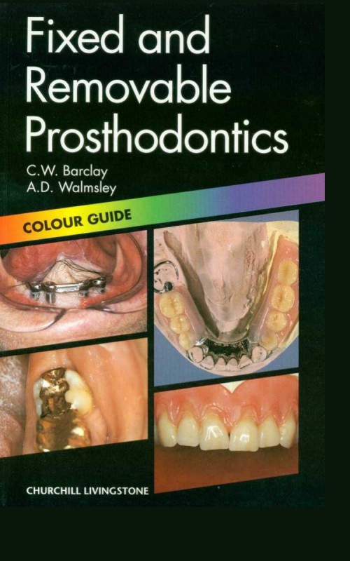
Fixed and Removable Prosthodontics 2nd Edition by Barclay, Damien Walmsley ISBN 044305813X 9780443058134


Original price was: $50.00.$25.00Current price is: $25.00.
Authors:C. W. Barclay; A. D. Walmsley , Series:Dentistry [340] , Tags:Medical; Dentistry; General , Author sort:Barclay, C. W. & Walmsley, A. D. , Ids:Google; 9780443058134 , Languages:Languages:eng , Published:Published:Sep 1998 , Publisher:Churchill Livingstone , Comments:Comments:A concise pictorial overview of fixed and removable prosthodontics, in the handy pocket-size Colour Guide format. Topics are presented in double-page spreads, with text on the left and illustrations on the right. Prosthetics is the area of dentistry which involves the fitting of crowns, bridges, implants and dentures to the patient, and it is one of the most important areas of day to day work for the average dentist. A wide variety of different cases are covered in the explanatory text which accompanies the high quality colour illustrations.Coverage of one of the most important areas of day-to-day restorative dental work in the proven Colour Guide format High quality colour illustrations of complete dentures, partial dentures, crowns, bridges, implants Concise explanatory text covers a wide variety of different cases
Fixed and Removable Prosthodontics 2nd Edition by C. W. Barclay, A. Damien Walmsley – Ebook PDF Instant Download/Delivery. 044305813X, 978-0443058134
Full download Fixed and Removable Prosthodontics 2nd Edition after payment

Product details:
ISBN 10: 044305813X
ISBN 13: 978-0443058134
Author: C. W. Barclay, A. Damien Walmsley
A concise pictorial overview of fixed and removable prosthodontics, in the handy pocket-size Colour Guide format. Topics are presented in double-page spreads, with text on the left and illustrations on the right. Prosthetics is the area of dentistry which involves the fitting of crowns, bridges, implants and dentures to the patient, and it is one of the most important areas of day to day work for the average dentist. A wide variety of different cases are covered in the explanatory text which accompaniesthe high quality colour illustrations.
- Coverage of one of the most important areas of day-to-day restorative dental work in the proven Colour Guide format
High quality colour illustrations of complete dentures, partial dentures, crowns, bridges, implants
- Concise explanatory text covers a wide variety of different cases
Fixed and Removable Prosthodontics 2nd Table of contents:
1. Soft Tissue Examination
-
Squamous Cell Carcinoma
- Aetiology and Pathology
- Diagnosis
- Management
- Figures:
- Fig. 1: Squamous cell carcinoma in the cheek.
- Fig. 2: Close-up of lesion.
- Fig. 3: Microscopic appearance of squamous cell carcinoma.
-
Denture-induced Hyperplasia
- Aetiology and Pathology
- Diagnosis
- Management
- Figures:
- Fig. 4: Denture hyperplasia of the upper ridge.
- Fig. 5: Denture hyperplasia of the lower ridge.
- Fig. 6: Causative denture in place.
-
Assorted Soft Tissue Disorders
- Pregnancy Epulis
- Figure: Fig. 7: Pregnancy epulis.
- Fibroepithelial Polyp
- Figure: Fig. 8: Fibroepithelial polyp.
- Wegener’s Granulomatosis
- Figure: Fig. 9: Wegener’s granulomatosis.
- Diagnosis and Management
- Pregnancy Epulis
-
Denture-induced Stomatitis
- Aetiology and Pathology
- Diagnosis
- Management
- Figures:
- Fig. 10: Denture stomatitis in the upper arch of a complete denture patient.
- Fig. 11: Denture stomatitis under a cobalt/chromium partial denture.
- Fig. 12: Smear from the denture surface showing fungal hyphae.
-
Cotton Wool Burn
- Aetiology and Pathology
- Diagnosis
- Management
- Figures:
- Fig. 13: Cotton wool burn on the lower lip.
- Fig. 14: Cotton wool burn in the labial sulcus.
- Fig. 15: Bonjela burn.
-
Leukaemia
- Aetiology and Pathology
- Diagnosis
- Management
- Figures:
- Fig. 16: Leukaemia—appearance of right cheek.
- Fig. 17: Leukaemia—appearance of the tongue.
- Fig. 18: Leukaemia—appearance of the forearm.
2. Hard Tissue Examination
-
Keratocyst
- Aetiology and Pathology
- Diagnosis
- Management
- Figures:
- Fig. 19: Keratocyst—extraoral view.
- Fig. 20: Keratocyst—external oblique view.
- Fig. 21: Keratocyst—panoramic view.
-
Torus Palatinus and Torus Mandibularis
- Aetiology and Pathology
- Diagnosis
- Management
- Figures:
- Fig. 22: Palatine torus.
- Fig. 23: Palatine torus.
- Fig. 24: Mandibular torus.
-
Severe Resorption
- Aetiology and Pathology
- Diagnosis
- Management
- Figures:
- Fig. 25: Severe mandibular atrophy—extraoral view.
- Fig. 26: Severe mandibular atrophy—lateral cephalometric view.
- Fig. 27: Severe mandibular atrophy—panoramic view.
-
Clinical Appearance of Severe Mandibular Resorption
- Aetiology and Pathology
- Diagnosis
- Management
- Figures:
- Fig. 28: Early mandibular resorption.
- Fig. 29: Mandibular resorption.
- Fig. 30: Prominent genial tubercles.
-
Periodontal Disease/Root Caries
- Aetiology and Pathology
- Diagnosis
- Management
- Figures:
- Fig. 31: Loss of periodontal attachment.
- Fig. 32: Radiographic appearance of periodontal bone loss.
- Fig. 33: Root caries.
-
Periodontal Acrylic Veneer
- Definition, Advantages, Disadvantages, Procedure
- Figures:
- Fig. 34: Severe gingival recession.
- Fig. 35: Periodontal acrylic veneer masking recession.
- Fig. 36: Periodontal acrylic veneer out of the mouth.
-
Root Resection
- Definition, Indications, Management
- Figures:
- Fig. 37: Root resection of the distal root of a mandibular first molar.
- Fig. 38: Root resection of the mesial root of a mandibular first molar.
- Fig. 39: Root resection of the distobuccal root of a maxillary first molar.
-
Tetracycline Staining
- Aetiology and Pathology
- Diagnosis
- Management
- Figures:
- Fig. 40: Severe tetracycline staining.
- Fig. 41: Tetracycline staining.
- Fig. 42: Tetracycline staining exacerbated by gingival recession.
-
Amelogenesis Imperfecta
- Aetiology and Pathology
- Diagnosis
- Management
- Figures:
- Fig. 43: Amelogenesis imperfecta.
- Fig. 44: Amelogenesis imperfecta.
- Fig. 45: Amelogenesis imperfecta.
3. Complete Dentures
-
Normal Anatomy
- Upper and Lower Denture Bearing Areas
- Figures:
- Fig. 46: Normal anatomy—upper edentulous arch.
- Fig. 47: Normal anatomy—lower edentulous arch.
- Fig. 48: Normal anatomy—anterior view of arches.
-
Impression Materials
- Types, Indications, and Properties
- Figures:
- Fig. 49: Impression materials—plaster of Paris.
- Fig. 50: Impression materials—zinc oxide/eugenol.
- Fig. 51: Impression materials—alginate.
-
Selective Compression Technique
- Aim and Procedure
- Figures:
- Fig. 52: Mucodisplacive impression with impression compound.
-
Mucostatic Impression Technique
- Aim and Procedure
- Figures:
- Fig. 54: Ridge being displaced.
- Fig. 55: Mucostatic using ZOE and Plaster of Paris.
-
Conventional Complete Dentures
- Definition, Indications, Advantages, Disadvantages, Procedure
- Figures:
- Fig. 56: Upper and lower wax rims sealed with bite registration paste and then separated.
- Fig. 57: (Figure missing)
- Fig. 58: Complete dentures inserted and the occlusion being refined.
-
Aesthetics of Complete Dentures
- Definition and Procedure
- Figures:
- Fig. 59: Aesthetics—relationship of anterior teeth to upper occlusal rim.
- Fig. 60: Arrangement of irregular teeth.
- Fig. 61: Gross irregularity of artificial teeth.
4. Copy Dentures
-
Techniques and Procedures
- Definition, Indications, Advantages, Disadvantages
- Figures:
- Fig. 62: Use of reversible hydrocolloid to copy dentures.
- Fig. 63: Murray/Woolland technique.
- Fig. 64: Replica record block technique.
-
Standard Copy Technique
- Definition, Indications, Advantages, Disadvantages, Procedure
- Figures:
- Fig. 65: Standard copy denture technique—template.
- Fig. 66: Templates placed on average movement articulator.
- Fig. 67: Set-up of copy denture templates.
-
Copy Denture for Severe Tooth Wear
- Definition, Indications, Advantages, Disadvantages
- Figures:
- Fig. 68: Severe tooth wear copy denture—presentation.
- Fig. 69: Copy denture after modification with original dentures.
- Fig. 70: Final clinical result.
People also search for Fixed and Removable Prosthodontics 2nd:
mclindent fixed and removable prosthodontics
difference between fixed and removable prosthodontics
university of manchester fixed and removable prosthodontics
periodontal considerations in preparation for fixed and removable prosthodontics
what is a removable prosthodontics
You may also like…
eBook PDF
Fundamentals of Fixed Prosthodontics 3rd Edition by Herbert Shillingburg 086715201X 9780867152012
eBook PDF
British Dental Journal Volume 228 Issue 11 June 2020 1st Edition by Damien Walmsley ISSN 14765373