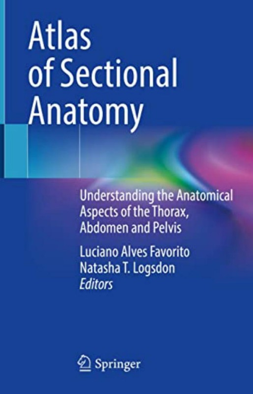
Atlas of Sectional Anatomy Understanding the Anatomical Aspects of the Thorax Abdomen and Pelvis 1st edition by Luciano Alves Favorito, Natasha Logsdon ISBN 3030916871 Â 978-3030916879
$50.00 Original price was: $50.00.$25.00Current price is: $25.00.
Authors:Luciano Alves Favorito, Natasha T. Logsdon , Series:Anatomy [297] , Author sort:Luciano Alves Favorito, Natasha T. Logsdon , Languages:Languages:eng , Publisher:Springer
Atlas of Sectional Anatomy Understanding the Anatomical Aspects of the Thorax, Abdomen & Pelvis 1st edition by Luciano Alves Favorito, Natasha Logsdon – Ebook PDF Instant Download/Delivery. 3030916871 978-3030916879
Full download Atlas of Sectional Anatomy Understanding the Anatomical Aspects of the Thorax, Abdomen & Pelvis 1st edition after payment
Product details:
ISBN 10: 3030916871
ISBN 13: 978-3030916879
Author: Luciano Alves Favorito, Natasha Logsdon
Sectional anatomy is a valuable resource for understanding and interpreting imaging exams, specially computed tomography (CT) and magnetic resonance imaging (MRI). Thus, health professionals should have a solid anatomical knowledge to properly evaluate such exams during clinical assessments of cardiac, thoracic, abdominal, proctologic, gynecological and urological diseases.
The chapters in this book describe the thoracic anatomy, the abdominal wall, retroperitoneal space, and the male and female pelvis. Sectional images of cadaveric material illustrate the thoracic and the abdominal cavities, kidney, ureter, prostate, penis and other male and female organs. The images and descriptions build familiarity with the anatomical traits and can be applied in the fields of urology, gynecology, proctology, radiology and surgery.
This work appeals to a wide range of readers, from health professionals to residents and students of different medical specialties.
Atlas of Sectional Anatomy Understanding the Anatomical Aspects of the Thorax, Abdomen & Pelvis 1st Table of contents:
Part I: Introduction to Sectional Anatomy
-
Basic Concepts of Sectional Anatomy
-
The Principles of Cross-Sectional Imaging: CT, MRI, and Ultrasound
-
Planes of Section: Sagittal, Coronal, and Axial
-
Understanding Slice Thickness and Resolution
-
Orientation of Images and Terminology
-
-
Imaging Modalities in Sectional Anatomy
-
Computed Tomography (CT) in Sectional Imaging
-
Magnetic Resonance Imaging (MRI) and Its Application
-
Advantages and Limitations of Each Imaging Technique
-
Comparison of Different Modalities in Anatomical Imaging
-
Part II: Thorax (Chest)
-
Overview of Thoracic Anatomy
-
The Bony Thorax: Ribs, Sternum, and Vertebrae
-
The Pleura and Lungs: Structures and Functions
-
The Heart and Pericardium
-
The Mediastinum and Its Subdivisions
-
-
Axial Sections of the Thorax
-
Cross-Sectional Views of the Chest
-
Detailed Imaging of the Lungs and Bronchi
-
Imaging the Heart: Chambers, Valves, and Major Vessels
-
The Thoracic Aorta and Its Branches
-
Imaging of the Diaphragm and Upper Abdominal Organs
-
-
Coronal and Sagittal Views of the Thorax
-
Coronal Sections of the Lungs and Heart
-
Sagittal Views of the Thoracic Organs
-
The Role of Sectional Imaging in Thoracic Surgery
-
Part III: Abdomen
-
Overview of Abdominal Anatomy
-
Abdominal Wall and Peritoneum
-
The Digestive System: Stomach, Small and Large Intestines
-
The Liver, Gallbladder, and Pancreas
-
The Kidneys, Ureters, and Bladder
-
The Spleen and Adrenal Glands
-
-
Axial Sections of the Abdomen
-
Cross-Sectional Imaging of the Abdominal Cavity
-
The Gastric and Intestinal Systems
-
Detailed Views of the Liver, Spleen, and Pancreas
-
Renal and Urological Structures in Sectional Views
-
-
Coronal and Sagittal Views of the Abdomen
-
Coronal Sectional Views of the Abdominal Organs
-
Sagittal Imaging of the Gastrointestinal Tract
-
Imaging of Vascular Structures: Abdominal Aorta, Iliac Arteries
-
The Role of Imaging in Abdominal Pathologies
-
Part IV: Pelvis
-
Overview of Pelvic Anatomy
-
Pelvic Bones and Pelvic Cavity
-
The Reproductive Organs: Male and Female Anatomy
-
The Urinary Bladder and Rectum
-
The Pelvic Vessels and Nerves
-
-
Axial Sections of the Pelvis
-
Cross-Sectional Views of the Pelvic Cavity
-
Imaging the Pelvic Organs: Bladder, Rectum, and Uterus
-
Detailed Imaging of Male and Female Reproductive Systems
-
The Pelvic Floor and Muscles
-
-
Coronal and Sagittal Views of the Pelvis
-
Coronal Sections of the Pelvis
-
Sagittal Views of the Pelvic Organs
-
Imaging the Pelvic Vasculature and Nerve Structures
-
Clinical Relevance of Pelvic Imaging in Surgery and Pathology
-
Part V: Clinical Applications of Sectional Anatomy
-
Anatomy in Clinical Diagnosis
-
The Role of Sectional Imaging in Diagnosis of Thoracic Pathologies
-
Abdominal Pathologies: Tumors, Trauma, and Infections
-
Pelvic Pathologies: Gynecological, Urological, and Proctological Disorders
-
Vascular Imaging and Its Clinical Applications
-
-
Sectional Anatomy in Surgical Planning
-
Preoperative Imaging for Thoracic Surgeries
-
Abdominal and Pelvic Surgery Planning Using Sectional Imaging
-
Image-Guided Procedures: Biopsies, Stent Placements, and Other Interventions
-
-
Advances in Sectional Imaging
-
3D Imaging and Its Role in Clinical Practice
-
Functional Imaging Techniques: PET, SPECT
-
Artificial Intelligence in Sectional Anatomy and Imaging
-
People also search for Atlas of Sectional Anatomy Understanding the Anatomical Aspects of the Thorax, Abdomen & Pelvis 1st:
what is the atlas in anatomy
atlas anatomy definition
what are the 5 branches of anatomy
which atlas is best for anatomy
atlas cross sectional anatomy

