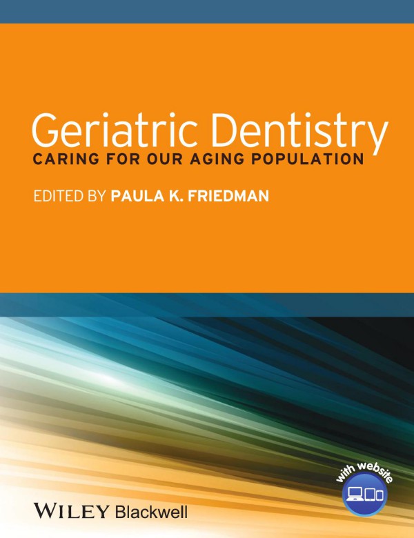
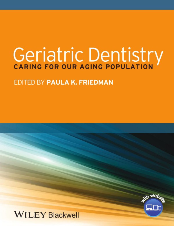
Geriatric Dentistry 1st Edition by Paula K Friedman ISBN 1118935349 9781118935347
$50.00 Original price was: $50.00.$25.00Current price is: $25.00.
Authors:Friedman, Paula K. , Author sort:Friedman, Paula K. , Published:Published:Jul 2014
Geriatric Dentistry 1st Edition by Paula K. Friedman – Ebook PDF Instant Download/Delivery. 1118935349, 9781118935347
Full download Geriatric Dentistry 1st Edition after payment
Product details:
ISBN 10: 1118935349
ISBN 13: 9781118935347
Author: Paula K. Friedman
Geriatric Dentistry: Caring for Our Aging Population provides general practitioners, dental students, and auxiliary members of the dental team with a comprehensive, practical guide to oral healthcare for the aging population.
Geriatric Dentistry 1st Table of contents:
Part 1 Underlying Principles of Aging
Chapter 1 Aging: Implications for the Oral Cavity
Aging of the US population
Ethnic diversity
Trends in oral health in older adults
Table 1.1 Trend of edentulism by racial/ethnic groups (1999–2008) (%) (weighted)*
Figure 1.1 Predicted rate of edentulism.
Oral health disparities in older adults
Functional status and oral health
Xerostomia, medications, and oral health
Case study 1
Cognitive function and oral health
Case study 2
Clinical and policy implications
DISCUSSION QUESTIONS
References
Chapter 2 Palliative Care Dentistry
Introduction
Mucositis and stomatitis
Figure 2.1 Palliative care team treating the patient and family.
Table 2.1 Impact of oral problems in palliative care
Box 2.1 Index for mucositis
National Cancer Institute common terminology criteria for mucositis severity*
Nutrition
Figure 2.2 Oral problems in palliative care.
Dysphagia
Nausea and vomiting
Delirium
Xerostomia and salivary gland hypofunction
Table 2.2 The pH of artificial saliva agents
Candidiasis
Figure 2.3 Formulation for sugar-free nystatin solution.
Figure 2.4 Nystatin popsicles.
Cancer and quality of life
Figure 2.5 Nystatin suspension plus lubricant.
Herpes and palliative care
Depression and the oral cavity
Caries prevention
Taste disorders
Treatment planning
Box 2.2 CARE anagram
Communication
Case study 1
Dental appraisal and approach
Case study 2
Figure 2.6 Radiograph of jaw (see Case study 2).
Dental management
Figure 2.7 Oral fistula resulting from radiotherapy (see Case study 2).
DISCUSSION QUESTIONS
References
Part 2 Clinical Practice
Chapter 3 Living Arrangements for the Elderly: Independent Living, Shared Housing, Board and Care Facilities, Assisted Living, Continuous Care Communities, and Nursing Homes
Introduction
Figure 3.1 Summary of the results of an American Association of Retired Persons’ survey on seniors’ choices for living arrangements.
Independent living/age in place
Congregate housing/retirement communities
Figure 3.2 (a,b) Theresa Dewar, aged 83, leaving her unassisted living facility to move to a nursing home with the help of Lois Halligan.
Assisted living
Table 3.1 Sample of week’s activities in an assisted living facility
Board and care home
Continuing care retirement communities
Figure 3.3 (a,b) Photographs from McMinnville, Oregon Continuing Care Retirement Community.
Nursing homes
Figure 3.4 Theresa Dewar now in her new Florida nursing home.
Table 3.2 Comparison and cost of different living options*
Long-term care insurance
Conclusion
Case study 1
Case study 2
DISCUSSION QUESTIONS
References
Chapter 4 Palmore’s Facts on Aging Quiz: Healthcare Providers’ Perceptions of Facts and Myths of Aging
Introduction
Tests of knowledge about older adults
Table 4.1 Palmore’s Facts on Aging Quiz*
Scores of recent dental graduates on the FAQ1
Table 4.2 Mean Facts on Aging Quiz scores by country of training
Table 4.3 Mean Facts on Aging Quiz scores by gender
Table 4.4 Changes in Facts on Aging Quiz scores with time by selected variables
What is the relationship between results of tests of knowledge about older adults and good care?
Summary
DISCUSSION QUESTIONS
References
Chapter 5 The Senior-Friendly Office
Case study
Mrs. Gonzalez
Case study questions
Introduction
Sensory impairments
Vision in older adults
Presbyopia
Cataracts
Figure 5.1 Example of vision affected by cataracts (a) as compared to normal (b).
Glaucoma
Figure 5.2 Example of vision affected by glaucoma (a) as compared to normal (b).
Diabetic retinopathy
Figure 5.3 Example of vision affected by diabetic retinopathy (a) as compared to normal (b).
Age-related macular degeneration
Figure 5.4 Example of vision affected by age-related macular degeneration (a) as compared to normal (b).
Hemianopia or hemianopsia
Figure 5.5 Example of vision affected by hemianopia or hemianopsia (a) as compared to normal (b).
Creating a functional senior-friendly office for patients with vision impairments
Box 5.1 Recommended strategies for patients with vision impairments
Figure 5.6 Examples of good contrast (left) versus not so good contrast (right).
Hearing loss
Presbycusis
Communication strategies
Mobility
Six steps to a safe wheelchair transfer
Step 1: Determine the patient’s needs
Step 2: Prepare the dental operatory
Step 3: Prepare the wheelchair
Step 4: Perform the two-person transfer (Fig. 5.7)
Step 5: Position the patient after the transfer
Step 6: Transfer from the dental chair to the wheelchair
Figure 5.7 The two-person transfer. (a) First clinician stands behind the patient. (b) Second clinician initiates the lift.
Osteoarthritis or degenerative joint disease
Managing mobility issues
Figure 5.8 An example of a bad waiting area design for older patients. This waiting area has deep chairs that are low to the ground. While there is good ambient light there is no task lighting for reading. The floors are highly reflective and will be slippery if they get wet. There is no place to put down any papers. The only small table has a plant on it. There is much room for improvement in this waiting area.
Figure 5.9 An example of a good waiting area design for older patients. Notice the overhead lighting and the task lighting. The chairs have arms and although cushioned for comfort but are not overstuffed and difficult to get up from. There are no throw rugs and there is emergency lighting should power be lost. One thing to notice is the glass table tops that have sharp corners. Either a bumper around the edge of the table or a rounded edge may be more friendly to older adults should someone bump into the table.
Management strategies for patients with mobility limitations
Electronic health records
Case study
Revisiting Mrs. Gonzalez
Conclusion
References
Part 3 Decision Making and Treatment Planning
Chapter 6 Geriatric Patient Assessment
Introduction
Case study 1
Communication status
Physical status
Figure 6.1 Percentage of persons with limitations in activities of daily living by age group: 2009.
Mobility status
Get Up and Go Gait Assessment
Mental status
Case study 2
The three-minute Mini-cog Assessment
Figure 6.2 Abnormal Clock Drawing Test (CDT) results from Mini-cog Assessment.
Nutritional status
Social support
Case study 3
Medical status
Case study 4 (Fig. 6.3)
Figure 6.3 Dental images from patient case demonstrating the characteristics of disease presentation in the elderly (see Case study 4 and text for more details).
Figure 6.4 Illustration of homeostenosis.
KEY POINTS OF THE MEDICAL CONSULTATION
Summary
DISCUSSION QUESTIONS (for Case Study 5, p. 69)
Case study 5
“Mrs. F.”
References
Chapter 7 Treatment Planning and Oral Rehabilitation for the Geriatric Dental Patient
Introduction
KEY POINTS
Diagnostic studies to facilitate planning and treatment
Planning for dental treatment in the older adult
Can the patient chew foods of the desired consistency?
Does the patient choke or have swallowing problems?
Can the patient manage a prosthesis?
Case study
Can the patient tolerate the prosthesis?
Can the caregiver(s) manage prosthesis?
Which teeth are most strategic for patient to maintain?
Teeth in occlusion
Teeth structure that can provide proprioception
At least one tooth on either side of each arch, preferably in same coronal plane
Teeth with periodontal support
Teeth requiring restoration of coronal structure
Maxillary and mandibular central or lateral incisor
Cuspids
Bicuspids, especially with opposing tooth contact
The lone standing molar tooth
Maxillary anterior roots that oppose mandibular anterior teeth
Can a patient function with a shortened dental arch?
What is the patient’s disease trajectory and can I maintain his or her oral status along this trajectory?
Treatment planning: issues and approaches
Global issues and approaches
Table 7.1 Common global issues and some possible approaches
Dental issues and approaches
Table 7.2 Common dental issues and possible approaches
Treatment planning: important goals
Important areas to be addressed for treatment goals and decisions
Implant restorations for the elderly
Figure 7.1 (a,b) Single implants used as root form anchors for porcelain fused to metal crowns (PFMs).
Figure 7.2 (a,b) Single implant placed on the upper right to retain and support a partial denture.
Figure 7.3 Multiple implants support an implant fixed bridge, upper right, and crowns. Note the anterior cantilever pontics at tooth sites no. 6 and no. 11, which are kept out of occlusion to minimize lateral forces.
Figure 7.4 (a,b) Two implants with a bar and clip attachment system.
Figure 7.5 (a,b) Two maxillary implants to retain a maxillary complete denture. Note that the full palate will provide tissue support and that the denture flanges have been constructed to help maximize conventional denture retention.
Figure 7.6 (a,b) Four implants in the maxillary arch and four implants in the mandibular arch. Implant overdentures were constructed that are totally implant supported with no tissue support. (c) Four implants in each arch with retentive ball attachments and gold caps used to retain and support maxillary and mandibular overdentures.
Treatment planning: important goals
Conclusion
DISCUSSION QUESTIONS
References
Chapter 8 Informed Consent for the Geriatric Dental Patient
Informed consent
Background
Informed consent for geriatric patients
Case study 1
Case study 1 questions
Case study 2
Case study 2 questions
Case study 3
Case study 3 questions
References
Chapter 9 Evidence-Based Decision Making in a Geriatric Practice
Introduction
KEYPOINT
The process
Sites for online EBDM tutorials
Sources of evidence
KEYPOINT
Sources for systematic reviews
Critical appraisal of the evidence
KEYPOINT
Table 9.1 Journals of interest to a geriatric practice sorted by impact factor*
The following questions would be most applicable for a SR
Implementation of an evidence-based geriatric dentistry practice
KEYPOINT
KEYPOINT
EBDM in practice
EBDM case study 1
Case study 1: The situation
Case study question
Communications containing the current guidelines/information statements
Cardiac
Reference no. 1
Orthopedics
Reference no. 2
Systematic review of studies relevant to post-dental procedure infective endocarditis
Reference no. 3
Objective
Search strategy, data collection, and analysis for this SR
Study inclusion criteria
The intervention
Outcomes of interest
Data collection and analysis
Results
Authors’ conclusions
Clinical implications
Position papers relevant to post-dental procedure joint infections
Reference no. 4
Methods
Results
Authors’ conclusions
Discussion
Reference no. 3
Discussion
EBDM case study 2
Case study 2 the situation
Case study question
Systematic review relevant to the management of “burning mouth syndrome”
Reference no. 1
Authors’ objective
Search strategy, data collection and analysis for this review
Study inclusion criteria
The intervention
Outcomes of interest
Data collection and analysis
Results
Authors’ conclusions
Position paper relevant to the management of BMS
Reference no. 2
Discussion
Conclusion
References
Part 4 Common Geriatric Oral Conditions and their Clinical Implications
Chapter 10 Root Caries
Introduction
Figure 10.1 Primary root caries under heavy plaque accumulation: teeth nos. 22–27.
Prevalence and risk factors
Caries risk assessment
Figure 10.2 Tooth no. 11 shows secondary caries apical to a root carious lesion previously restored with amalgam.
Pathologic factors versus protective factors
Diet
Genetic susceptibility
Saliva
Fluoride
Chlorhexidine
Silver diamine fluoride
Clinical decision making
Caries removal
Box 10.1 Clinical tip
Steps in treating root caries with partial caries removal
Restorative materials: amalgam, composite, glass ionomer
Box 10.2 Clinical tip
Atraumatic restorative treatment
Box 10.3 Future directions
Case study
Figure 10.3 Root caries are clinically detectable on most remaining teeth. The clinical crown of tooth no. 11 is completely missing due to caries. Arrow shows an example of root caries.
Figure 10.4 Radiographs taken to determine the extent of the carious lesions (see case study for details).
DISCUSSION QUESTIONS
References
Chapter 11 Periodontal Disease
Introduction
Figure 11.1 Dependency ratios for the USA, 2010–2050.
Box 11.1 Age-related risk factors of periodontal disease
Epidemiology of periodontal disease
Box 11.2 Myths about periodontal disease
The role of periodontal disease in oral health/overall health
Senescence of tissue
Identifying periodontal disease
Figure 11.2 Patient with inflamed gingiva and plaque and tartar (calculus) build-up.
Figure 11.3 Patient displaying inflamed gingiva with plaque and tartar (calculus) build-up
Figure 11.4 Patient displaying gingival recession and root exposure.
Table 11.1 Medications and symptoms as risk factors for periodontal disease
Box 11.3 Health issues associated with periodontal disease and older adults
Cancer, cancer therapy, and periodontal disease
Cognitive functioning
Dexterity/functional issues
Osteoporosis
Menopausal status
Implants and periodontal disease
Aspiration pneumonia
Diet and nutritional changes
Figure 11.5 Patient with root caries.
Psychologic considerations
DISCUSSION QUESTIONS
References
Further reading
Chapter 12 Endodontic Management of the Aging Patient
Introduction
The dental pulp
Figure 12.1 (a) Histologic section of the dental pulp complex of a young tooth (15years old). Note the dense odontoblast and cell-rich zones. (b) Histologic section of the dental pulp complex of a 59-year-old patient. Note the lesser number of odontoblasts present.
Figure 12.2 (a) Histologic section of the dental pulp complex of a young tooth (15years old). Note the highly vascularized tissue. (b) Histologic section of the dental pulp complex of a second 59-year-old patient. Note the lesser number of odontoblasts present. Also note the calcifications found in the pulp tissue.
Dentin and the odontoblasts
Figure 12.3 (a) A 9-year-old patient with typically young incisors. Note the size of the root canal systems and the incomplete root end development of the three teeth pictured. (b) A 44-year-old patient with restored maxillary central incisors. Note the great change in the size of the root canal spaces, partially attributed to the mesial and distal deposition of tertiary dentin due to composite placement. (c) Maxillary right lateral incisor and central incisor of a 73-year-old patient. Note the absence of restorations with no history of trauma. When compared to Fig. 12.8, the physiologic deposition of secondary dentin is evident.
Figure 12.4 Periapical radiographs of a premolar taken of a male patient at three times over a 40-year period. (a) Mandibular first premolar at age 33. The root canal system can be seem to the apex of the tooth. (b) The same tooth at age 52. (c) The same tooth at age 73 years. The same pattern is seen in a second patient at the same time frames (d, e, f).
Figure 12.5 (a) Low power photomicrograph of an unstained root canal space (ground section) of an upper central incisor typical of a 26–30-year-old individual. Note the size of the root canal space (Original mag. ×20). (b) Low power photomicrograph of an unstained root canal space (ground section) of an upper central incisor typical of a over 71-year-old individual. Secondary dentin fills the entire pulp chamber of the root canal space. Note that the secondary dentin formation is tubular as opposed to atubular dentin. Its formation is due to age and not to caries or placement of restorations.
Sensory mechanisms
Vascularity
Management considerations
Diagnosis
Tooth testing
Radiographs
Treatment methods
Figure 12.6 (a) Radiograph of a lower left first molar with little or any coronal structure remaining in a 67-year-old male patient. The patient’s dentist suggests the tooth’s removal and placement of a crown-restored implant. Note the thinness of the root canal systems, probably due to tertriary dentin formation. (b) Radiograph of the same tooth after endodontic therapy and placement of a crown.
Figure 12.7 (a) Radiograph of a lower right first molar in a 71-year-old male patient. Note the large carious lesion at the distal gingival margin below the restoration with successful root canal therapy in the second molar. (b) The caries has been removed and packed with amalgam and the tooth has a second root canal treatment. This case was seen by a periodontist who told the patient that it could not be retained and a crown restored implant would be a better treatment.
Endodontic therapy of aging patients
Appointments
Diagnosis
Medical history
Treatment plan
Endodontic treatment
Figure 12.8 (a,b) Radiographs of a 73-year-old male patient. Note the almost complete calcification of the root canal systems of the maxillary second premolar and first and second molars. The premolar became sensitive to percussion and biting (Tooth Sleuth®). Endodontic surgery was carried out with placement of a reverse fill amalgam rather than remove the crown.
Access openings and cleaning and shaping
Obturation
Retreatment
Vital pulp therapy
Regenerative endodontics
Periodontal considerations
Conclusion
Case study 1
Complaints
Case study questions: diagnosis and treatment planning
DISCUSSION QUESTIONS
References
Further reading
Chapter 13 Oral Mucosal Lesions
Introduction
Burning mouth syndrome
Etiology
Clinical presentation
Diagnosis
Treatment
Candidiasis (see also Chapter 2)
Etiology
Clinical presentation
Pseudomembraneous candidiasis (Fig. 13.1)
Figure 13.1 Pseudomembranous candidiasis.
Erythematous (atrophic) candidiasis (Fig. 13.2)
Figure 13.2 Erythematous candidiasis.
Figure 13.3 Denture stomatitis.
Figure 13.4 Angular chelitis.
Figure 13.5 Median rhomboid glossitis.
Hyperplastic candidiasis (candida leukoplakia) (Fig. 13.6)
Figure 13.6 Hyperplastic candidiasis.
Diagnosis
Treatment
Table 13.1 Topical antifungal agents
Table 13.2 Systemic antifungal agents
Epulis fissuratum (inflammatory fibrous hyperplasia, denture-induced fibrous hyperplasia, denture granuloma)
Etiology
Clinical presentation
Diagnosis
Treatment
Geographic tongue (benign migratory glossitis)
Etiology
Clinical appearance
Figure 13.7 Geographic tongue.
Diagnosis
Treatment
Hairy tongue
Etiology
Clinical presentation
Figure 13.8 Hairy tongue.
Treatment
Herpes simplex (recurrent)
Etiology
Clinical presentation
Figure 13.9 Recurrent intraoral herpes.
Diagnosis
Treatment
Table 13.3 Medications used to treat recurrent herpetic infections
Table 13.4 Topical analgesics
Herpes zoster (shingles)
Etiology
Clinical presentation
Diagnosis
Treatment
Irritation fibroma
Etiology
Clinical presentation
Figure 13.10 Irritation fibroma.
Diagnosis
Treatment
Leukoplakia
Etiology
Clinical presentation
Figure 13.11 Leukoplakia.
Diagnosis
Treatment
Lichen planus
Etiology
Clinical presentation
Figure 13.12 (a) Reticular lichen planus. (b) Erosive lichen planus.
Diagnosis
Treatment
Table 13.5 Medications (in order of increasing potency) for treatment of ulcerative lesions
Mucous membrane pemphigoid (cicatricial pemphigoid)
Etiology
Clinical presentation
Diagnosis
Treatment
Papillary hyperplasia
Etiology
Clinical presentation
Figure 13.13 Papillary hyperplasia.
Diagnosis
Treatment
Pemphigus
Etiology
Clinical presentation
Diagnosis
Treatment
Recurrent aphthous ulcers
Etiology
Clinical presentation
Figure 13.14 (a) Minor aphthous ulcers; (b) Major aphthous ulcers; (c) Herpetiform aphthous ulcers.
Diagnosis
Treatment
Traumatic ulcerations
Etiology
Clinical presentation
Diagnosis
Treatment
Varices (varix)
Etiology
Clinical presentation
Diagnosis
Treatment
Case study
Figure 13.15 (a) Angular chelitis; (b) poor oral hygiene with debris present; (c) denture stomatitis. See case study for further details.
References
Chapter 14 Xerostomia
Introduction
Role of saliva and functions of salivary components
Defining and recognizing xerostomia and SGH
Table 14.1 Subjective symptoms and clinical findings associated with xerostomia and/or salivary gland hypofunction
Figure 14.1 Dry, cracked (arrows) lips in an elderly patient with medication-induced xerostomia.
Figure 14.2 Dry, red and depapillated, fissured tongue.
Figure 14.3 Root caries, plaque accumulation and dry erythematous oral mucosa due to medication-induced dry mouth.
Figure 14.4 Erythematous, dry palatal mucosa (mucositis) with white plaques indicative of an opportunistic fungal infection caused by Candida albicans.
Prevalence
Effect of aging on salivary glands and flow rates
Etiology of xerostomia and/or SGH
Medication use
Table 14.2 Examples of frequently used medications causing xerostomia
Radiotherapy to head and neck
Systemic disorders
Treatment modalities
Preventing dental caries and mucosal diseases
Table 14.3 Antifungal agents for treatment of oral candidiasis
Interventions to stimulate salivary flow
Systemic saliva stimulants
Saliva substitutes
Table 14.4 Examples of saliva replacement products (salivary substitutes) commercially available
Future directions
Case study 1
Oral examination
Figure 14.5 (a,b) Posterior bitewing radiographs illustrating multiple primary and secondary caries lesions in a 64-year-old woman with dry mouth and hyposalivation.
Figure 14.6 Maxillary periapical radiograph illustrating multiple interproximal recurrent caries on anterior teeth in a 64-year-old woman with dry mouth and hyposalivation.
Figure 14.7 Mandibular periapical radiograph illustrating multiple recurrent caries lesions on anterior teeth in a 64-year-old woman with dry mouth and hyposalivation.
Case study questions
Case study 2
Figure 14.8 Panoramic radiograph demonstrating the status of dentition at the initial visit.
Figure 14.9 Mandibular periapical radiograph illustrating the anterior teeth after delivery of dentures and prior to radiotherapy for head and neck cancer.
Oral examination
Figure 14.10 Dry palatal mucosa with localized erythema and soreness secondary to radiation and medication-induced xerostomia.
Figure 14.11 Mandibular periapical radiograph illustrating gross caries and retained root tips of anterior teeth in a 59-year-old man with radiation and medication-induced xerostomia. Patient received radiation for treatment of SCCA of the tongue and throat a year ago. Note white plaques on the tongue that are indicative of candidiasis (arrow).
Figure 14.12 Root caries on the remaining mandibular teeth in a 59-year-old man who a year ago received radiation therapy for oral cancer. The teeth were caries free prior to radiotherapy.
Case study questions
Case study 3
Oral examination
Figure 14.13 Dry, pale mucosa in a patient with xerostomia and hyposalivation.
Figure 14.14 Gross coronal caries on mandibular anterior teeth in a patient with xerostomia and hyposalivation.
Figure 14.15 Multiple caries and heavily restored dentition in a patient with xerostomia and hyposalivation.
Case study questions
MULTIPLE CHOICE QUESTIONS
References
Chapter 15 Prosthetic Considerations for Frail and Functionally Dependent Older Adults
Introduction
The aging population
Decision making in prosthodontics
Case study 1
Functionally independent older adult
What are the impacts of the following medical problems on treatment?
Dental treatment
Treatment plan no. 1
Maxilla
Mandible
Comment
Treatment plan no. 2
Maxilla
Mandible
Comment
Treatment plan no. 3
Maxilla
Mandible
Comment
Maxilla
Mandible
Evaluation of treatment
Figure 15.1 Worn complete maxillary denture and worn mandibular teeth with ill fitting removable prosthesis (RPD) see Case study 1 for more details).
Figure 15.2 Orthopantomograph showing resorbed anterior maxilla and worn mandibular teeth (see Case study 1 for more details).
Figure 15.3 Periapical radiographs showing worn dentition and periapical lesion on no. 25 (see Case study 1 for more details).
Figure 15.4 (a–d) The restored dentition (see Case study 1 for more details).
Sociodemographic information
Health history
Tips and techniques for treating frail or functionally dependent elders
Time of the appointment
Length of the appointment
Patient positioning
Need for antibiotic prophylaxis
Use of local anesthetic with vasoconstrictor (epinephrine)
Level of cognitive impairment
Case study 2
Frail older adult
Case study questions
What is the impact of his medical problems on his dental treatment?
Hypertension
Diabetes
Stroke
Cataracts
Dental treatment
Treatment plan no. 1 (emergency care – pain and infection)
Comment
Treatment plan no. 2 (limited care – restoration of function)
Comment
Treatment plan no. 3 (comprehensive care)
Comment
Final treatment plan
Evaluation of treatment
Figure 15.5 Maxillary arch, showing the patient’s fixed partial denture, which is removable, and the extensive caries of the abutments (see Case study 2 for more details).
Figure 15.6 Mandibular arch, showing the loss of crowns on teeth nos. 27 and 28 (see Case study 2 for more details).
Figure 15.7 Orthopantomograph showing the remaining dentition (see Case study 2 for more details).
Figure 15.8 Shows the immediate maxillary complete denture and the immediate mandibular interim resin removable prosthesis (RPD) in occlusion which restored the patient’s esthetics and function (see Case study 2 for more details).
Figure 15.9 Shows the patient’s healed arches after a year. He has not had any more caries on the abutments and states that he is using the PreviDent® 5000 gel (see Case study 2 for more details).
Evaluations for prosthodontic rehabilitation
Systematic evaluation of the dentition
Designing removable partial dentures for frail and functionally dependent older adults
Figure 15.10 The terminal dentition flowchart for decision making.
Do the least possible harm by preserving the existing dentition
RPDS should be easy to insert and remove
Design of the RPD should be simple so that maintenance is easy
Design of the RPD should allow for potential failure of some of its units
Flexible dentures – a new approach to RPDs
Complete dentures
Overdentures
New or replacement complete dentures
Healthy tissues
Motivation to learn how to use dentures
A need for adequate neuromuscular skills
Treatment of patients with neuromuscular deficits
Conclusion
References
Chapter 16 Medical Complexities
Introduction
Prescription and natural drug use as the window to systemic health
Polypharmacy
Tips for maximizing optimal medication compliance
Systematic review of the medication list
Emergency drugs for emergency situations
Table 16.1 Polymedicine checklist
Drugs that suggest potential risk
Hypoglycemic drugs and insulin
Anticoagulants
Table 16.2 Diabetes and associated diseases: impact on oral health care
Bisphosphonates
Immunosuppressants, chemotherapeutics, and radiation
Antidepressants
Over-the-counter and natural medications
Conclusion: an interdisciplinary approach
Case study 1
Mr. Hernandez
Case study questions
Case study 2
Mrs. Andrews
Case study questions
Case study 3
Mr. Sullivan
Case study questions
Case study 4
Mrs. Tsang
Case study questions
Clinical examples
Clinical example 1
78-year-old man
Treatment plans to consider
Clinical example 2
A 67-year-old man
Suggested treatment
Phase 1: immediate needs
Phase 2: post 6 months CVA
Bibliography
Part 5 Care Delivery
Chapter 17 Delivery Systems
NURSING HOMES
Definitions
Assisted living facility
Intermediate care facility
Long-term care
Nursing home
Skilled nursing facility
Contracts and affiliation agreements
Record keeping
Medical charts
Patient information or ‘face” sheet (Fig. 17.1)
Advanced directives section
History and physical section
Progress notes
Figure 17.1 Patient information sheet.
Doctors’ orders section
Consult section (Fig. 17.2)
Laboratory result section
Dental chart
Treatment delivery options
On-site operatory
Figure 17.2 Consultation form.
Bedside
Table 17.1 Alert and oriented (A&O)
Multi-purpose room/beauty salon
Note
Patient referrals to outside resources
Billing
Summary
Box 17.1 Basic principles that can enhance the overall experience for provider and patient
Case study
Past medical history
Case study question
HOME VISITS
Preparation for the home visit
Figure 17.3 Algorithm for case study. MCD, Medicaid; MD, medical doctor; SNF, skilled nursing facility.
The visit
Record keeping
Billing/house/extended care facility call
MULTIPLE CHOICE QUESTIONS
References
Chapter 18 Portable Dentistry
Introduction
Radiographs
Suction
Figure 18.1 An over 500lb patient in an assisted living facility. (Aribex NOMAD-Pro® and DEXIS® images.)
Figure 18.2 A contagious patient in an isolation room setting. (Aribex NOMAD-Pro® and DEXIS® images.)
Portable and mobile carts
Figure 18.3 Ergonom-X® self-developing Dental Film.
Figure 18.4 Aseptico portable cart with suction, single canister, and single water bottle.
Portable carts
Figure 18.5 Mobile carts: DNTL Works Port-Op II (left); Forest Dental Cart model 5910 (right).
Mobile carts (Fig. 18.5)
Figure 18.6 Head lights.
Lighting
Patient chairs (Fig. 18.7a,b)
Figure 18.7 Patients can sit in their own wheelchair (a) or chair (b).
Lathe
Computer equipment
Wraps and props
Figure 18.8 Rainbow Wrap® (a) and Rainbow® Knee Belts (b).
Figure 18.9 Molt mouth gag.
Headrests
Figure 18.10 Rainbow® wrap and Jennings prop.
Figure 18.11 Isolite® 5-in-1.
Figure 18.12 Assistant serving as patient’s headrest.
Maintenance and repairs
Vans
Sedation
Figure 18.13 Nitrous oxide mask and Open Wide® mouth rest.
Level of care
Figure 18.14 At the very least, we aim for pain-free and infection-free treatment.
Figure 18.15 Portable hygiene kit with hygienists treating an Alzheimer’s patient in a nursing home.
Inventory control
Fees
Opportunity
Figure 18.16 Bed-bound patient having a denture fabricated and delivered at a facility.
Case study 1
Ella Mae
Figure 18.17 Ella Mae’s tooth no. 6 is extruded and acutely painful. She joked that she was very “long in the tooth.” (See Case study 1 for more details)
Figure 18.18 DEXIS® instant digital imaging with NOMAD® handheld portable X-ray unit and a laptop computer. (See Case study 1 for more details)
Figure 18.19 The tooth is anesthetized and extracted bedside, providing relief of pain, and restoring normal function to Ella Mae’s quality of life. (See Case study 1 for more details)
Case study 2
Marguerite
Figure 18.20 Two teeth supporting a bridge fracture, causing pain upon closure. (See Case study 2 for more details)
Figure 18.21 The teeth to be extracted are anesthetized. (See Case study 2 for more details)
Figure 18.22 The fractured teeth are extracted bedside. (See Case study 2 for more details)
Figure 18.23 The bridge and fracture teeth are removed. (See Case study 2 for more details)
DISCUSSION QUESTIONS
Bibliography
Appendix: Resources
ADSA
Aribex NOMAD Handheld X-ray
Aseptico
Colgate-Palmolive
Dental Elite
DentalEZ Group
866 DTEINFO
DEXIS X-Ray Systems
DOCS Education
DUX Dental
Ergonom-X Dental Film
Hu-Friedy
Isolite Systems
Lexi-Comp
Porter Instrument – Nitrous Oxide
Septodont
Special Care Dentistry Association
Specialized Care Co.
Ultralight Optics
Walter Lorentz Surgical/Biomet 3-i
Chapter 19 Promoting Oral Health Care in Long-Term Care Facilities
Introduction
Figure 19.1 An oral health promotion model with application to a variety of long-term care (LTC) settings. Created by Wener, Bertone, and Yakiwchuk, 2012
Assess strengths and challenges
Collaborate
Collaboration
Use personnel effectively
Table 19.1 Roles in long-term care (LTC) can extend far beyond clinical care
Apply best practices
Standards (health promotion ring 1)
Table 19.2 Terminology
Table 19.3 Regulatory requirements direct and shape practice
Oral health in LTC legislation
Oral care guidelines
Elders access to care legislation
Commitment (health promotion ring 2)
Table 19.4 Assessing your readiness to be an oral health champion
Getting to know the players
Collaborating and partnering for a common ground approach
Listening to learn and build trust
Figure 19.2 Training slide that helps to establish “common ground.” Created by Wener, Bertone, and Yakiwchuk, 2012.
Figure 19.3 Training slide that translates “invisible” oral disease into a visible wound to which caregivers can relate.
Figure 19.4 Training slide that changes the perception of daily mouth care from that of grooming to preventing infection.
Caregiver education and training (health promotion ring 3)
Venues and timing
Figure 19.5 Tailor content to participants needs based on their role in supporting oral health in long-term care. Photographs depict training using a preclinical dental laboratory and reusable product kits.
Content
Figure 19.6 Training slide that introduces caregivers to new products helpful for dependent older adults.
Strategies that work
Assessment and clinical care: health promotion ring 4
Figure 19.7 Training slide to emphasize the impact of effective daily mouth care for both residents and caregivers.
Table 19.5 Training lessons learned
Table 19.6 Oral assessments in long-term care (LTC)*
Timing
Table 19.7 Preventive oral health strategies for dependent older adults
Daily mouth care: health promotion ring 5
Individualized daily mouth care plans
Figure 19.8 Training slide that reinforces individualizing mouth care based on level of independence.
Daily mouth care tip 1
Daily mouth care tip 2
Daily mouth care tip 3
Daily mouth care strategies
Daily mouth care tip 4
Supporting front-line caregivers
Figure 19.9 Training slide that provides helpful strategies for individuals that exhibit care resistant behavior (CRB).
Figure 19.10 Poster providing the message that oral health is part of total health.
Case study
Mrs. Smith
Daily mouth care plan for Mrs. Smith
Conclusion
DISCUSSION QUESTIONS
References
Appendix 19.1 Resources for Promoting Oral Health in Long-Term Care*
Comprehensive oral care education and training programs and materials for caregivers
Other educational materials
Geriatric textbooks
Continuing education training program for oral health professionals
Documents for oral health promotion guidelines in LTC
Australia
Canada
UK
USA
Continuous care community examples
Appendix 19.2 An Example of Oral Health Promotion Guidelines for a Long-Term Care Facility
Purpose
Policy
Operational procedures
Education
Professional dental care
Appendix 19.3 University of Manitoba Oral Health Assessment Worksheet
Appendix 19.4 University of Manitoba Fact Sheet
Basic Mouth Care: Caring for Those With Natural Teeth
Appendix 19.5 University of Manitoba Fact Sheet: Basic Mouth Care – Caring For Those With Dentures/False Teeth/No Teeth
Chapter 20 Dental Professionals as Part of an Interdisciplinary Team
Introduction
THE ORAL HEALTH–OVERALL HEALTH RELATIONSHIP
Historical retrospective: focal infection theory of disease
Current understanding: the mouth–body connection
Oral and system conditions – interrelationships
Periodontal disease
Tooth loss and edentulism
Aspiration pneumonia
Peptic ulcer disease
Therapeutics and treatments affecting oral health, systemic health, or both
Table 20.1 Common oral side effects associated with drugs or drug classes
Drug-induced salivary hypofunction
Figure 20.1 Clinical manifestations of xerostomia. Rampant root caries and coronal caries resulting in unsupported and fractured enamel of mandibular anterior teeth. Note the dry, fissured tongue in the background.
Herbal supplements
Zinc-containing denture adhesives
Head and neck radiation therapy
Antiresorptive drugs and osteonecrosis of the jaw
Systemic diseases affecting oral health
Diagnostic uses of oral fluids
Case study 1
Ms. Joanne W.
Case study 1: resolution
Figure 20.2 The dentition of Ms. W. Note the severity of the gingival inflammation and the supperative fluids accumulating in the buccal vestibules, as well as the flaring teeth.
Figure 20.3 A close up of Ms. W.’s marginal gingiva and the purulent suppuration from the gingival sulci.
Figure 20.4 Ms. W.’s mouth after extractions, alveoloplasty, and suture placement.
Figure 20.5 Ms. W.’s extracted teeth. Observe the many granulomas clinging to several of the teeth roots.
INTERPROFESSIONAL CARE
Table 20.2 Potential interprofessional collaboration for dental professionals
Dental–medical collaboration
Dental–rehabilitation collaboration
Dental–nursing collaboration
Dental–pharmacy collaboration
Case study 2
Mr. John S.
Case study 2: resolution
Dental–dietician collaboration
Dental–social worker collaboration
Interprofessional geriatric dental care
Strategies for successful interprofessional consultations
Tips for optimizing the outcomes of a request for a medical consultation
Table 20.3 Dental professionals’ guide to interprofessional consultation
EXPANSION OF THE DENTAL WORKFORCE
Access to care for older adults
The impetus behind the initiative to expand the dental workforce
Table 20.4 Written consultation content and example
Table 20.5 Links and resources
Historical perspective
From planning to action: Minnesota
Dental workforce expansion status in other states
Table 20.6 Required content of a dental therapy CMA in Minnesota*
Table 20.7 Delegated duties of Minnesota dental therapists (DTs) and advanced dental therapists (ADTs)*
Case study 3
Ms. Louise F.
Case study 3: Resolution
Summary
DISCUSSION QUESTIONS
References
Part 6 Future Vision
Chapter 21 Planning for the Future
Introduction
The continuum of aging
Figure 21.1 Older population by age from 1900 and projected through 2050. Projections for 2010 through 2050 are from US Census Bureau (2008), Table 12. The source of the data for 1900–2000 is US Census Bureau (2002), Table 5. This table was compiled by the US Administration on Aging using the Census data noted.
Table 21.1 Prevalence of chronic diseases and disability or limitations by age group, 2006*
Health and social policies for older adults
“Medically necessary” oral health care
Table 21.2 Medicare coverage of dental services as specified in statute or by the Health Care Financing Administration*
Expanding the evidence base
Focus on the future: innovation to improve the oral health of vulnerable elders
Innovative models of care delivery
Integrated models of care
Redefining roles
Use of new technology
PACE program: interdisciplinary geriatric care in action
Figure 21.2 The PACE model of integrated and team-managed care for older patients. DME, durable medical equipment; OT/PT, occupational therapy/physical therapy.
Figure 21.3 Steps in clinical decision making for geriatric dental patients.
Advancing a policy agenda to improve the oral health of vulnerable elders
Case study 1
Heart transplantation for 77-year-old man
Case study questions
Case study 2
Mrs. Ellie King’s dentures
Past medical history
Dental history
Health behaviors
Case study questions
Case study 3
Mr. Ellis’s toothache
Case study questions
DISCUSSION QUESTIONS
Summary
References
Back Matter
Answer Section
Chapter 14: Multiple choice questions
Chapter 17: Multiple choice questions
Chapter 18: Discussion questions
Chapter 20: Discussion questions
People also search for Geriatric Dentistry 1st:
geriatric dentistry
geriatric dentistry near me
ubc geriatric dentistry
textbook of geriatric dentistry
textbook of geriatric dentistry pdf
You may also like…
eBook PDF
Bone Regulators and Osteoporosis Therapy 1st editon by Paula Stern ISBN 303057377X 978-3030573775

