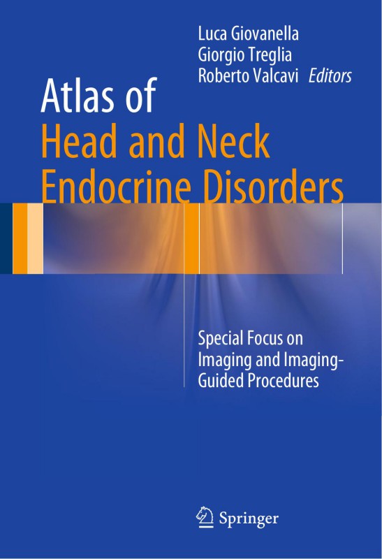
Atlas of Head and Neck Endocrine Disorders Special Focus on Imaging and Imaging Guided Procedures 1ts Edition by Luca Giovanella, Giorgio Treglia, Roberto Valcavi ISBN 3319222767 9783319222769
$50.00 Original price was: $50.00.$25.00Current price is: $25.00.
Authors:Unknown , Author sort:Unknown , Published:Published:Oct 2015
Atlas of Head and Neck Endocrine Disorders Special Focus on Imaging and Imaging Guided Procedures 1ts Edition by Luca Giovanella, Giorgio Treglia, Roberto Valcavi – Ebook PDF Instant Download/Delivery. 3319222767, 9783319222769
Full download Atlas of Head and Neck Endocrine Disorders Special Focus on Imaging and Imaging Guided Procedures 1ts Edition after payment
Product details:
ISBN 10: 3319222767
ISBN 13: 9783319222769
Author: Luca Giovanella, Giorgio Treglia, Roberto Valcavi
This atlas draws on a multidisciplinary approach to provide a comprehensive overview on endocrine disorders of the head and neck, with particular emphasis on the role of imaging and image-guided procedures. The first section discusses the basic characteristics of the imaging methods and other techniques used for evaluation and diagnosis. The remainder of the book focuses on application of these methods in thyroid, parathyroid, and other endocrine disorders of the head and neck. The coverage is wide ranging, encompassing Graves’ disease, toxic multinodular goiter, toxic adenoma, thyroiditis, non-toxic goiter, benign nodules, and the different forms of thyroid carcinoma, as well as parathyroid adenoma, hyperplasia, and carcinoma and paragangliomas. Informative, high-quality images are provided by international experts in endocrine disorders, including endocrinologists, pathologists, radiologists, nuclear medicine physicians, and surgeons, who also discuss sample cases and provide syntheses of the relevant scientific literature.
Atlas of Head and Neck Endocrine Disorders Special Focus on Imaging and Imaging Guided Procedures 1ts Table of contents:
Part I: Basic Characteristics of Imaging Methods and Other Techniques for Evaluation of Neck Endocri
1: Ultrasonography
1.1 Thyroid Ultrasonography
1.1.1 General Aspects
1.1.2 Normal Ultrasound Presentation of Thyroid Gland
1.1.3 Ultrasound Evaluation of Thyroid Lesions
1.1.4 The Dilemma of Small Non-palpable Thyroid Nodules Incidentally Discovered by US
1.1.5 Neck Ultrasonography in the Follow-Up of Thyroid Cancer Patients
1.1.6 Use of Ultrasound to Guide Fine-Needle Aspiration Cytology (FNAC) or Core Needle Biopsy (CN
1.1.6.1 Fine-Needle Aspiration Cytology (FNAC)
1.1.6.2 Core Needle Biopsy (CNB)
1.1.7 Thyroid Ultrasound Reports
1.2 Ultrasonography of Parathyroid Glands
References
2: Nuclear Medicine Techniques
2.1 Introduction
2.2 Thyroid Diseases: Radioactive Tracers and Nuclear Medicine Methods
2.2.1 Radiotracers Describing the Function of Follicular Cells
2.2.2 Radiotracers Mapping Cellular Proliferative Activity
2.2.3 Thyroid Diseases: Nuclear Medicine Imaging Methods
2.3 Parathyroid Nuclear Medicine Imaging
2.4 Nuclear Medicine Imaging in Head and Neck Neuroendocrine Tumors
References
3: Computed Tomography and Magnetic Resonance Imaging
3.1 CT and MRI in Thyroid Diseases
3.2 CT and MRI in Parathyroid Diseases
3.3 CT and MRI in Head and Neck Paragangliomas
References
4: Percutaneous Minimally Invasive Techniques
4.1 Introduction
4.2 Percutaneous Minimally Invasive Techniques
4.3 Summary of indications
References
5: Pathology
5.1 Thyroid
5.1.1 Graves’ Disease
5.1.2 Thyroid Autonomy
5.1.3 Thyroiditis
5.1.4 Goiter
5.1.5 Well-Differentiated Thyroid Carcinomas
5.1.6 Poorly Differentiated and Undifferentiated Thyroid Carcinomas
5.1.7 Medullary Thyroid Carcinoma
5.2 Parathyroid Glands and Related Diseases
5.3 Paragangliomas
References
Part II: Thyroid Diseases
6: Graves’ Disease
6.1 Introduction
6.2 Diagnosis
6.3 Therapy
References
7: Thyroid Autonomy
7.1 Introduction
7.2 Diagnosis
7.3 Therapy
References
8: Thyroiditis
8.1 Introduction
8.2 Painful Thyroiditis
8.2.1 Subacute Thyroiditis
8.2.2 Radiation-Induced Thyroiditis
8.2.3 Suppurative Thyroiditis
8.3 Painless Thyroiditis
8.3.1 Autoimmune Thyroiditis
8.3.2 Painless Sporadic Thyroiditis and Postpartum Thyroiditis
8.3.3 Drug-Induced Thyroiditis
8.3.4 Riedel’s Thyroiditis
8.4 Diagnosis and Follow-Up of Thyroiditis
References
9: Nontoxic Uninodular Goiter
9.1 Introduction
9.2 Position
9.3 Extracapsular Relationships
9.4 Internal Content
9.5 Shape
9.6 Echogenicity
9.7 Echotexture
9.8 Calcifications
9.9 Margins
9.10 Vascularity
9.11 Size
9.12 Elastosonography
9.13 Combination of US and Scintigraphic Findings
References
10: Nontoxic Multinodular Goiter
10.1 Introduction
10.2 Diagnosis and Management of Nontoxic Multinodular Goiter
References
11: Differentiated Thyroid Carcinoma
11.1 Introduction
11.2 Management of DTC
References
12: Medullary Thyroid Carcinoma
12.1 Diagnosis
12.2 Therapy and Follow-Up
References
13: Anaplastic Carcinoma and Other Tumors
13.1 Introduction
13.2 Anaplastic Thyroid Carcinoma
13.3 Lymphomas
13.4 Thyroid Metastases
References
Part III: Parathyroid Diseases
14: Primary Hyperparathyroidism
14.1 Introduction
14.2 Preoperative Localization Studies
14.2.1 Neck Ultrasound
14.2.2 Selective Venous Sampling
14.2.3 Planar Scintigraphy and SPECT/CT in Combination with Single or Dual Isotopes
14.2.4 PET/CT
References
Part IV: Other Endocrine Diseases of the Neck
15: Head and Neck Paragangliomas
15.1 Introduction
15.2 Imaging Methods in HNPGL
References
16: Other Neuroendocrine Tumors of Head and Neck Region
16.1 Introduction
16.2 Imaging for NETs of Head and Neck Region
People also search for Atlas of Head and Neck Endocrine Disorders Special Focus on Imaging and Imaging Guided Procedures 1ts:
atlas of head and neck endocrine
atlas of head and neck surgery
atlas in head and neck anatomy
atlas of endocrinology for hormone therapy
anatomy of head and neck pdf
You may also like…
eBook PDF
Head and Neck Imaging Case Review Series 4th Edition by David Yousem ISBN 1455776297 9781455776290

