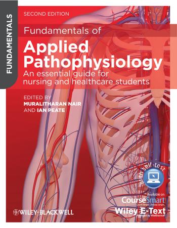
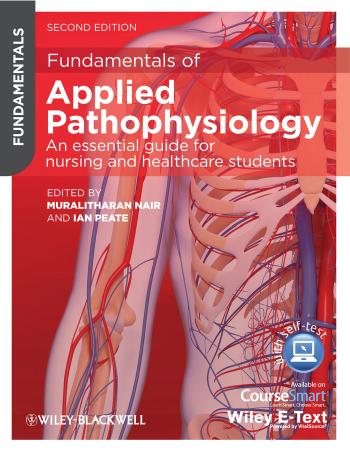
Fundamentals of Applied Pathophysiology 2nd edition by Muralitharan Nair Ian Peate 9781118502372 111850237X
$50.00 Original price was: $50.00.$25.00Current price is: $25.00.
Authors:Nair, Muralitharan, Peate, Ian , Series:Physiology [85] , Tags:Physiology , Author sort:Nair, Muralitharan, Peate, Ian , Languages:Languages:eng , Published:Published:Dec 2012 , Publisher:Wiley , Comments:Comments:All rights reserved. No part of this publication may be reproduced, stored in a retrieval system, or transmitted, in any form or by any means, electronic, mechanical, photocopying, recording or otherwise, except as permitted by the UK Copyright, Designs and Patents Act 1988, without the prior permission of the publisher
Fundamentals of Applied Pathophysiology 2nd edition by Muralitharan Nair Ian Peate- Ebook PDF Instant Download/Delivery.9781118502372,111850237X
Full download Fundamentals of Applied Pathophysiology 2nd edition after payment
Product details:
ISBN 10:111850237X
ISBN 13:9781118502372
Author:Muralitharan Nair Ian Peate
Fundamentals of Applied Pathophysiology is designed specifically for nursing and healthcare students, providing a straightforward, jargon-free, accessible introduction to pathophysiology. Highly visual and written specifically for students, the second edition of this best-selling textbook provides clear explanations of the anatomy of the human body, and the effects of disease or illness on normal physiology. To make study easier, the book includes learning outcomes, a range of activities to test learning, key words, end-of-chapter glossaries, and clinical case scenarios, and is supported by an online resource centre with further activities and exercises.
Key Features:
- Superb full colour illustrations, bringing this subject to life
- Full of extra features to help improve the learning process, including key words, test-your-knowledge, exercises, further reading and learning outcomes
- New case studies throughout to help you understand how to apply the knowledge in clinical practice
- Supported by an online resource centre at www.wiley.com/go/fundamentalsofappliedpathophysiology with fantastic extras for both lecturers and students, including an image bank, interactive multiple choice questions, true/false exercises, word-searches, glossary flash-cards, label-the diagram activities, and more!
Fundamentals of Applied Pathophysiology 2nd Table of contents:
1 Cell and body tissue physiology
Contents
Key words
Test your prior knowledge
Learning outcomes
Introduction
Anatomy of the cell
Figure 1.1 Simplified structure of a cell.
The cell membrane
Figure 1.2 The cell membrane.
Functions
Figure 1.3 Endocytosis.
Transport across the cell membrane
Figure 1.4 Osmosis.
Figure 1.5 Osmosis and movement of solute.
Cytoplasm
Role of cytoplasm
Nucleus
Figure 1.12 Types of cells.
Mitosis and meiosis
Mitosis
Prophase
Figure 1.6 Prophase.
Metaphase
Figure 1.7 Metaphase.
Anaphase
Figure 1.8 Anaphase.
Telophase
Figure 1.9 Telophase.
Cell cycle
Figure 1.10 Cell cycle.
Meiosis
Table 1.1 Stages of meiosis.
First meiotic stage
Prophase I
Metaphase I
Anaphase I
Telophase I
Second meiotic stage
Fusion of the gametes
The organelles
Endoplasmic reticulum
Figure 1.11 Endoplasmic reticulum (ER).
Golgi apparatus
Lysosomes
Peroxisomes
Mitochondria (single = mitochondrion)
Cytoskeleton
Microfilaments
Microtubules
Intermediate filaments
Centrioles, cilia and flagella
Centrioles
Cilia and flagella
Types of cells
Tissues
Epithelial tissue
Figure 1.13 Simple epithelium.
Figure 1.14 Stratified epithelium.
Figure 1.15 Epithelial cells classified according to shape.
Glandular epithelium
Connective tissue
Bone
Cartilage
Dense connective tissue
Loose connective tissue
Areolar tissue
Adipose tissue
Reticular connective tissue
Blood
Muscle tissue
Skeletal muscle
Cardiac muscle
Smooth muscle
Figure 1.16 Peristalsis.
Nervous tissue
Tissue repair
Conclusion
Test your knowledge
Activities
Fill in the blanks
Label the diagram
Word search
Glossary of terms
References
2 Cancer
Contents
Key words
Test your prior knowledge
Learning outcomes
Introduction
Biology of cancer
Figure 2.1 Apoptosis (Cell Migration Lab, University of Reading, https://www.reading.ac.uk/cellmigration/apoptosis.htm).
Figure 2.2 Molecular biology model (after King, 2000).
Figure 2.3 Clinical model (after King, 2000).
Causes of cancer
Genes
Environmental factors
Radiation
Smoking
Diet
Alcohol
Sexual and reproductive behaviour
Environmental pollution
Occupation
Hormones
Oral contraceptives
Male hormones
Viruses
Staging of cancers
Figure 2.4 Common sites and symptoms of cancer metastasis.
Signs and symptoms of cancer
Treatment of cancer
Drug therapy
Side effects
Radiation therapy
Immunotherapy
Surgical removal
Hormonal therapy
Photodynamic therapy
Gene therapy
Prevention of cancer
Examples of cancers
Acute lymphoblastic leukaemia
Case study
Treatment and prognosis
Lung cancer
Case study
Breast cancer
Figure 2.5 Diagram of breast showing lobules and ducts.
Figure 2.6 Symptoms of breast cancer.
Conclusion
Test your knowledge
Activities
Fill in the blanks
Label the diagram
Word search
Further resources
BMC Cancer
British Cancer Journal
Cancer Research UK
Cancer Symptoms
Inside Cancer
Glossary of terms
References
3 Inflammation, immune response and healing
Contents
Key words
Test your prior knowledge
Learning outcomes
Introduction
Infectious micro-organisms
Spread of infection
Droplet spread
Air currents (airborne transmission)
Aerosol transmission
Water transmission
Contact transmission
Soil
Inoculation
Faecal-oral route
Vector transmission
Contaminated intermediates
Case study
Types of infectious micro-organisms
Bacteria
Figure 3.1 Shapes of bacteria.
Cocci
Bacilli
Spiral
Bacterial reproduction
Figure 3.2 Bacterial reproduction – simple fission.
Viruses
Infection of host cells
Figure 3.3 Viral replication.
Fungi
Figure 3.4 Typical fungi shapes. (a) Cryptococcus neoformans, (b) Candida albicans, (c) Microsporum canis, (d) Histoplasma capsulatum, (e) Epidermophyton floccosum, (f) Trichophyton mentagrophytes, (g) Aspergillus fumigatus, (h) ringworm infection of the hair.
Protozoa
Rickettsiae, chlamydiae and mycoplasmas
Rickettsiae
Chlamydiae
Mycoplasmas
Helminths
Transmission
Case study
The immune system
Organs, cells and proteins of the immune system
The lymphatic system
Figure 3.5 The lymphatic system.
Lymphoid tissue
Figure 3.6 Lymph node.
Other lymphoid organs
Types of immunity
Non-specific immunity
Physical barriers
Mechanical barriers
Chemical barriers
Blood cells
Phagocytic cells
Figure 3.7 Phagocytosis.
Mediator cells
Specific immunity
Immune problems
Inflammatory response
Figure 3.8 The inflammatory process.
Summary of inflammation
Conclusion
Test your knowledge
Activities
Fill in the blanks
Label the diagram
Word search
Further resources
AVERT
CELLS alive!
National Resource for Infection Control (NRIC)
The Biology Project
The Department of Health (DH)
HIV tutorial (University of Utah)
Department of Health – E. coli outbreak
Glossary of terms
References
4 Shock
Contents
Key words
Test your prior knowledge
Learning outcomes
Introduction
Case study
Types of shock
Table 4.1 Types of shock and common causative factors (Docherty and Hall, 2002).
Hypovolaemic shock
Figure 4.1 Hypovolaemic shock.
Cardiogenic and obstructive shock
Distributive shock
Figure 4.2 Distributive shock.
Case study
Anaphylactic shock
Septic shock
Neurogenic (vasogenic) shock
Pathophysiology of shock
Table 4.2 A summary of the clinical presentation of different types of shock.
Stages of shock
Stage 1: Compensatory (non-progressive) stage of shock
Neural compensatory mechanisms
Hormonal compensatory mechanisms
Figure 4.3 Renin–angiotensin mechanism.
Chemical compensatory mechanisms
Stage 2: Progressive (decompensated) stage of shock
Stage 3: Irreversible (refractory) stage of shock
Table 4.3 Physiological changes that occur at each stage of shock (adapted from Sole et al., 2008).
Care of the patient in shock
Pharmacological management of shock
Table 4.4 Common drugs used to manage shock (adapted from Sole et al., 2008).
Table 4.5 Additional management of shock according to type.
Conclusion
Test your knowledge
Activities
Fill in the blanks
Word search
Further resources
Resuscitation Council (UK)
NHS Clinical Knowledge Summaries
Trauma and shock factsheet
Toxic shock syndrome
Food Allergy & Anaphylaxis Network (FAAN)
British Society for Allergy and Clinical Immunology
Glossary of terms
References
5 The heart and associated disorders
Contents
Key words
Test your prior knowledge
Learning outcomes
Introduction
Location of the heart
Figure 5.1 Location of the heart.
Structures of the heart
Figure 5.2 The cardiac muscle.
Chambers of the heart
Figure 5.3 The chambers and the valves of the heart.
Valves of the heart
Vessels of the heart
Figure 5.4 The vessels of the heart.
Table 5.1 Summary of the vessels and their functions.
Blood flow through the heart
Figure 5.5 Blood flow through the heart. The arrows indicate the direction of blood flow.
Conducting systems of the heart
Figure 5.6 The conducting system of the heart.
The SA node
The AV node
Bundle of His
Right and left bundle branches
Purkinje fibres
Nerve supply of the heart
Figure 5.7 The cardioregulatory centre.
Diseases of the heart
Learning outcomes
Case study
Myocardial infarction
Figure 5.8 Myocardial infarction.
Aetiology
Investigations
Pathophysiology
Signs and symptoms
Care and management
Pharmacological and non-pharmacological treatment
Heart failure/congestive heart failure
Case study
Aetiology
Pathophysiology
Investigations
Pathophysiology of right heart failure
Signs and symptoms of right heart failure
Pathophysiology of left heart failure
Left heart failure (backward effects)
Left heart failure (forward effects)
Signs and symptoms of left heart failure
Care and management
Pharmacological and non-pharmacological treatment
Cardiogenic shock
Aetiology
Investigations
Pathophysiology
Figure 5.9 Vicious cycle of cardiogenic shock.
Signs and symptoms
Care and management
Pharmacological interventions
Angina
Investigations
Pathophysiology
Figure 5.10 Blockage of the coronary arteries.
Types of angina
Signs and symptoms
Care and management
Non-pharmacological interventions
Pharmacological interventions
Conclusion
Test your knowledge
Activities
Fill in the blanks
Label the diagram
Word search
Further resources
Online nutritional information and facts
NHS Choices
BUPA
National Institute for Health and Clinical Excellence (NICE)
Department of Health – Policy, guidance and publications for NHS and social care professionals
Glossary of terms
References
6 The vascular system and associated disorders
Contents
Key words
Test your prior knowledge
Learning outcomes
Introduction
Figure 6.1 Blood flow.
Overview of blood vessels
Structure of the blood vessels
Figure 6.2 Structures of an artery and a vein.
Arteries
Figure 6.3 Elastic recoil of the aorta.
Figure 6.4 Capillaries.
Capillaries
Venules
Veins
Figure 6.5 Bicuspid valves of a vein.
Blood pressure
Factors that can affect blood pressure
Diseases of the blood vessels
Case study
Atherosclerosis/arteriosclerosis
Aetiology
Investigations
Pathophysiology of atherosclerosis
Figure 6.6 Atheroma build-up in an artery.
Table 6.1 Effects of atherosclerosis on different sites.
Signs and symptoms of atherosclerosis
Care and management
Pharmacological interventions for atherosclerosis
Hypertension
Note
Aetiology
Investigations
Common presenting symptoms
Pathophysiology
Primary hypertension
Secondary hypertension
Malignant hypertension
Isolated systolic hypertension
Non-pharmacological interventions
Pharmacological interventions
Aneurysm
Figure 6.7 (a) A dissecting aneurysm; (b) a fusiform aneurysm; and (c) a saccular aneurysm.
Aetiology
Investigations
Symptoms
Care and management
Figure 6.8 Dacron graft for an abdominal aortic aneurysm. Synthetic graft used for surgery. Reproduced with permission from Vascutek.
Pharmacological interventions
Case study
Peripheral vascular disease
Aetiology
Investigations
Arterial insufficiency
Pathophysiology
Table 6.2 Comparison between an arterial and venous insufficiency.
Signs and symptoms
Care and management
Figure 6.9 Dacron graft for peripheral vascular disease. Synthetic graft used for surgery. Reproduced with permission from Vascutek.
Pharmacological interventions
Venous insufficiency/varicose veins
Figure 6.10 Varicose veins.
Figure 6.11 Calf-muscle pump.
Veins affected with varicosity
Figure 6.12 Veins of the leg susceptible to varicosity.
Signs and symptoms
Complications
Figure 6.13 (a) Venous ulcer; (b) venous eczema; and (c) lipodermatosclerois.
Care and management
Deep vein thrombosis
Figure 6.14 Formation of thrombosis.
Aetiology
Pathophysiology
Figure 6.15 Formation of a platelet plug.
Figure 6.16 Formation of a clot.
Signs and symptoms
Care and management
Pharmacological interventions
Conclusion
Test your knowledge
Activities
Fill in the blanks
Label the diagram
Word search
Further resources
Patient UK
Scottish Intercollegiate Guidelines (SIGN)
National Institute for Health and Clinical Excellence (NICE)
Department of Health (DH)
British Hypertension Society
British Heart Foundation
Glossary of terms
References
7 The blood and associated disorders
Contents
Key words
Test your prior knowledge
Learning outcomes
Introduction
Figure 7.1 Components of blood.
Composition of blood
Figure 7.2 Cells of the blood.
Properties of blood
Functions of blood
Transportation
Regulation
Protection
Plasma
Water in plasma
Plasma proteins
Albumin
Globulins
Fibrinogen
Plasma electrolytes
Gases
Nutrients and waste products of metabolism
Formed elements of blood
Red blood cells
Figure 7.3 Red blood cell.
Haemoglobin
Figure 7.4 Haemoglobin molecule.
Formation of red blood cells
Figure 7.5 Formation of blood cells.
Transport of respiratory gases
Destruction of red blood cells
Figure 7.6 Haemolysis of red blood cells.
White blood cells
Neutrophils
Eosinophils
Basophils
Monocytes
Lymphocytes
Platelets
Haemostasis
Vasoconstriction
Platelet aggregation
Coagulation
Table 7.1 Blood clotting factors.
Blood groups
Figure 7.7 ABO blood groups.
Table 7.2 Blood groups.
Rhesus factor
Diseases of the blood
Learning outcomes
Case study
Anaemia
Microcytic anaemia
Iron deficiency anaemia
Aetiology
Investigations
Pathophysiology
Signs and symptoms
Care and management
Pharmacological interventions
Macrocytic anaemia
Aetiology
Folate deficiency
Aetiology
Symptoms
Vitamin B12 deficiency
Aetiology
Symptoms
Care and management of macrocytic anaemia
Normocytic anaemia
Aplastic anaemia
Aetiology
Pathophysiology
Symptoms
Care and management
Pharmacological interventions
Haemolytic anaemia
Aetiology
Pathophysiology
Signs and symptoms
Care and management
Sickle cell anaemia
Figure 7.8 Sickled red blood cell.
Pathophysiology
Figure 7.9 Sickle cell in microcirculation.
Symptoms
Care and management
Leukaemia
Case study
Aetiology
Pathophysiology
Symptoms
Care and management
Pharmacological and non-pharmacological interventions
Thrombocytopenia
Aetiology
Pathophysiology
Signs and symptoms
Care and management
Pharmacological and non-pharmacological interventions
Conclusion
Test your knowledge
Activities
Fill in the blanks
Label the diagram
Word search
Further resources
Contact a Family
National Institute for Health and Clinical Excellence (NICE) – Treatment for chronic myeloid leukaemia
National Institute for Health and Clinical Excellence (NICE) – Treatment of anaemia in patients with chronic kidney disease
Sheffield NHS
Biomedical Central
National Heart, Lung and Blood Institute
Glossary of terms
References
8 The renal system and associated disorders
Contents
Key words
People also search for Fundamentals of Applied Pathophysiology 2nd :
applied pathophysiology for the advanced practice nurse 2021
fundamentals of applied pathophysiology
gould’s pathophysiology 6th edition apa citation
applied pathophysiology chapter 2 quizlet
understanding pathophysiology 6th edition pdf free download
You may also like…
eBook PDF
Caring for Children and Families 1st Edition by Ian Peate, Lisa Whiting 0470019700 9780470019702
eBook PDF
MENS HEALTH The Practice Nurses Handbook 1st Edition by Ian Peate 0470035552 9780470035559

