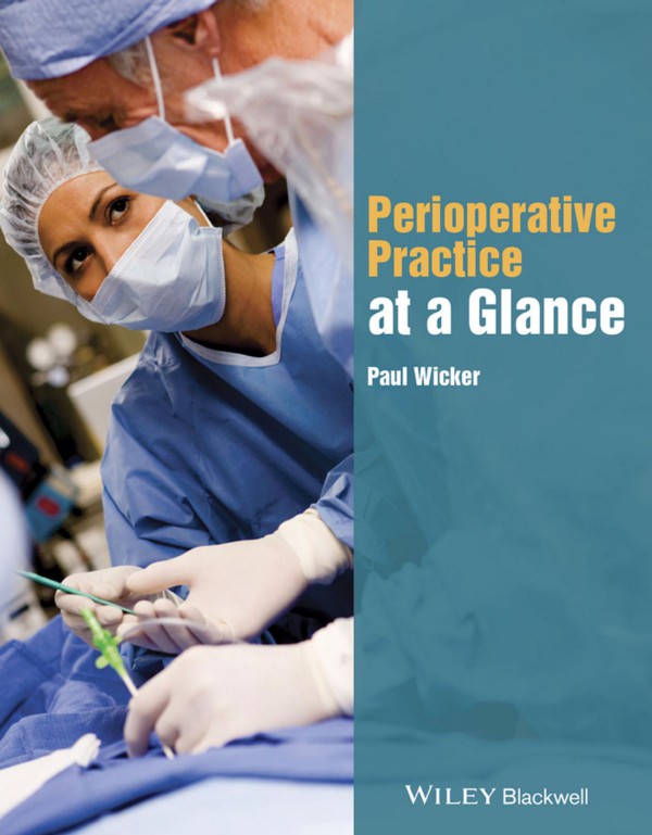
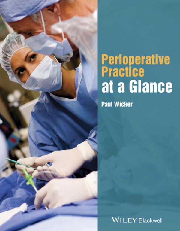
Perioperative Practice at a Glance 1st Edition by Paul Wicker 1118842154 9781118842157
$50.00 Original price was: $50.00.$25.00Current price is: $25.00.
Authors:Paul Wicker , Tags:Medical; Nursing; Critical & Intensive Care; Critical Care; (Nursing and Healthcare) , Author sort:Wicker, Paul , Ids:Goodreads; Google; 9781118842157 , Languages:Languages:eng , Published:Published:May 2015 , Publisher:Wiley-Blackwell , Comments:Comments:From the publishers of the market-leadingat a Glanceseries comes this new title on all aspects of caring for patients in the perioperative environment. From pre-operative care, through the anaesthetic and surgical phases to post-operation and recovery, this easy-to-read, quick-reference resource uses the uniqueat a Glanceformat to quickly convey need-to-know information in both images and text, allowing vital knowledge to be revised promptly and efficiently.Brings together all aspects of perioperative practice in one easy-to-read book Moves through the patient journey, providing support to perioperative practitioners in all aspects of their role Covers key information on perioperative emergencies Includes material on advanced skills to support Advanced Practitioners Each topic is covered in two pages, allowing for easy revision and reference This is a must-have resource for operating department practitioners and students, theatre nurses and nursing students, and trainee surgeons and anaesthetists.
Perioperative Practice at a Glance 1st Edition by Paul Wicker – Ebook PDF Instant Download/Delivery. 1118842154, 9781118842157
Full download Perioperative Practice at a Glance 1st Edition after payment
Product details:
ISBN 10: 1118842154
ISBN 13: 9781118842157
Author: Paul Wicker
A highly visual learning consolidator covering everything healthcare practitioners in the surgical and operating departments need to know about care of patients in the perioperative environment.
Perioperative Practice at a Glance 1st Table of contents:
Part 1 Introduction to perioperative practice
1 Preoperative patient preparation
Table 1.1 Normal physiological lab values
Figure 1.1 Checking patient’s wrist band on entry to the operating department
Figure 1.2 Checking patient’s care plan on entry into the operating department
Figure 1.3 Practitioner in reception checks the patient’s notes and confirms status
Preoperative visiting
Preoperative assessment
Preoperative investigations
Reducing postoperative complications
2 Theatre scrubs and personal protective equipment (PPE)
Figure 2.1 A practitioner prepared for cleaning the operating room and protected by personal protective equipment, including hat, gloves, mask, face shield and apron
Theatre scrubs
Headwear
Footwear
Surgical masks
Patient dress
Theatre scrubs outside theatre
3 Preventing the transmission of infection
Figure 3.1 Key elements of standard precautions to help prevent infection in patients
Operating room cleaning
Personal protection while cleaning
Cleaning equipment
Cleaning between cases
Cleaning at the end of cases
Risks to practitioners
Blood-borne viruses
Sharps and splash injuries
MRSA patients
4 Preparing and managing equipment
Figure 4.1 Equipment checklist This is an example of a potential checklist for equipment. The checklist in each operating theatre depends on the surgical speciality and the equipment that is present.
Initial equipment checks
Anaesthetic equipment
Surgical equipment
5 Perioperative patient care
Figure 5.1 Sample of patient care plan
Patient outcomes
Care pathways
6 Surgical Safety Checklist – Part 1
Figure 6.1 Countries where the Safe Surgery Checklist (SSCL) was piloted, resulting in lower incidences of surgery-related deaths and complications
Figure 6.2 Undertaking the SSCL
Figure 6.3 Example of a Safe Surgery Checklist
The Surgical Safety Checklist (Figure 6.3)
Introducing the checklist into the operating department
Adjusting the checklist
7 Surgical Safety Checklist – Part 2
Figure 7.1 Surgical Safety Checklist This document can be updated and amended to conform to hospital rules and regulations within the NHS
SIGN IN
TIME OUT
SIGN OUT
8 Legal and professional accountability
Figure 8.1 Legal and professional accountability
Figure 8.2 The anaesthetic practitioner checks the patients details and discusses the actions to be taken during anaesthesia
Modes of accountability
Legal issues
Defences against negligence
Assault and consent
Hospital requirements
Accountability and professional practice
9 Interprofessional teamworking
Figure 9.1 Better Training Better Care This poster refers to a pilot project entitled ‘Better Training Better Care’, which is being supported by Health Education England. The pilot projects involve using cadaveric workshops to train surgeons and perioperative practitioners, and also to increase collaboration between all the professions in perioperative care
Interprofessional conflicts
Part 2 Anaesthesia
10 Preparing anaesthetic equipment
Figure 10.1 Basic anaesthetic machine, for use in the anaesthetic room
Figure 10.2 Anaesthetic machine, ECG monitor, capnograph, ventilator etc., used during surgery
Figure 10.3 Guedal (oral) airways These devices may be used in association with a face mask or postoperatively until the patient wakes up
Figure 10.4 ET tubes, laryngoscope, oral airways, laryngeal mask airways (LMA)
Figure 10.5 Cuffed tracheostomy tube
Airway tubes
Consumable items
Monitoring devices
Medical gas cylinders
Fluid warmers
11 Checking the anaesthetic machine
Box 11.1 Checking anaesthetic equipment
Figure 11.1 Checklist for anaesthetic equipment
Basic steps for checking an anaesthetic machine
12 Anatomy and physiology of the respiratory and cardiovascular systems
Figure 12.1 Respiratory anatomy
Figure 12.2 Gas exchange in the alveoli
Figure 12.3 Cardiovascular anatomy (external)
Figure 12.4 Cardiovascular anatomy (internal)
Figure 12.5 Cardiovascular blood flow
Figure 12.6 ECG
Respiratory system
Mechanics of breathing
Physiology of gas exchange
Cardiovascular system
The ECG
The coronary circulation
13 Anaesthetic drugs
Figure 13.1 Action of inhalational agents
Figure 13.2 Neuromuscular junction
Figure 13.3 Action of muscle relaxants
Figure 13.4 Action of opioids
Induction agents
Inhalation agents
Muscle relaxants
Analgesics
14 Perioperative fluid management
Figure 14.1 Compartmental distribution of total body water in a 70 kg male
Figure 14.2 Osmosis Osmosis is the movement of fluid across a membrane into an area where there is a high concentration of solutes. Osmotic pressure is pressure that is applied to the chamber with the highest ratio of solutes, in order to prevent fluid moving from the chamber with the lowest level of solutes to the chamber with the highest number of solutes.
Figure 14.3 Crystalloids Samples of crystalloid solutions
Figure 14.4 Colloids Samples of colloid solutions
Figure 14.5 Intravenous cannula inserted in hand ready for anaesthetic drugs or intravenous fluids
Effects of crystalloids
Hartmann’s solution
Normal saline (NS)
5% dextrose
Hypertonic saline (2%, 3% and 7.5%)
Effects of colloids
Blood and blood byproducts
15 Monitoring the patient
Figure 15.1 CARESCAPE Monitor B850 The CARESCAPE Monitor B850 can be used for patients both preoperatively and postoperatively. The monitor is a high-acuity monitor that can help practitioners manage their patients by providing a dependable level of data continuity and integration across care areas
Electrocardiogram (ECG)
Pulse oximeters
Temperature probes
Vapour analyser
Capnography
Non-invasive blood pressure (NIBP) measurement
16 General anaesthesia
Figure 16.1 Anaesthetic drugs cupboard in anaesthetic room
Figure 16.2 Anaesthetic drugs prepared for anaesthetic, including Propofol, muscle relaxants and analgesics
Figure 16.3 Patient anaesthetised in the anaesthetic room
Signs and stages of anaesthesia (Guedel’s signs)
Induction, maintenance and extubation
Administration of anaesthetic agents
Total intravenous anaesthesia (TIVA)
Complications of general anaesthesia
17 Local anaesthesia
Figure 17.1 Local anaesthetic being injected prior to insertion of an intravenous cannula
Figure 17.2 Local anaesthetic injected into a knee
Figure 17.3 EMLA
Clinical use of local anaesthetics
Topical anaesthetics
Infiltrative local anaesthetics
18 Regional anaesthesia
Figure 18.1 Vertebral column anatomy
Figure 18.2 Vertebral column anatomy detail
Figure 18.3 Caudal epidural block
Figure 18.4 Epidural injection
Anatomy of the spine
Effects of local anaesthetics
Spinal technique
Epidural technique
Intravenous regional techniques
Part 3 Surgery
19 Roles of the circulating and scrub team
Figure 19.1 The circulating and scrub team
Figure 19.2 Practitioners cleaning operating room following surgery and preparing for the next patient’s arrival
Circulating practitioners
Scrub practitioners
Before surgery
During surgery
After surgery
20 Basic surgical instruments
Figure 20.1 An array of surgical instruments including: needle holders, retractors, scissors, tissues forceps, curettes, towel clips, bone holders, suction devices etc.
Figure 20.2 Instrument trays being set up for gynaecological surgery
Types of instruments
Cutting and dissecting
Grasping and holding
Clamping and occluding
Exposing and retracting
Suturing and stapling
Suctioning and aspirating
Microinstrumentation
Powered surgical instruments
21 Surgical scrubbing
Figure 21.1 Scrub procedures
Figure 21.2 Using scrub solution to wash hands and arms
Figure 21.3 Rinsing hands and arms at end of scrubbing
Figure 21.4 Drying hands and arms carefully to prevent contamination
Figure 21.5 Donning gloves and gown ready for surgery
Surgical scrub procedure
Side effects of surgical scrubbing
22 Surgical positioning
Figure 22.1 Patient positioning
Equipment requirements for positioning
Surgical positions
23 Maintaining the sterile field
Figure 23.1 Sterile and unsterile members of the team and the sterile field
Sterile procedures
Preparing a sterile tray
Surgical draping
24 Sterilisation and disinfection
Figure 24.1 Procedures for disinfection and sterilisation
Figure 24.2 Sterile instruments which are stored safely within two coverings to keep them sterile.
Methods of sterilisation
Physical agents
Chemical agents
Mechanical agents
25 Swab and instrument counts
Figure 25.1 Practitioners completing the swab board following counting of swabs and instruments Swab boards vary between hospitals although practitioners are always trained how to complete the boards according to hospital guidelines, rules and regulations.
Figure 25.2 Counting instruments following surgery, using an instrument count sheet
Rationale behind the swab and instrument count
Principles of the swab and instrument count
Checking swabs and other items
Checking instruments
26 Working with electrosurgery
Figure 26.1 Monopolar electrosurgery
Figure 26.2 Bipolar electrosurgery
Figure 26.3 Surgical effects of electrosurgery
Figure 26.4 Electrosurgery used to cut tissues The tip of the pencil is very thin which allows careful cutting of tissues and avoiding damage to other tissues.
How does electrosurgery work?
The three surgical effects of electrosurgery (Figure 26.3)
Monopolar electrosurgery (Figure 26.1)
Bipolar electrosurgery (Figure 26.2)
The return electrode
Safety checks
Hazards of electrosurgery
Conclusion
27 Tourniquet management
Figure 27.1 Attaching the velband
Figure 27.2 Wrapping the tourniquet
Figure 27.3 Fixing the tourniquet in place, and securing it
Figure 27.4 Applying an Eschmarch bandage
Figure 27.5 Applying the Rys-Davies Exsanguinator to remove blood from the arm. Following this the machine is switched on and the tourniquet inflates
Figure 27.6 Applying an Eschmarch bandage
Figure 27.7 Tourniquet machine
Applying the tourniquet
Pneumatic tourniquets
Side effects
28 Wounds and dressings
Figure 28.1 Applying Opsite © dressing to small wounds that have been sutured
Figure 28.2 Covering an open wound with melolin dressings
Figure 28.3 Wrapping soffban © wool around dressings to hold them in place
Figure 28.4 Applying a crepe bandage to the open leg wound to secure all dressings
Modes of healing
Types of dressings
Bandages
Complications
Part 4 Recovery
29 Introducing the recovery room
Figure 29.1 Patient being looked after in recovery by a recovery practitioner
Caring for the postoperative patient
Patients who have undergone spinal anaesthesia
Patients who have undergone general anaesthesia
Discharge from the recovery room
30 Patient handover
Figure 30.1 Patient handover in the recovery room following surgery
Admission into the recovery unit
Initial assessment
Documentation
Discharge
31 Postoperative patient care – Part 1
Figure 31.1 Entering recovery
Figure 31.2 Patient feeling unwell
Figure 31.3 Checking the patient’s notes and surgical procedure
Box 31.1 Respiratory status
Box 31.2 Cardiovascular status
Respiratory status (Box 31.1)
Cardiovascular status (Box 31.2)
32 Postoperative patient care – Part 2
Figure 32.1 Example of a postoperative patient care plan
Temperature status
General patient care
33 Monitoring in recovery
Figure 33.1 Checking the patient’s temperature postoperatively to check for any problems related to hypothermia or hyperthermia
Figure 33.2 Complex selection of monitoring devices used to care for patients safely.
Respiratory monitoring
Cardiovascular monitoring
Neuromuscular monitoring
Psychological monitoring
Temperature monitoring
Pain monitoring
Monitoring nausea and vomiting
Fluid monitoring
Urine output and voiding
Drainage and bleeding
34 Maintaining the airway
Figure 34.1 Oral airway and face mask
Figure 34.2 Tracheostomy
Figure 34.3 Nasopharyngeal airway
Figure 34.4 Endotracheal tube
Physiology of the airway
Circulation
Monitoring of the airway
Postoperative airway complications
Discharge criteria
35 Common postoperative problems
Table 35.1 Postoperative complications The prevalence of postoperative complications in a research study into postoperative patients who had undergone cardiac surgery (Lobo et al. 2008), which showed that the patient population had an overall major complication rate of 38.3% and 90-day mortality rate of 20.3%
Allergy
Haemorrhage
Septic shock
Intravascular catheters
Urinary complications
Wound dehiscence
Other issues
36 Managing postoperative pain
Figure 36.1 A pain rating scale
Figure 36.2 A PCAM® syringe pump which monitors the patients use of pain killers. The patient administers the pain killer by pressing the button attached to the black cable
Concept of pain
Effects of pain
Pain physiology
Managing pain
Systemic opioids
Nonsteroidal anti-inflammatory drugs (NSAIDS)
Regional techniques
Non-pharmacologic techniques
37 Managing postoperative nausea and vomiting
Figure 37.1 Physiology of vomiting
Figure 37.2 Act of vomiting
Physiology of vomiting
Risk factors
Prevention of vomiting
Anti-emetic therapy
Conclusion
Part 5 Perioperative emergencies
38 Caring for the critically ill
Box 38.1 Principal recommendations for caring for the critically ill
Figure 38.1 A recovery practitioner caring for a high-risk patient following major surgery
Critical risk areas
Improvements in patient care
High-risk patients in recovery
Conclusion
39 Airway problems
Figure 39.1 Insertion of oral airway The airway is first inserted upside down, and then reversed to insert into the trachea
Figure 39.2 Ventilation using a mask and bag Two practitioners make ventilation more effective and easier to control
Assessing the airway
Airway control
Tracheostomy
40 Rapid sequence induction
Box 40.1 Sedation
Box 40.2 Neuromuscular blocking (NMB) agents
Figure 40.1 Patient undergoing rapid sequence induction The anaesthetist is inserting the endotracheal tube while the practitioner is applying cricoid pressure to prevent gastric re ux from the stomach into the lungs
Preparation (T(time) − 10 minutes)
Preoxygenation (T − 5 minutes)
Pretreatment (T − 3 minutes)
Paralysis with induction (zero)
Placement of endotracheal tube (T + 30 seconds)
Postintubation management
41 Bleeding problems
Figure 41.1 The management of critical bleeding in surgical patients
Managing rapid blood loss in surgery
Managing rapid blood loss in recovery
42 Malignant hyperthermia
Box 42.1 EMHG guidelines: Recognising an MH crisis
Box 42.2 EMHG guidelines: Managing an MH crisis
Diagnosis
Symptoms
Prevention
Expected duration
Treatment (Box 42.2)
43 Cardiovascular problems
Figure 43.1a Cardiovascular system
Figure 43.1b Ischaemic heart attack
Figure 43.2 Replacement of blocked coronary arteries using blood vessels from arms or legs
Preoperative evaluation
Hypertension
Arrhythmias
Aortic stenosis
Congestive heart failure
44 Electrosurgical burns
Figure 44.1 Diathermy burns Forearm and hand – 50 days after the surgery
Figure 44.2 Diathermy burns Atrophy and contracture of the forearm and hand
Figure 44.3 An electrosurgical burn This 38-year-old patient underwent a circumcision. Instead of using bipolar electrosurgery, the surgeon used monopolar electrosurgery. As the current returned up the penis towards the return electrode, the vessels in the penis became coagulated, leading to necrosis. After two weeks the penis was amputated. This event occurred about 9 years ago. Bipolar electrosurgery would have prevented this from occurring, as the current would only have passed through the tissues held between the tines of the bipolar forceps
Thermal burns
Explosion and fire
45 Venous thromboembolism
Figure 45.1 Action of intermittent pneumatic compression (IPC) on blood vessels
Figure 45.2 Action of IPC on blood and lymphatic supplies
Physiology of DVT
Treatment options
Mechanical prophylaxis
Intermittent compression devices (ICD)
Precautions
46 Latex allergy
Figure 46.1 Some of the tissues affected following an allergic reaction The early phase of the allergic reaction typically occurs within minutes, or even seconds, following allergen exposure and is also commonly referred to as the immediate allergic reaction or Type I allergic reaction. The reaction is caused by the release of histamine and mast cell granule proteins by a process called degranulation, as well as the production of leukotrienes, prostaglandins and cytokines, by mast cells following the crosslinking of allergenspecific IgE molecules bound to mast cell FcεRI receptors. These mediators affect nerve cells, causing itching, smooth muscle cells causing contraction (leading to the airway narrowing seen in allergic asthma), goblet cells causing mucus production, and endothelial cells causing vasodilatation and oedema
Reducing problems with latex
Dangers of latex
Treatment for severe allergic reaction
Managing the perioperative environment
Postoperative management
Part 6 Advanced surgical practice
47 Assisting the surgeon
Figure 47.1 A patient undergoing wound closure following a fasciotomy due to trauma damage to his leg The surgical care practitioner is assisting the surgeon by attaching loops to the skin to help close the wound. The wound will not close completely because of the expanded leg muscles
Surgical First Assistant
Roles and responsibilities
Surgical Care Practitioner
Roles and responsibilities
48 Shaving, marking, prepping and draping
Figure 48.1 Shaving the patient prior to surgery
Figure 48.2 Disposal of swabs and instruments following prepping of skin
Figure 48.3 Common draping for major abdominal cases
Figure 48.4 Draping the patient’s leg using a drape with a central hole for the leg to go through
Figure 48.5 Draping the patient by covering their top, bottom and side – here the leg is being wrapped by a drape
Figure 48.6 Marking the patient
Shaving
Skin marking
Skin preparation
Draping
49 Retraction of tissues
Figure 49.1 (a) Deavers retractors, (b) Langenbecks retractors, (c) rake retractor, (d) Balfour self-retaining retractor and (e) Travers self-retaining retractor
Figure 49.2 (a) Transanal surgical retractor, for hip surgery, (b) table-mounted Omni-flex® retractor for hip surgery
Figure 49.3 Retraction of tissues using a self-retaining retractor and a Hohmann’s retractor
Figure 49.4 Curved Travers retractor
Figure 49.5 Gelpi self-retaining retractor
Holding retractors
Managing retractors
Hand-held retractors
Self-retaining retractors
Table-mounted retractors
50 Suture techniques and materials
Figure 50.1 Methods for releasing clips attached to suture materials
Figure 50.2 Examples of suture techniques
Figure 50.3 Suturing a damaged blood vessel and ligating other blood vessels in close proximity
Assisting the surgeon
Suture techniques
Suture materials
51 Haemostatic techniques
Figure 51.1 Coagulation of blood The coagulation of blood is dependent on the actions of enzymes and activity by blood cells. The end result, following the activation of coagulation by thrombin, is the formation of a blood clot that acts as a plug to stop further bleeding. During the healing phase, as the vessel repairs itself, the clot starts dissolving in a phase known as fibrinolysis During this phase the clots dissolve, releasing fibrin fragments.
Figure 51.2 Use of diathermy to stop bleeding
Assisting with haemostasis
Minor bleeding
Major bleeding
Haemostatic agents
52 Laparoscopic surgery
Figure 52.1 Laparoscopic surgery – placement of equipment for optimum viewing
Figure 52.2 Laparoscopic surgery in gynaecology
Figure 52.3 Laparoscopic cholecystectomy
Figure 52.4 Laparoscopic colorectal surgery
Before surgery
During surgery
Training and development
53 Orthopaedic surgery
Figure 53.1 Arthroscopy
Figure 53.2 Hip replacement
Figure 53.3 Knee-replacement surgery
Figure 53.4 Tibial surgery requiring insertion of nail
Specific factors in orthopaedic surgery
Arthroscopic knee surgery
Elective joint replacement
Trauma orthopaedics
54 Cardiac surgery
Figure 54.1 Aortic valve replacement
Figure 54.2 Mechanical bicuspid aortic valve
Figure 54.3 Aortic valve from a pig (porcine bioprosthesis
Role of the surgeon’s assistant
55 Things to do after surgery
Figure 55.1 Surgeon and assistant shaking hands after successfully completing surgery
Back Matter
References and further reading
1 Preoperative patient preparation
References
Further reading
Websites
Videos
2 Theatre scrubs and personal protective equipment
References
Further reading
Websites
Videos
3 Preventing the transmission of infection
References
Further reading
Websites
Videos
4 Preparing and managing equipment
References
Further reading
Websites
Videos
5 Perioperative patient care
References
Further reading
Websites
Videos
6–7 Surgical safety checklist
References
Further reading
Websites
Videos
8 Legal and professional accountability
References
Further reading
Websites
Videos
9 Interprofessional teamworking
References
Further reading
Websites
Videos
10 Preparing anaesthetic equipment
References
Further reading
Websites
Videos
11 Checking the anaesthetic machine
References
Further reading
Websites
Videos
12 Anatomy and physiology of the respiratory and cardiovascular systems
References
Further reading
Websites
Videos
13 Anaesthetic drugs
References
Further reading
Websites
Videos
14 Perioperative fluid management
References
Further reading
Websites
Videos
15 Monitoring the patient
References
Further reading
Websites
Videos
16 General anaesthesia
References
Further reading
Websites
Videos
17 Local anaesthesia
References
Further reading
Websites
Videos
18 Regional anaesthesia
References
Further reading
Websites
Videos
19 Roles of the circulating and scrub team
References
Further reading
Websites
Videos
20 Basic surgical instruments
References
Further reading
Websites
Videos
21 Surgical scrubbing
References
Further reading
Websites
Videos
22 Surgical positioning
References
Further reading
Websites
Videos
23 Maintaining the sterile field
References
Further reading
Websites
Videos
24 Sterilisation and disinfection
References
Further reading
Websites
Videos
25 Swab and instrument counts
References
Further reading
Websites
Videos
26 Working with electrosurgery
References
Further reading
Websites
Videos
27 Tourniquet management
References
Further reading
Websites
Videos
28 Wounds and dressings
References
Further reading
Websites
Videos
29 Introducing the recovery room
References
Further reading
Websites
Videos
30 Patient handover
References
Further reading
Websites
Videos
31 Postoperative patient care – Part 1
References
Further reading
Websites
Videos
32 Postoperative patient care – Part 2
References
Further reading
Websites
Videos
33 Monitoring in recovery
References
Further reading
Websites
Videos
34 Maintaining the airway
References
Further reading
Websites
Videos
35 Common postoperative problems
References
Further reading
Web sites
Videos
36 Managing postoperative pain
References
Further reading
Websites
Videos
37 Managing postoperative nausea and vomiting
References
Further reading
Websites
Videos
38 Caring for the critically Ill
References
Further reading
Websites
Videos
39 Airway problems
References
Further reading
Websites
Videos
40 Rapid sequence induction
References
Further reading
Websites
Videos
41 Bleeding problems
References
Further reading
Websites
Videos
42 Malignant hyperthermia
References
Further reading
Websites
Videos
43 Cardiovascular problems
References
Further reading
Website
Videos
44 Electrosurgical burns
References
Further reading
Websites
Videos
45 Venous thromboembolism
References
Further reading
Websites
Videos
46 Latex allergy
References
Further reading
Websites
Videos
47 Assisting the surgeon
References
Further reading
Websites
Videos
48 Shaving, marking, prepping and draping
References
Further reading
Websites
Videos
49 Retraction of tissues
References
Further reading
Websites
Videos
50 Suture techniques and materials
References
Further reading
Websites
Videos
51 Haemostatic techniques
References
Further reading
Websites
Videos
52 Laparoscopic surgery
References
Further reading
Websites
Videos
53 Orthopaedic surgery
References
Further reading
Websites
Videos
54 Cardiac surgery
References
Further reading
Websites
Videos
55 Things to do after surgery
References
Further reading
Websites
People also search for Perioperative Practice at a Glance 1st:
perioperative practice questions
rr perioperative area
ucla perioperative medicine
perioperative ati template
You may also like…
eBook PDF
Dental Materials at a Glance 2nd edition by Anthony von Fraunhofer 9781118684580 111868458
eBook PDF
Pre Clinical Dental Skills at a Glance 1st edition by James Field 9781118766620 1118766628

