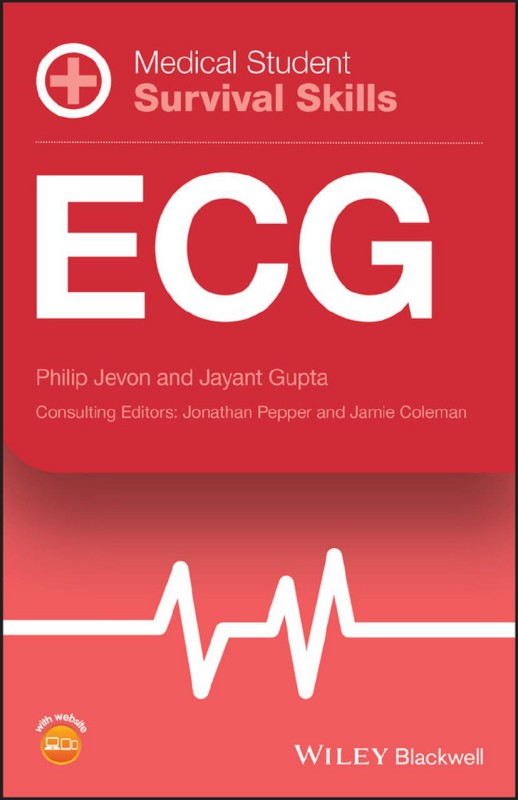
Medical Student Survival Skill ECG 1st Edition by Philip Jevon, Jayant Gupta 1118818172 9781118818176
$50.00 Original price was: $50.00.$25.00Current price is: $25.00.
Authors:Philip Jevon; Jayant Gupta , Series:Medicine [158] , Author sort:Jevon, Philip & Gupta, Jayant , Languages:Languages:eng , Published:Published:Apr 2019 , Publisher:Wiley Blackwell
Medical Student Survival Skill ECG 1st Edition by Philip Jevon, Jayant Gupta – Ebook PDF Instant Download/Delivery. 1118818172, 9781118818176
Full download Medical Student Survival Skill ECG 1st Edition after payment
Product details:
ISBN 10: 1118818172
ISBN 13: 9781118818176
Author: Philip Jevon, Jayant Gupta
Medical students encounter many challenges on their path to success, from managing their time, applying theory to practice, and passing exams. The Medical Student Survival Skills series helps medical students navigate core subjects of the curriculum, providing accessible, short reference guides for OSCE preparation and hospital placements. These guides are the perfect tool for achieving clinical success.
Medical Student Survival Skills: ECG is an indispensable resource for students new to ECG interpretation and cardiac arrhythmia recognition and treatment. Integrating essential clinical knowledge with practical OSCE advice, this portable guide provides concise and user-friendly coverage of all aspects of ECG monitoring, including atrial and ventricular fibrillation, myocardial infarction, and 12 lead ECG interpretation. Easy-to-find information, plentiful illustrations, OSCE checklists and expert discussions of actual ECG trace examples help medical students and junior doctors quickly get up to speed with ECG interpretation skills.
Medical Student Survival Skill ECG 1st Table of contents:
1 Introduction to ECG monitoring
Conduction system of the heart (figure 1.1)
PQRST complex (figure 1.2)
Sinus rhythm (figure 1.3)
2 Principles of ECG monitoring
Indications
ECG monitoring: Three ECG cable system (figure 2.1)
ECG monitoring: Five ECG cable system (figure 2.2)
ECG monitoring: Trouble shooting
Mechanisms of cardiac arrhythmias
3 Six stage approach to ECG interpretation
Electrical activity present?
QRS rate?
QRS rhythm: Regular or irregular?
QRS width: Normal or broad?
P present?
Relationship between P and QRS?
4 Sinus tachycardia
Causes
Identifying ECG features
Effects on patient
Treatment
5 Sinus bradycardia
Causes
Identifying ECG features
Effects on patient
Treatment
6 Sinus arrhythmia
Identifying ECG features
Effects on patient
Treatment
7 Atrial ectopic beats
Causes
Identifying ECG features
Effects on patient
Treatment
8 Atrial tachycardia
Causes
Identifying ECG features
Effects on patient
Treatment
9 Atrial flutter
Causes
Identifying ECG features
Effects on patient
Treatment
10 Atrial fibrillation
Causes
Identifying ECG features
Effects on patient
Treatment
11 AV junctional ectopics
Causes
Identifying ECG features
Effects on patient
Treatment
12 AV junctional escape rhythm
Causes
Identifying ECG features
Effects on patient
Treatment
13 Junctional tachycardia
Causes
Identifying ECG Features
Effects on patient
Treatment
14 Ventricular ectopics
Ventricular ectopic terminology
Causes
Identifying ECG features
Effects on patient
Treatment
15 Idioventricular rhythm
Causes
Identifying ECG features
Effects on patient
Treatment
16 Ventricular tachycardia
Causes
Identifying ECG features
Fusion and capture beats
Effects on patient
Treatment
17 Torsades de pointes
Causes
Identifying ECG features
Effects on patient
Treatment
18 First degree AV block
Causes
Identifying ECG features
Effects on patient
Treatment
19 Second degree AV block Mobitz type I (Wenckebach phenomenon)
Causes
Identifying ECG features
Effects on patient
Treatment
20 Second degree AV block Mobitz type II
Causes
Identifying ECG features
Effects on patient
Treatment
21 Third degree (complete) AV block
Causes
Identifying ECG features
Effects on patient
Treatment
22 Ventricular fibrillation
Causes
Identifying ECG features
Effects on patient
Treatment
23 Ventricular standstill
Causes
Identifying ECG features
Effects on patient
Treatment
24 Asystole
Causes
Identifying ECG features
Effects on patient
Treatment
25 Recording a 12 lead ECG
Common indications
Procedure
Locating the intercostal spaces for the chest leads
Alternative chest lead placements
Labelling the 12 lead ECG
Standardisation
26 What the standard 12 lead ECG records
Limb leads
Chest leads
Limb and chest leads and their relation to the surface of the heart
Configuration of the ECG waveform
27 Interpretation of a 12 lead ECG
P waves
PR interval
QRS rate
QRS rhythm
QRS complexes
Q waves
T waves
ST segment
QT interval
U waves
Association between the P waves and QRS complexes
Cardiac axis (figure )
Previous ECGs
28 ECG changes associated with myocardial infarction
Within minutes of myocardial infarction
Hours to days following myocardial infarction
Days to weeks following myocardial infarction
Localising the myocardial infarction from the ECG
Reciprocal changes
Diagnosing myocardial infarction in the presence of left bundle branch block
ECG examples of myocardial infarction
29 ECG changes associated with myocardial ischaemia
ST segment depression
T wave changes
30 ECG changes associated with bundle branch block
Left bundle branch block (figure 30.1)
Right bundle branch block (figure 30.2)
Left anterior fascicular block
Left posterior fascicular block
Bifascicular block (figure 30.3)
Trifascicular block (figure 30.4)
31 Wolff–Parkinson–White syndrome
Identifying ECG features
Classification
Complications
Treatment
Appendix A: Resuscitation council (UK) bradycardia algorithm
Appendix B: Resuscitation council (UK) tachycardia algorithm
Appendix C: Resuscitation council (UK) advanced life support (ALS) algorithm
Appendix D: Vagal manoeuvres
Appendix E: Synchronised electrical cardioversion
Synchronisation with the R wave
Shock energies
Procedure
Appendix F: External (transcutaneous) pacing
Indications
Advantages
Appendix G: Procedure for transcutaneous pacing
Appendix H: Definitions
People also search for Medical Student Survival Skill ECG 1st:
7 second ecg
6 second ecg skillstat
6 second ecg strip skillstat
6 second ecg strip simulator skillstat

